
Erythroplakia for oral pathology 10 mark acc to shafers
erythroplakia oral cavity red lesion
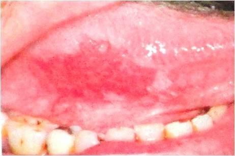
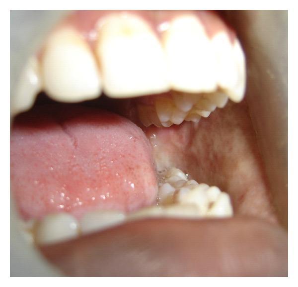
This clinical photograph displays a high-magnification view of the oral cavity, specifically the lateral border of the tongue. The primary finding is a well-demarcated, bright red, erythematous patch known as erythroplakia. The lesion exhibits a soft, velvety texture and lacks the keratinized surface characteristic of leukoplakia. This atrophic appearance results from epithelial thinning, which makes the underlying microvasculature more visible. Adjacent to the tongue, mandibular teeth with visible dental restorations are present. From a clinical perspective, erythroplakia is a significant precancerous condition, as it is histologically associated with severe epithelial dysplasia, carcinoma in situ, or invasive squamous cell carcinoma in approximately 90% of cases. The image serves as a key diagnostic example for oral pathology and oncology, emphasizing the importance of identifying non-ulcerated red lesions that cannot be attributed to other inflammatory or traumatic causes.
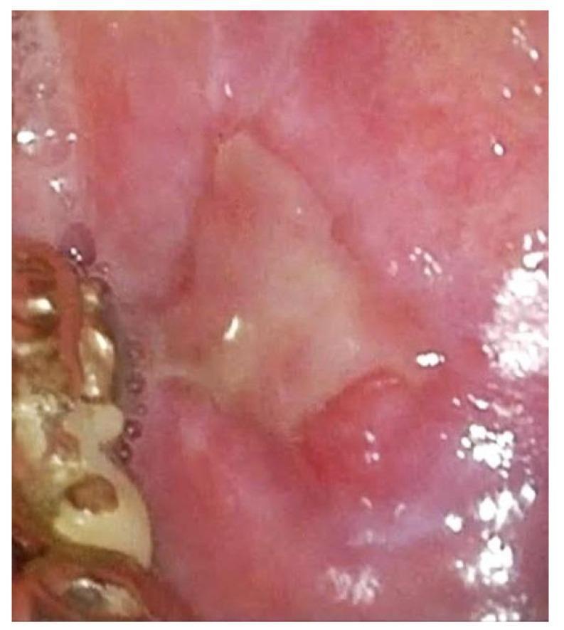
This clinical photograph, captured via white light imaging (WLI) oral endoscopy, demonstrates an ulcerated erythroplasic lesion on the oral mucosa. The central portion of the lesion features an irregular, yellowish-white fibrinous base characteristic of ulceration, surrounded by a distinct area of intense red discoloration (erythroplakia). The adjacent peripheral mucosa exhibits diffuse erythema, suggesting associated inflammation or increased vascularity. On the left side of the frame, orthodontic hardware or a dental appliance is visible, providing anatomical context within the oral cavity. This image is used to illustrate premalignant or malignant mucosal changes, such as squamous cell carcinoma or severe dysplasia, highlighting the importance of recognizing combined red and white (erythroleukoplakia) patterns during clinical examination.
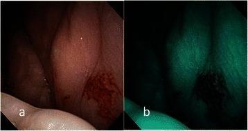
This composite diagnostic image displays an endoscopic examination of oral erythroplakia on the left buccal mucosa. Figure (a) utilizes conventional broadband white light (BWL), revealing a well-demarcated, flat, bright red plaque with a velvety and slightly glistening surface texture. Figure (b) shows the same lesion under Narrow-Band Imaging (NBI), which enhances the visualization of the microvascular architecture. The NBI view demonstrates Type IV intraepithelial papillary capillary loop (IPCL) destruction, characterized by irregular angiogenesis, loss of normal capillary loop structure, and a disorganized vascular arrangement. This microvascular pattern is a significant clinical indicator for advanced pathology. The clinical significance of this finding is demonstrated by the corresponding pathological diagnosis of invasive squamous cell carcinoma, highlighting the role of NBI in identifying high-risk premalignant and malignant lesions during oral cavity screening.
Erythroplakia - Oral Pathology (10 Marks, Shafer's Style)
Definition
Synonyms / Related Terms
- Erythroplasia of Queyrat (when on the glans penis)
- Speckled erythroplakia (erythroleukoplakia) - mixed red and white lesion; the red component carries the highest malignant risk
Epidemiology
-
Prevalence: 0.02% to 0.83% (varies by geographic region; higher in South and Southeast Asia due to tobacco/areca nut habits)
-
The least common of all oral potentially malignant disorders (PMDs), but the most dangerous
-
Age: mainly middle-aged individuals (40-70 years)
-
Sex: Males > Females (2:1), likely due to higher tobacco/alcohol exposure
-
Particularly described in chutta smokers (reverse cigar smokers) of India
-
Fitzpatrick's Dermatology, Vol. 1-2, p. 1908
Etiology and Risk Factors
- Tobacco - in all forms (cigarettes, bidis, cigars, smokeless/chewing tobacco, reverse smoking)
- Alcohol - acts synergistically with tobacco; causes mucosal dehydration and allows carcinogen penetration
- Areca nut / betel quid - contains arecoline and other carcinogens
- Poor oral hygiene
- Nutritional deficiencies (iron, vitamins A, B, C)
- Chronic sun exposure (for lip lesions)
- HPV (high-risk types 16, 18) - implicated in some cases
- Genetic factors: High mutation rate of the p53 tumor suppressor gene has been demonstrated in erythroplakic lesions, reflecting their high malignant potential
Clinical Features
| Feature | Description |
|---|---|
| Appearance | Fiery red, velvety, sharply demarcated patch or plaque |
| Surface | Smooth and homogeneous; may be pebbled or stippled; sometimes slightly eroded |
| Level | Usually level with or slightly depressed below surrounding mucosa |
| Palpation | Soft, velvety feel; induration is highly suspicious for invasive SCC |
| Symptoms | Usually asymptomatic |
| Size | Typically < 1.5 cm; occasionally up to 4 cm in widest diameter |
| Number | Usually solitary |
Sites of Predilection (Shafer's classic "high-risk" floor-of-mouth triad):
- Soft palate (most common)
- Floor of the mouth
- Buccal mucosa
- Also: ventral tongue, lateral tongue, gingiva
Histopathological Features
Shafer and Waldron resected erythroplakic lesions in 58 patients and found that 91% had evidence of dysplasia (mild, moderate, or severe) or carcinoma in situ within specimens. - Cummings Otolaryngology, p. 1656
- Severe epithelial dysplasia - most common finding (~40%)
- Carcinoma in situ (CIS) - (~40%)
- Invasive squamous cell carcinoma - present focally in many cases (~10%)
- Orderly epithelial maturation (no dysplasia) - rare (<10%)
Microscopic characteristics:
- Marked epithelial atrophy - fewer cell layers, explaining the red clinical appearance
- Nuclear hyperchromatism, increased nuclear-cytoplasmic ratio
- Loss of normal polarity
- Atypical mitotic figures, especially in upper epithelial layers
- Subepithelial chronic inflammatory infiltrate with lymphocytes and macrophages
- Vascular dilatation in the underlying connective tissue
Clinical Image
Differential Diagnosis
- Erythematous (atrophic) candidiasis - responds to antifungals
- Atrophic lichen planus - bilateral, reticular pattern at margins, Wickham's striae
- Lupus erythematosus - butterfly pattern, systemic features, ANA positive
- Pemphigus vulgaris / Cicatricial pemphigoid - nikolsky sign, bulla formation
- Kaposi's sarcoma - violaceous color, HIV/immunosuppression background
- Squamous cell carcinoma - induration, ulceration, fixation
- Amelanotic melanoma - rare, requires biopsy
- Chronic mechanical/thermal/contact trauma - history resolves on removal
- Erythroleukoplakia - mixed lesion; the red components are the high-risk areas
Malignant Transformation Potential
- >50% of cases undergo malignant transformation (Robbins & Kumar Basic Pathology)
- 91% show dysplasia or CIS at initial biopsy (Shafer & Waldron)
- In erythroleukoplakia, the red component carries the greatest risk
- Lesions exceeding 80 mm² after excision have significantly higher recurrence rates
- Much higher risk than leukoplakia (5-25% transformation rate)
Diagnosis
- Clinical diagnosis of exclusion after ruling out all known causes
- Biopsy is mandatory - incisional biopsy from the most indurated or deeply red area
- Multiple biopsies recommended for large lesions to sample the highest-grade area
- Toluidine blue staining and brush cytology may guide biopsy site selection
- Histopathological examination is the gold standard
Treatment and Management
- Elimination of risk factors - cessation of tobacco and alcohol is mandatory
- Surgical excision with adequate margins - treatment of choice
- CO2 laser excision - equally effective, preferred for accessible lesions (Fitzpatrick's)
- Photodynamic therapy (PDT) - used for extensive or multifocal lesions
- Long-term follow-up - mandatory due to high recurrence and transformation risk
- Recurrence is more common in larger lesions (>80 mm²)
- Lesions with invasive SCC are treated as full-blown oral carcinoma (surgery + radiotherapy)
Summary Table
| Feature | Erythroplakia |
|---|---|
| Definition | Red patch, diagnosis of exclusion |
| Prevalence | 0.02-0.83% (least common PMD) |
| Sex | Males > Females (2:1) |
| Sites | Soft palate, floor of mouth, buccal mucosa |
| Appearance | Fiery red, velvety, soft |
| Histology | 90% show severe dysplasia/CIS/invasive SCC |
| Malignant risk | Highest of all PMDs (>50%) |
| Treatment | Surgery / CO2 laser + risk factor elimination |
Erythroplakia for oral pathology 10 mark
Erythroplakia - Oral Pathology (10 Marks)
Definition
Classification / Types
- Homogeneous erythroplakia - uniformly red, velvety patch
- Speckled erythroplakia (erythroleukoplakia) - mixed red and white areas; the red component carries the highest malignant risk
- Granular erythroplakia - red patch with a granular or pebbled surface
Epidemiology
- Prevalence: 0.02% to 0.83% (least common of all oral potentially malignant disorders)
- Age: 40-70 years (middle-aged adults predominantly)
- Sex: Males > Females (2:1) due to higher tobacco/alcohol exposure
- Particularly prevalent in chutta smokers (reverse cigar smokers) of India
- Higher prevalence in South and Southeast Asia
Etiology and Risk Factors
- Tobacco - all forms: cigarettes, bidis, cigars, reverse smoking, smokeless tobacco
- Alcohol - synergistic with tobacco; dehydrates mucosa, enhances carcinogen penetration
- Areca nut / betel quid - contains arecoline and nitrosamines
- Poor oral hygiene
- Nutritional deficiencies - vitamins A, B, C; iron deficiency
- HPV (high-risk types 16, 18) - implicated in a subset
- Genetic factors - high mutation rate of p53 tumor suppressor gene demonstrated in erythroplakic lesions, explaining their high malignant potential
Clinical Features
| Feature | Description |
|---|---|
| Appearance | Fiery red, velvety, sharply demarcated patch |
| Surface | Smooth and homogeneous; may be pebbled or stippled |
| Level | Level with or slightly depressed below surrounding mucosa |
| Palpation | Soft, velvety; induration = warning sign for invasive SCC |
| Symptoms | Usually asymptomatic |
| Size | Usually < 1.5 cm; rarely up to 4 cm |
| Number | Typically solitary |
Sites of Predilection (Shafer's high-risk triad):
- Floor of the mouth
- Soft palate and fauces
- Buccal mucosa
- Also: ventral and lateral tongue, gingiva
Histopathological Features
Shafer and Waldron studied erythroplakic lesions in 58 patients and found that 91% showed dysplasia (mild, moderate, or severe) or carcinoma in situ at initial biopsy.
| Finding | Approximate Frequency |
|---|---|
| Severe epithelial dysplasia | ~40% |
| Carcinoma in situ (CIS) | ~40% |
| Invasive SCC (focal) | ~10% |
| No dysplasia (orderly maturation) | <10% |
Microscopic details:
- Marked epithelial atrophy - reduced cell layers (explains the red clinical colour)
- Loss of normal polarity and stratification
- Nuclear hyperchromatism, increased N:C ratio
- Atypical mitotic figures in upper epithelial layers
- Drop-shaped rete ridges
- Dense subepithelial chronic inflammatory infiltrate - lymphocytes, macrophages
- Vascular dilatation in connective tissue papillae
Clinical Image
Differential Diagnosis
- Erythematous candidiasis - responds to antifungals; Candida demonstrated on smear/culture
- Atrophic lichen planus - bilateral, reticular white striae at margins, Wickham's striae
- Lupus erythematosus - systemic features, ANA positive, butterfly pattern
- Pemphigus vulgaris - Nikolsky's sign positive, intraepithelial bulla
- Cicatricial pemphigoid - subepithelial bulla, scarring
- Kaposi's sarcoma - violaceous, HIV/immunosuppression background
- Squamous cell carcinoma - induration, ulceration, fixation to deep structures
- Amelanotic melanoma - requires biopsy for diagnosis
- Thermal / traumatic erythema - history present, resolves on removing cause
- Erythroleukoplakia - mixed lesion; red areas most at risk
Malignant Transformation Potential
- >50% undergo malignant transformation overall
- 91% show dysplasia or CIS at initial biopsy (Shafer & Waldron)
- In erythroleukoplakia, the red patches are most prone to transformation
- Lesions exceeding 80 mm² after excision have significantly higher recurrence risk
- Transformation rate far exceeds leukoplakia (5-25%)
Diagnosis
- Clinical diagnosis of exclusion - all other known causes ruled out
- Biopsy is mandatory for every erythroplakic lesion - no exceptions
- Incisional biopsy from the most indurated, deeply red, or suspicious area
- Multiple biopsies for large lesions to sample highest-grade zone
- Toluidine blue staining - guides biopsy site (retains dye in dysplastic areas)
- Brush cytology / exfoliative cytology - as an adjunct
- Histopathological examination - gold standard
Treatment and Management
| Modality | Details |
|---|---|
| Risk factor elimination | Cessation of tobacco and alcohol - mandatory first step |
| Surgical excision | Treatment of choice; wide local excision with clear margins |
| CO2 laser excision | Equally effective; preferred for accessible lesions, less bleeding |
| Photodynamic therapy (PDT) | For extensive or multifocal lesions |
| Long-term follow-up | Mandatory - minimum 6-monthly review |
| For invasive SCC | Treated as oral squamous cell carcinoma (surgery ± radiotherapy) |
Summary / Key Points
| Feature | Erythroplakia |
|---|---|
| Definition | Red patch, diagnosis of exclusion |
| Prevalence | 0.02-0.83% (least common PMD) |
| Sex | Males > Females (2:1) |
| Colour | Fiery red, velvety |
| Sites | Floor of mouth, soft palate, buccal mucosa |
| Histology | 90-91% - severe dysplasia / CIS / invasive SCC |
| Malignant risk | HIGHEST of all PMDs (>50%) |
| Treatment | Surgery / CO2 laser + risk factor elimination |
Oral submucous fibrosis op 10 mark (def, etiopathogenesis, classification of any, clinical features, histopathology, investigation, treatment)
oral submucous fibrosis buccal mucosa fibrotic bands trismus
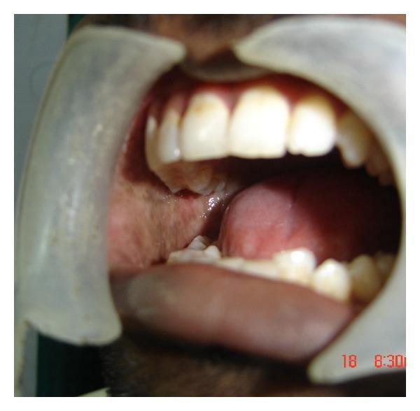
This clinical photograph shows an intraoral view of a patient with oral submucous fibrosis (OSMF). The image features the buccal mucosa, which appears markedly pale and blanched, with visible whitish, fibrotic bands replacing the healthy pink mucosal tissue. These changes are characteristic of chronic, progressive scarring. Retractors are used to expose the oral cavity, revealing a significantly restricted mouth opening (trismus), indicated by the limited interincisal distance between the maxillary and mandibular teeth. While the dentition and tongue are visible, their movement and the overall aperture are constrained by the underlying submucosal fibrosis. This image demonstrates the preoperative clinical presentation of OSMF, highlighting the loss of mucosal elasticity and restricted oral motility associated with this condition.
This clinical photograph shows an intraoral view of a patient with oral submucous fibrosis (OSMF). The primary feature is the presence of pale, blanched, and fibrotic buccal mucosa, most notable on the left side of the image. The healthy pink color is replaced by whitish fibrous bands and a marble-like appearance, indicating significant submucosal collagen deposition and reduced vascularity. The oral cavity exhibits restricted mouth opening (trismus), as evidenced by the limited interincisal distance and the close proximity of the tongue and teeth. The tongue is positioned low in the oral cavity, showing normal dorsal papillae, but its mobility is clinically constrained by the surrounding fibrotic tissues. This image serves as a classic educational example of the clinical presentation of OSMF, a premalignant condition often associated with areca nut or tobacco use, characterized by progressive stiffness of the oral mucosa and significant functional impairment in jaw movement.
Oral Submucous Fibrosis (OSMF) - Oral Pathology (10 Marks)
Definition
- First described by Schwartz (1952) as "atrophia idiopathica (tropica) mucosae oris"
- The term "Oral Submucous Fibrosis" was coined by Pindborg and Sirsat (1966)
- WHO definition (1994): "A chronic, insidious disease affecting any part of the oral cavity and sometimes the pharynx, although occasionally preceded by and/or associated with vesicle formation, is always associated with juxta-epithelial inflammatory reaction followed by a fibroelastic change of the lamina propria."
- Classified as an Oral Potentially Malignant Disorder (OPMD)
Epidemiology
- Predominantly affects people from South and Southeast Asia (India, Pakistan, Sri Lanka, Bangladesh, Taiwan)
- Prevalence in India: 0.2% to 0.5% of the population
- Over 2.5 million people affected worldwide
- Age: Usually 20-40 years (young adults); increasingly seen in teenagers due to commercially packaged gutkha
- Males > Females (historically; now equalizing due to gutkha use in both sexes)
- Malignant transformation rate: 7.6% over 17 years (India data); approximately 4.5-7.6% overall
Etiology and Pathogenesis
Etiological Factors
-
Areca nut (betel nut) - PRIMARY cause
- Chewed as betel quid (paan), paan masala, gutkha, mawa
- Active alkaloid: Arecoline - directly stimulates fibroblasts to increase collagen production
- Tannins in areca nut stabilize collagen by cross-linking, preventing degradation
- Catechins stimulate fibroblast proliferation and collagen synthesis
-
Tobacco - acts synergistically
-
Slaked lime (calcium hydroxide) - in betel quid; causes mucosal irritation
-
Chili peppers (capsaicin) - proposed co-factor; causes mucosal irritation
-
Nutritional deficiencies - vitamins A, B complex, C; iron deficiency; anemia
-
Genetic predisposition - HLA-A10, HLA-B7, HLA-DR3 associations reported
-
Autoimmune mechanisms - elevated serum IgG, IgA, IgM; autoantibodies reported
-
Copper - areca nut is rich in copper; copper stimulates lysyl oxidase, which cross-links collagen
Pathogenesis (Step-by-step)
Areca nut chewing
        ↓
Arecoline + Tannins → Direct mucosal irritation → Initial inflammatory response
        ↓
Arecoline stimulates fibroblasts → Upregulation of collagen synthesis (Types I, III, VI)
        ↓
Tannins stabilize and cross-link collagen → Reduced collagen degradation
        ↓
Copper (in areca nut) → activates Lysyl oxidase → further collagen cross-linking
        ↓
Failure of collagen remodeling (reduced collagenase / MMP activity)
        ↓
Collagen accumulates in lamina propria → Hyalinization of connective tissue
        ↓
Progressive fibrosis → Fibrotic bands in buccal mucosa, lips, tongue, palate
        ↓
Ischemia from fibrosis → Epithelial atrophy → Dysplasia → Risk of SCC
- TGF-β1 (Transforming Growth Factor beta-1) - major pro-fibrotic cytokine; upregulated by arecoline
- Inducible Nitric Oxide Synthase (iNOS) - elevated in early OSMF; contributes to erythema
- p53 mutations - found in dysplastic epithelium
- Reduced collagenase / MMP levels - key mechanism for collagen accumulation
- Altered epithelial-mesenchymal interactions
Classification
1. Pindborg and Sirsat Classification (1966) - Based on Histopathology
| Stage | Description |
|---|---|
| Very early | Finely fibrillar collagen in lamina propria with no hyalinization |
| Early | Fine, slightly wavy collagen fibers with early hyalinization; mild chronic inflammation |
| Moderately advanced | Dense, hyalinized collagen; moderate chronic inflammation; epithelial atrophy |
| Advanced | Complete hyalinization; dense, glass-like collagen; epithelial atrophy; minimal inflammation; fibrotic bands |
2. Khanna and Andrade Classification (1995) - Based on Clinical Features (Mouth Opening / Interincisal Distance)
| Group | Interincisal Distance | Clinical Features |
|---|---|---|
| Group I | > 35 mm | No trismus; burning sensation; erythema; early cases |
| Group II | 26-35 mm | Mild trismus; pale mucosa; fibrotic bands palpable |
| Group III | 15-25 mm | Moderate trismus; classic marble-white mucosa; restricted tongue protrusion |
| Group IV A | < 15 mm | Severe trismus; extensive fibrosis; additional features |
| Group IV B | < 15 mm | Severe trismus + premalignant/malignant changes (leukoplakia, SCC) |
3. Haider Classification (2000) - Based on Clinical Features
| Stage | Features |
|---|---|
| Stage I (Faucial) | Only fauces involved; burning sensation |
| Stage II (Faucial + Oral) | Fauces + oral mucosa; mild trismus |
| Stage III (Oral + Pharyngeal) | Oral + pharynx ± esophagus; severe trismus |
Clinical Features
Symptoms (in order of disease progression)
- Burning sensation in mouth (worsened by spicy food) - often the first symptom
- Increased salivation
- Mucosal vesiculation - small blisters that may ulcerate
- Erythema of buccal mucosa - related to elevated iNOS
- Altered taste sensation
- Gradual blanching and pallor of oral mucosa (marble-white appearance)
- Palpable fibrotic bands in buccal mucosa, lips, tongue
- Progressive reduction in mouth opening (trismus)
- Restricted tongue protrusion
- Difficulty chewing and swallowing
- Severe trismus (interincisal distance < 15 mm)
- Severe dysphagia and speech difficulty
- Leathery, inelastic, marble-white mucosa
- Shrunken uvula - "bud-like" uvula
- Fibrosis of soft palate and fauces
- Depapillation of tongue
- Hearing loss - due to involvement of Eustachian tubes
- Development of leukoplakia on fibrotic mucosa
- Malignant transformation to SCC (~7.6% over 17 years)
Signs:
- Reduced mouth opening (measured by interincisal distance)
- Fibrotic bands visible and palpable in buccal mucosa
- Blanched, inelastic mucosa
- "Marble-white" or "papyrus-white" colour
Clinical Image
Histopathological Features
Epithelial Changes:
- Epithelial atrophy - reduced rete ridges, thinned epithelium
- Epithelial dysplasia - variable; mild in 46%, moderate in 52%, severe in 2% (Cummings)
- Hyperkeratosis or parakeratosis may be present
- Loss of rete ridges (flattening)
Connective Tissue Changes (by stage):
| Stage | Connective Tissue Appearance |
|---|---|
| Early | Edematous, loosely arranged fine collagen fibers; dilated capillaries |
| Intermediate | Dense, hyalinized collagen bands; reduced vascularity; moderate chronic inflammation |
| Advanced | Complete hyalinization - glass-like, homogeneous, avascular collagen replacing normal connective tissue; minimal inflammation |
Other Features:
- Chronic inflammatory infiltrate - lymphocytes and plasma cells (variable)
- Reduced vascularity - explaining the pale colour
- Muscle degeneration - degeneration of buccinator and other muscles in advanced cases
- Fibrosis of minor salivary gland ducts
- Variable degrees of dysplasia in the overlying epithelium
Investigations
1. Biopsy and Histopathological Examination
- Gold standard for diagnosis and staging
- Incisional biopsy from representative area
- Confirms fibrosis, dysplasia, rules out SCC
2. Clinical Examination
- Measurement of interincisal distance (mouth opening) - for staging and monitoring
- Tongue protrusion measurement
3. Exfoliative Cytology / Brush Biopsy
- Screening tool to detect dysplastic cells
4. Blood Investigations
- CBC - to detect anemia (iron deficiency common)
- Serum iron, ferritin, B12, folate
- Elevated ESR
- Serum immunoglobulins (IgG, IgA, IgM may be elevated - autoimmune component)
5. Imaging
- MRI - gold standard for evaluating extent of fibrosis, muscle involvement, depth of bands
- Ultrasound - to assess thickness of fibrotic bands
- OPG / CBCT for jaw assessment if needed
6. Toluidine Blue Staining
- Highlights dysplastic areas; guides biopsy site
7. Molecular / Research Investigations
- Serum TGF-β1 levels - elevated
- MMP / TIMP ratio - reflects collagen metabolism
- p53 immunostaining on biopsy - to detect dysplastic transformation
Treatment and Management
A. General Measures (All Stages)
- Cessation of areca nut, tobacco, and alcohol - mandatory; disease stabilizes but fibrosis may not reverse
- Nutritional supplementation - vitamins A, B complex, C, E; iron
- Physiotherapy - mouth-opening exercises (forceful opening, tongue depressors, TheraBite device)
- High-protein diet
B. Medical Treatment
| Agent | Mechanism / Use |
|---|---|
| Intralesional corticosteroids (triamcinolone acetonide, dexamethasone) | Anti-inflammatory; reduce collagen synthesis; given weekly for 10-12 weeks |
| Hyaluronidase (intralesional) | Breaks down hyaluronic acid in ground substance; increases tissue permeability; enhances steroid effect |
| Pentoxifylline (oral) | Reduces TGF-β, inhibits fibroblast proliferation; improves microcirculation |
| Collagenase (intralesional) | Directly degrades accumulated collagen |
| Interferon-γ | Anti-fibrotic cytokine; reduces collagen synthesis |
| Lycopene (oral antioxidant) | Reduces oxidative stress; shown to improve mouth opening |
| Spirulina | Antioxidant; shown to improve symptoms |
| Aloe vera | Anti-inflammatory; used as adjunct |
| Turmeric / Curcumin | Anti-inflammatory, antioxidant |
| Placental extracts | Used in some Indian centers |
C. Surgical Treatment (for Groups III and IV / Advanced OSMF)
| Procedure | Indication / Details |
|---|---|
| Surgical release of fibrotic bands | Incision/excision of fibrotic bands via intraoral approach; modest success alone |
| Reconstruction with split-thickness skin graft | Post-excision defect coverage |
| Nasolabial flap | Pedicled flap for buccal mucosal defects; commonest reconstruction |
| Buccal fat pad (BFP) flap | Used for posterior buccal mucosa defects post band release |
| Tongue flap | For larger defects |
| Temporalis muscle / coronoidectomy | Severe trismus; mandibular coronoid process reduction to improve opening |
| CO2 laser / KTP laser | Ablation of fibrous bands with less bleeding and better healing |
| Radiosurgery | Newer modality for band release |
D. Treatment of Malignant Transformation
- Biopsy confirmation of SCC
- Managed as oral squamous cell carcinoma - surgery (wide excision + neck dissection) ± radiotherapy ± chemotherapy
Summary Table
| Feature | OSMF |
|---|---|
| First described by | Schwartz (1952) |
| Term coined by | Pindborg & Sirsat (1966) |
| Chief cause | Areca nut (arecoline) |
| Key mechanism | Increased collagen synthesis + reduced degradation |
| Hallmark clinical feature | Trismus + marble-white mucosa |
| Histopathology | Epithelial atrophy + submucosal hyalinization |
| Malignant transformation | ~7.6% over 17 years |
| Classification | Pindborg (histological); Khanna & Andrade (clinical) |
| Treatment | Cessation of habit + corticosteroids + surgery |
- OSMF is a chronic, progressive, irreversible disease if the habit continues
- The areca nut alkaloid arecoline is the primary pathogenic agent
- Histopathologically, it progresses from loose collagen → hyalinization → glass-like avascular collagen
- The atrophic epithelium overlying dense fibrosis is the diagnostic hallmark
- It carries a 7.6% malignant transformation rate - regular follow-up is essential
- Treatment is primarily palliative in advanced stages; habit cessation is the only preventive measure
Carcinoma in situ
carcinoma in situ oral epithelium histology full thickness dysplasia
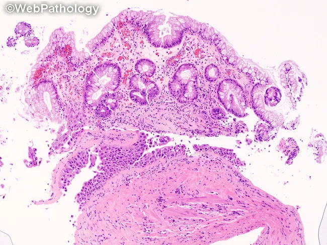
Histopathology (light microscopy) of bladder diverticulum mucosa in H&E-stained sections, with two fragmentary regions illustrating distinct epithelial phenotypes. The lower fragment demonstrates urothelial carcinoma in situ (CIS): full-thickness high-grade urothelial dysplasia with marked nuclear enlargement, pleomorphism, hyperchromasia, loss of polarity, and increased mitotic activity, while the basement membrane remains intact, indicating non-invasive disease. The surrounding stroma shows preserved lamina propria without invasion. The upper fragment reveals intestinal metaplasia within the urothelium, characterized by intestinal-type columnar epithelium with goblet cells and mucin-producing cells; this metaplastic epithelium is discontinuous from the CIS and sites of goblet cell formation are evident. Together, these findings indicate divergent differentiation within the same diverticular mucosa: flat CIS coexisting with intestinal metaplasia. Clinically relevant interpretation includes the diagnosis of high-grade non-muscle-invasive urothelial carcinoma confined to the mucosa (CIS) and associated metaplastic change, a potential premalignant/metaplastic process that can complicate histologic interpretation and influence risk stratification. Diagnostic significance: CIS confers high risk for progression to muscle-invasive disease; management considerations include intravesical therapy (BCG) or cystoscopic surveillance with repeat sampling; intestinal metaplasia may predispose to glandular neoplasia and warrants careful follow-up. This composite histology underscores the need for meticulous sampling of bladder diverticula and adjunct immunohistochemistry in equivocal cases.
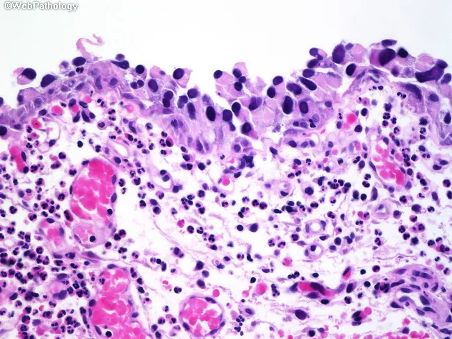
Light microscopy of an hematoxylin-and-eosin (H&E) stained bladder mucosa section showing urothelial carcinoma in situ (CIS). The specimen depicts urothelial epithelium with full-thickness high-grade atypia: markedly pleomorphic, hyperchromatic nuclei with prominent nucleoli and abundant eosinophilic cytoplasm, increased nuclear-to-cytoplasmic ratio, and conspicuous architectural disarray with loss of polarity. Basal and superficial layers demonstrate uniform dysplasia across the entire thickness, while the basement membrane remains intact, consistent with non-invasive disease. An acute inflammatory infiltrate is present in the underlying lamina propria, yet invasion is not evident. The histologic pattern is diagnostic of CIS and correlates with high-grade urothelial carcinoma in situ, a precursor to invasive urothelial carcinoma. Clinically, CIS carries a high risk of recurrence and progression and mandates intravesical therapy consideration (e.g., BCG) and close surveillance with cystoscopy and urine cytology. This image is valuable for diagnostic education, differential diagnosis with reactive urothelial changes, high-grade dysplasia, and invasive carcinoma, and for correlating histology with ancillary tests. The description supports precision in pathology reporting, guiding staging, treatment planning, and research on bladder cancer pathobiology. The image supports educational objectives in surgical pathology, cytology correlations, and multidisciplinary tumor boards; it also enhances database annotations for machine-assisted detection of non-invasive urothelial carcinoma and related premalignant lesions.
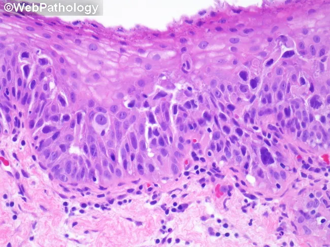
Histopathology examination of a urinary bladder urothelium specimen stained with Hematoxylin and Eosin reveals urothelial carcinoma in situ with pagetoid spread into areas of non-keratinizing squamous metaplasia. The epithelium shows full-thickness dysplasia characterized by marked cellular pleomorphism, enlarged hyperchromatic nuclei, prominent nucleoli, increased mitotic activity, and loss of polarity. Individual malignant cells are scattered singly and in small clusters within the urothelial layer and, notably, within metaplastic squamous foci, consistent with pagetoid spread. The squamous metaplasia itself demonstrates non-keratinizing features without evident keratin pearl formation. Basal layer continuity appears preserved, with no overt invasion into the underlying stroma evident in this field; however, CIS is a high-grade intraepithelial neoplasm with a risk of progression to infiltrative disease if not treated. The background shows mild chronic inflammatory infiltrate. Diagnosis: Urothelial carcinoma in situ with pagetoid spread into non-keratinizing squamous metaplasia of urothelium. Clinical relevance: high-grade intraepithelial lesion requiring staging and management decisions, including intravesical therapy or surgical consideration; differential includes high-grade urothelial carcinoma with squamous differentiation; correlation with cystoscopic findings and imaging advised; monitor for multifocal disease. Significance: recognizing pagetoid spread in CIS has prognostic implications, guiding surveillance intervals and intravesical therapy decisions. This image is valuable for pathology education and tumor biology studies on urothelial-squamous differentiation.
squamous cell carcinoma in situ oral mucosa histopathology dysplasia spectrum
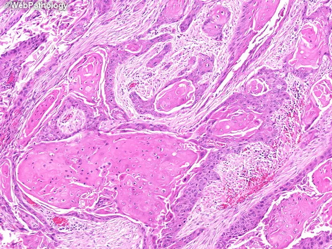
High-magnification histopathology image of oral mucosa showing keratinizing, well-differentiated squamous cell carcinoma with classic keratin pearl formation. The neoplasm displays nests and cords of malignant squamous cells invading the fibrous lamina propria, with abundant eosinophilic cytoplasm and intercellular bridges. Keratinization is evident both centrally within tumor nests and at the periphery, producing concentric keratin pearls of pale eosinophilic keratin. Tumor cells are relatively well differentiated, with recognizable squamous maturation and occasional mitotic figures. The adjacent mucosa demonstrates diverse epithelial alterations, including squamous metaplasia (keratinizing and nonkeratinizing variants), dysplasia, and areas consistent with squamous cell carcinoma in situ, reflecting field cancerization around the invasive focus. Inflammatory infiltrate and edema are present in the superficial lamina propria, while deeper stroma shows variable desmoplastic response. The surface epithelium may show thinning or ulceration over the lesion. No clear vascular invasion is evident at this magnification, though deeper planes could reveal invasion into connective tissue and perineural spread. This image provides valuable diagnostic features for distinguishing keratinizing SCC from nonkeratinizing variants and other mucosal neoplasms, and highlights the relevance of adjacent dysplasia/CIS as indicators of tumor field effect and oncologic margins. It serves as an educational reference for pathology, histology, and clinical tumor biology.
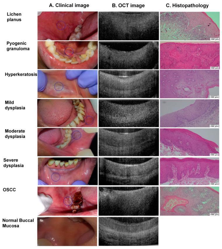
A multi-modal comparison chart displaying clinical photographs (column A), Optical Coherence Tomography (OCT) scans (column B), and histopathology slides (column C) for a spectrum of oral cavity conditions. The rows categorize findings into: non-dysplastic lesions (Lichen planus, Pyogenic granuloma, Hyperkeratosis), progressive stages of dysplasia (mild, moderate, and severe), Oral Squamous Cell Carcinoma (OSCC), and normal buccal mucosa. Clinical images show various presentations including white lacy patches, reddish nodules, and ulcerated masses. OCT cross-sections demonstrate progressive loss of tissue stratification and increased epithelial thickness or signal disruption associated with malignant transformation. Histopathology (Hematoxylin and Eosin stain, 100x magnification) provides the definitive diagnosis, showing structural changes such as epithelial thickening, cellular atypia, and basement membrane disruption in severe dysplasia and OSCC. This diagnostic panel illustrates the correlation between non-invasive imaging modalities and gold-standard biopsy results in the detection and grading of oral premalignant and malignant lesions.
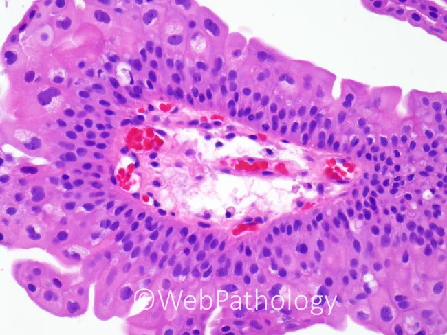
This is a hematoxylin and eosin stained histopathology micrograph of oral mucosa epithelium with underlying connective tissue (buccal/oral mucosa). Light microscopy reveals a stratified squamous epithelial layer with orderly cellular architecture and a preserved basement membrane. The surface epithelium shows uniform basal and parabasal cells, relatively normal cytoplasmic maturation, and no overt dysplasia or malignant epithelial proliferation. In the lamina propria, there is a mild inflammatory infiltrate consisting of lymphocytes and plasma cells, accompanied by scattered neutrophils and increased vascularity with dilated capillaries. The subepithelial stroma appears edematous but without frank ulceration. Red blood cells are visible within small vascular channels in the superficial lamina propria. Overall, the image demonstrates a benign inflammatory mucosal process with vascular congestion rather than neoplastic transformation. Clinically, these findings are compatible with acute or chronic mucositis, reactive/traumatic irritation, or mild infectious mucosal inflammation, depending on clinical context. Diagnostic significance lies in ruling out dysplasia or carcinoma in situ within this tissue plane and guiding management toward conservative therapy and etiologic assessment. This image is well-suited for educational purposes in histology, oral pathology, and pathology training, enabling recognition of epithelial maturation, inflammatory infiltrates, and vascular changes in mucosal tissue. Correlation with clinical symptoms strengthens interpretation.
Carcinoma In Situ (CIS) - Oral Pathology (10 Marks)
Definition
- Synonym: Intraepithelial carcinoma / Stage 0 carcinoma / Tis (Tumor in situ)
- CIS represents the most severe end of the epithelial dysplasia spectrum, just short of invasive carcinoma
- The distinguishing feature from invasive carcinoma is the intact basement membrane - once this is breached, the lesion becomes invasive SCC
- CIS is classified as an Oral Potentially Malignant Disorder (OPMD)
Synonyms and Related Terms
- Intraepithelial carcinoma
- Bowen's disease (when CIS occurs on skin)
- Erythroplasia of Queyrat (when on glans penis)
- High-grade intraepithelial neoplasia (Grade III / CIN III)
- Stage 0 squamous cell carcinoma (TisN0M0)
- In head and neck cancer staging: TisN0M0
Historical Perspective
- Concept of CIS first introduced by Broders (1932)
- Distinguished from invasive SCC by Warren (1943) based on basement membrane integrity
- Pindborg contributed extensively to understanding oral epithelial dysplasia and CIS in the context of oral PMDs
Relationship to Epithelial Dysplasia
Normal epithelium
        ↓
Hyperplasia (hyperkeratosis, acanthosis)
        ↓
Mild dysplasia (atypia confined to lower 1/3 of epithelium)
        ↓
Moderate dysplasia (atypia in lower 2/3 of epithelium)
        ↓
Severe dysplasia (atypia in >2/3 of epithelium)
        ↓
CARCINOMA IN SITU (full-thickness atypia, basement membrane INTACT)
        ↓
Invasive SCC (basement membrane BREACHED, stromal invasion)
Epidemiology / Incidence
- CIS is most often found in association with leukoplakia or erythroplakia rather than as a standalone lesion
- Found in ~15.2% of leukoplakia biopsies (severe dysplasia or CIS combined) - Cummings Otolaryngology
- Erythroplakia is the oral lesion most frequently associated with CIS (~40% of erythroplakic lesions are CIS at biopsy)
- Age: 40-70 years; Males > Females
- High-risk sites: floor of mouth, ventral/lateral tongue, soft palate
Etiology and Risk Factors
- Tobacco - smoking and smokeless forms; most significant risk factor
- Alcohol - synergistic with tobacco
- Areca nut / betel quid - especially in South Asia
- HPV (Human Papillomavirus) - high-risk types 16 and 18
- Chronic mucosal irritation - ill-fitting dentures, sharp teeth
- Nutritional deficiencies - vitamins A, C, E; iron
- Immunosuppression
- UV radiation - for lip CIS (actinic cheilitis)
- Genetic factors - p53 mutation, loss of heterozygosity (LOH)
Pathogenesis
- p53 mutations - most common molecular event; loss of tumor suppressor function
- Loss of heterozygosity (LOH) at chromosomes 3p, 9p (p16/CDKN2A), 17p, 13q
- Inactivation of Rb pathway - loss of cell cycle control
- Telomerase activation - cellular immortalization
- Upregulation of cyclin D1 - uncontrolled cell cycle entry
- Downregulation of E-cadherin - loss of cell adhesion
Clinical Features
| Presentation | Description |
|---|---|
| Erythroplakia | Most commonly associated; fiery red, velvety, sharply demarcated patch - CIS found in ~40% |
| Leukoplakia | White patch; CIS found in ~15% of high-grade cases |
| Erythroleukoplakia | Mixed red and white; red areas most likely to harbor CIS |
| Ulceration | Non-healing ulcer |
| Normal-appearing mucosa | CIS may occasionally occur in clinically unremarkable mucosa ("field cancerization") |
Clinical Clues Suggesting CIS (vs. Simple Dysplasia):
- Induration on palpation
- Fixation to underlying tissue
- Velvety, erythematous surface
- Lesion at a high-risk site (floor of mouth, ventral tongue, soft palate)
- Persistence despite removal of irritants
- Size > 2 cm
- Non-homogeneous appearance
Histopathological Features (KEY)
Hallmark: Full-thickness epithelial atypia with intact basement membrane
Cellular Features:
- Nuclear enlargement - large, hyperchromatic, irregular nuclei
- Increased nuclear-cytoplasmic (N:C) ratio
- Nuclear hyperchromatism - deeply stained, coarsely granular chromatin
- Prominent nucleoli - conspicuous, often multiple
- Nuclear pleomorphism - variation in size and shape
- Abnormal mitotic figures - tripolar, star-shaped, asymmetric mitoses
- Loss of normal polarity - cells no longer oriented perpendicular to the basement membrane
- Dyskeratosis - premature individual cell keratinization within the prickle cell layer
- Keratin pearl formation within the epithelium (without breaching basement membrane)
Architectural Features:
- Full-thickness involvement - atypical cells present from basal layer to surface
- Loss of normal stratification - the orderly basal → prickle → granular → keratin layer sequence is completely disrupted
- Drop-shaped rete ridges - bulbous, irregular rete pegs
- Basal cell hyperplasia - increased proliferative activity
- Loss of intercellular cohesion - "falling apart" appearance
- No invasion - basement membrane is intact on PAS stain or reticulin stain
Connective Tissue:
- Chronic inflammatory infiltrate - lymphocytes and plasma cells in lamina propria (host immune response)
- Blood vessels may be prominent (contributing to erythematous clinical appearance)
- No stromal desmoplasia (that is a feature of invasive SCC)
Staining Aids:
- PAS (Periodic Acid-Schiff) stain - highlights intact basement membrane
- Reticulin stain - demonstrates intact reticular basement membrane
- Ki-67 immunostaining - high proliferative index throughout full epithelial thickness
- p53 immunostaining - positive throughout epithelial thickness
Histological Image
Comparison: Dysplasia vs. CIS vs. Invasive SCC
| Feature | Severe Dysplasia | CIS | Invasive SCC |
|---|---|---|---|
| Atypia extent | >2/3 epithelial thickness | Full thickness | Full thickness |
| Basement membrane | Intact | Intact | Breached |
| Stromal invasion | Absent | Absent | Present |
| Desmoplasia | Absent | Absent | Present |
| Lymphovascular invasion | Absent | Absent | May be present |
| Treatment | Excision ± surveillance | Excision | Surgery ± RT/CT |
Grading of Oral Epithelial Dysplasia (WHO System)
| Grade | Atypia Extent | Features |
|---|---|---|
| Mild dysplasia | Lower 1/3 (basal layer) | Minimal nuclear atypia |
| Moderate dysplasia | Lower 2/3 | Moderate nuclear changes, mitoses in lower 2/3 |
| Severe dysplasia | >2/3 thickness | Marked atypia, atypical mitoses |
| CIS | Full thickness | All features of malignancy, basement membrane intact |
Investigations/Diagnosis
-
Biopsy - mandatory and gold standard
- Incisional biopsy from the most suspicious area
- Multiple biopsies for large lesions
- Toluidine blue guides site selection
-
Histopathological examination
- H&E staining for routine assessment
- PAS / reticulin for basement membrane integrity
- Immunohistochemistry: p53, Ki-67, p16
-
Exfoliative cytology / Brush biopsy
- Screening tool; not definitive
-
Toluidine blue vital staining
- Retained by dysplastic / CIS areas
- Guides biopsy site
-
Narrow-band imaging (NBI) / Autofluorescence
- Newer optical aids to detect high-grade lesions
-
Imaging (MRI/CT)
- To rule out any occult stromal invasion in advanced cases
Treatment and Management
1. Habit cessation
- Tobacco, alcohol, areca nut - mandatory
2. Surgical Excision
- Treatment of choice for CIS
- Wide local excision with adequate margins (5-10 mm clear margin)
- Confirm clear margins with frozen section intraoperatively
- Close interaction between surgeon and pathologist essential
3. CO2 Laser Excision
- Effective alternative; good healing, less morbidity
- Suitable for accessible lesions
4. Photodynamic Therapy (PDT)
- For extensive/multifocal CIS
- Uses photosensitizer + laser light activation
5. Radiotherapy
- Role controversial for CIS (TisN0M0)
- Used when:
- Multiple endoscopic resections fail
- Extensive involvement of multiple subsites
- Patient unfit for surgery
- Not generally recommended for high-grade dysplasia alone
- (Cummings Otolaryngology, p. 1585)
6. Surveillance
- Mandatory long-term follow-up - every 3-6 months
- 18.1% of patients with CIS/severe dysplasia at initial biopsy developed invasive SCC within 3 years (Cummings)
- Patients must be counselled about the high risk of progression
Prognosis
- CIS with no invasive component has an excellent prognosis after adequate excision
- 18.1% risk of progression to invasive SCC within 3 years if untreated (vs. 3.7% for no dysplasia)
- Recurrence is common if:
- Margins are not clear
- Habit is not discontinued
- Field cancerization is present
- Oral CIS is a marker of field cancerization - patients are at risk for second primary tumors elsewhere in the aerodigestive tract
Summary
| Feature | CIS |
|---|---|
| Definition | Full-thickness squamous atypia, basement membrane intact |
| Key distinguishing feature | Intact basement membrane (vs. invasive SCC) |
| Clinical appearance | Usually erythroplakia or leukoplakia |
| Most common associated lesion | Erythroplakia (~40% are CIS) |
| Histology hallmark | Full-thickness loss of polarity, atypical mitoses |
| Stain to confirm BM intact | PAS / reticulin |
| 3-year progression to SCC | ~18% |
| Treatment | Surgical excision / CO2 laser |
- CIS is the penultimate step before invasive SCC - the only difference is the intact basement membrane
- Erythroplakia is the clinical lesion most likely to be CIS - biopsy every erythroplakic lesion
- The histological diagnosis of CIS vs. severe dysplasia is sometimes subjective - close clinicopathological correlation is required (same lesion may be called Grade III intraepithelial neoplasia, high-grade dysplasia, or TisN0M0 by different pathologists)
- PAS and reticulin stains confirm basement membrane integrity - the defining criterion of CIS vs. invasive SCC
- CIS is a marker of field cancerization - the entire at-risk mucosa must be monitored
Papilloma (without pathogenesis)
oral squamous papilloma cauliflower exophytic lesion tongue palate
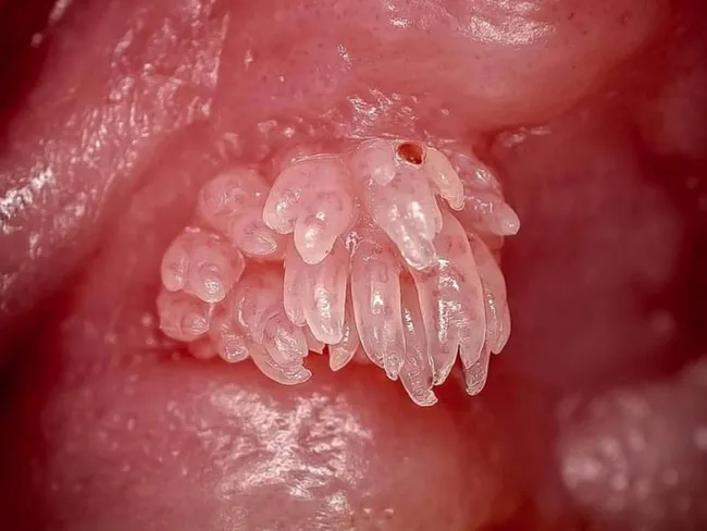
Imaging modality: Clinical photography of an intraoral exophytic lesion. Location: oral mucosa, most commonly tongue, soft palate, lips; this image shows a clustered, pedunculated, cauliflower-like eruption with finger-like projections. Size small, up to 0.5 cm; color white-pink depending on keratinization. The lesion is soft and non-tender. The appearance is characteristic of oral squamous papilloma, HPV-induced, most often HPV types 6 and 11. Pathology: benign proliferative lesion with papillary fronds lined by stratified squamous epithelium; hyperkeratosis; koilocytes may be present; fibrovascular cores. Etiology: human papillomavirus infection; transmitted via direct contact; common in pediatric and adult populations; slight male predominance. Imaging features: not radiographic; no imaging modality beyond clinical photography. Differential: verruca vulgaris, condyloma acuminatum, focal epithelial hyperplasia (heck disease), other exophytic mucosal lesions including papilloma; need histopathology to differentiate. Clinical significance: benign, low risk of malignant transformation; complete excision recommended to prevent recurrence; recurrences possible if HPV persists; counsel on HPV transmission. Utility: this image is useful for education, differential diagnosis of oral exophytic lesions, and correlating clinical appearance with HPV-related papillomas; could support cases in dental clinics, oral surgery, pathology teaching, and dermatology resources. Note: clinical correlation with examination and biopsy is advised for definitive diagnosis. HPV typing may guide management and counseling.
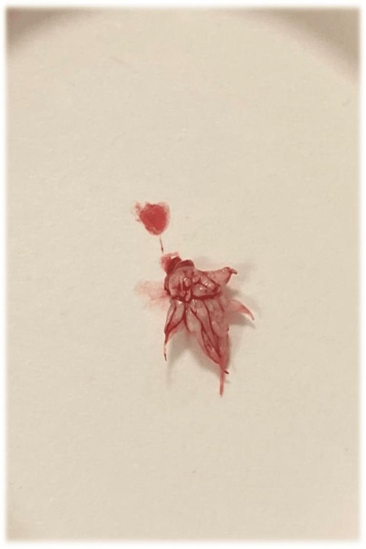
This clinical photograph displays a macroscopic view of a surgically excised intraoral specimen, identified as a squamous papilloma. The lesion measures approximately 0.9 cm x 0.5 cm and exhibits a characteristic exophytic, pedunculated morphology. Its most distinctive feature is a 'feather-like' or 'cauliflower-like' configuration, consisting of numerous branching, finger-like epithelial projections radiating from a central base. The specimen is predominantly white, indicating surface hyperkeratosis, with visible focal areas of red staining due to residual blood from the excision procedure. The irregular, warty surface texture is typical of benign epithelial neoplasms of the soft palate. This image serves as a clinical example of the macroscopic presentation of an oral squamous papilloma, highlighting the relationship between the pedunculated base and the characteristic projections used for diagnostic identification in oral and maxillofacial pathology.
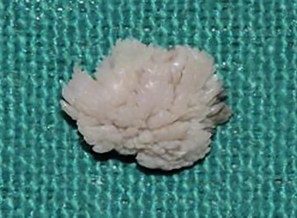
This clinical photograph displays a gross surgical specimen of an excised oral lesion against a green sterile backdrop. The specimen is a pale, pinkish-white, lobulated soft tissue mass measuring approximately 1.5 cm. Morphologically, the lesion exhibits a classic cauliflower-like or exophytic papillary architecture, characterized by numerous fine, finger-like (digitiform) projections and a fringed peripheral margin. These macroscopic features are highly characteristic of a squamous papilloma, often found on the hard palate. The image demonstrates the hallmark surface keratinization and papillary growth pattern used in the clinical identification of benign epithelial tumors of the oral cavity. Educational focus includes the recognition of verrucous and papillary lesions in oral pathology.
Oral Squamous Papilloma - Oral Pathology (10 Marks)
Definition
- It is the most common benign epithelial neoplasm of the oral cavity
- Classified as a benign tumor of epithelial origin
Etiology
- Human Papillomavirus (HPV) - primary etiological agent
- HPV types 6 and 11 - most common (low-risk types; rarely associated with malignancy)
- HPV types 2 and 4 - occasionally implicated (same types causing common warts)
- HPV types 16 and 18 - rarely, in immunocompromised patients
- Mode of transmission:
- Direct contact / person-to-person contact with HPV-infected tissue
- Autoinoculation from warts elsewhere (hands to mouth)
- Vertical transmission - maternal birth canal (relevant in recurrent respiratory papillomatosis in children)
- Sexual transmission (orogenital contact) - implicated in some oropharyngeal cases
Incidence and Epidemiology
- Most common benign oral mucosal neoplasm seen in oral pathology practice
- Accounts for approximately 2.5-8% of all oral tumors submitted for biopsy
- Occurs at any age - but most common between 3rd and 5th decades (30-50 years)
- Slight male predominance in adults
- In children - associated with maternal HPV infection (perinatal transmission) or recurrent respiratory papillomatosis
- No definitive racial or geographic predilection
Sites of Predilection
| Site | Frequency |
|---|---|
| Tongue (lateral border, ventral surface, dorsum) | Most common |
| Soft palate and uvula | Very common |
| Hard palate | Common |
| Lips | Common |
| Buccal mucosa | Less common |
| Gingiva / alveolar mucosa | Occasional |
| Floor of mouth | Occasional |
| Tonsils / oropharynx | Can occur |
Clinical Features
Gross Appearance:
| Feature | Description |
|---|---|
| Shape | Exophytic, cauliflower-like / warty / verrucous papillary growth |
| Surface | Rough, irregular, with multiple finger-like projections |
| Attachment | Pedunculated (narrow stalk) - most common; occasionally sessile (broad base) |
| Color | White to pink depending on degree of surface keratinization; heavily keratinized lesions appear white |
| Size | Usually small - 0.3 to 1.5 cm; rarely exceeds 2 cm |
| Number | Usually solitary; multiple lesions suggest condyloma acuminatum or focal epithelial hyperplasia |
| Consistency | Soft, non-tender, compressible |
| Borders | Well-demarcated |
| Induration | Absent - induration would suggest malignant change |
Symptoms:
- Usually asymptomatic - discovered incidentally or noticed as a painless growth
- Occasional mild discomfort if traumatised
- Rarely causes functional problems (speech, swallowing) unless very large
- No spontaneous bleeding in uncomplicated cases
Clinical Image
Histopathological Features (KEY)
Low-Power (Scanning) View:
- Multiple finger-like (digitiform) papillary projections radiating from a central base
- Each projection has a central fibrovascular core surrounded by stratified squamous epithelium
- The overall architecture resembles a "cauliflower" or "sea anemone"
High-Power (Microscopic) Features:
- Stratified squamous epithelium - orderly, well-differentiated covering each papillary frond
- Hyperparakeratosis or hyperorthokeratosis - prominent surface keratinization (explains white colour)
- Acanthosis - thickening of the prickle cell layer
- Koilocytes (koilocytic atypia) - pathognomonic of HPV infection
- Large squamous cells with perinuclear cytoplasmic vacuolization (halo)
- Pyknotic, raisin-like, hyperchromatic, irregular nuclei
- Found mainly in the upper spinous and granular layers
- Prominent granular cell layer - hypergranulosis
- Prominent rete ridges at the base
- No nuclear atypia or dysplasia in typical benign papilloma
- Mitotic figures may be present but are normal in appearance (never atypical)
- Fibrovascular stroma forming the core of each papillary projection
- Chronic inflammatory infiltrate - variable lymphocytes and plasma cells
- Dilated capillaries within the core
Summary of Histological Hallmarks:
Papillary fronds + fibrovascular cores + stratified squamous epithelium + koilocytes + hyperkeratosis + NO dysplasia
Variants / Related HPV Lesions (Differential Context)
| Lesion | HPV Type | Features |
|---|---|---|
| Squamous papilloma | 6, 11 | Solitary, pedunculated, cauliflower |
| Verruca vulgaris (common wart) | 2, 4 | Rough, raised; children; hyperkeratosis, no koilocytes in oral variant |
| Condyloma acuminatum | 6, 11 | Multiple, sessile, broader base; sexually transmitted; genital + oral |
| Focal epithelial hyperplasia (Heck's disease) | 13, 32 | Multiple flat/dome-shaped papules; autosomal recessive; indigenous populations |
| Recurrent respiratory papillomatosis | 6, 11 | Multiple; children (perinatal); airway (larynx mainly); recurs aggressively |
Differential Diagnosis
| Condition | Differentiating Feature |
|---|---|
| Verruca vulgaris | More heavily keratinized; children; HPV 2/4; viral inclusions in superficial epithelium |
| Condyloma acuminatum | Multiple, broader base, sexually transmitted, genital involvement |
| Focal epithelial hyperplasia (Heck's disease) | Multiple flat papules; HPV 13/32; indigenous populations |
| Irritation fibroma | Smooth surface, sessile, no papillary projections |
| Verrucous carcinoma | Older patients, larger, white, broad base; tobacco user; dysplasia on histology |
| Well-differentiated SCC (papillary variant) | Larger, dysplasia and atypia on histology; invasive |
| Sialadenoma papilliferum | Rare; ductal origin; near palate; glandular component on histology |
| Inflammatory papillary hyperplasia | Multiple nodular projections; under ill-fitting denture; no fibrovascular core pattern |
Investigations
- Clinical examination - appearance usually diagnostic
- Excisional biopsy - treatment and definitive diagnosis simultaneously
- Histopathological examination (H&E) - gold standard
- Immunohistochemistry (IHC):
- p16 immunostaining - surrogate marker for high-risk HPV
- Anti-HPV capsid antibody staining
- In situ hybridization (ISH) / PCR - for definitive HPV typing (research or equivocal cases)
- No imaging routinely required for small lesions
Treatment
Treatment of Choice: Surgical Excision
-
Conservative surgical excision - removal of lesion down to the base (stalk) with a small margin of normal tissue
- Base must be completely removed to reduce recurrence
- Excision should include the periosteum if the lesion is on gingiva/hard palate
- Excised tissue always sent for histopathological examination
-
CO2 Laser ablation/excision
- Equally effective; less bleeding; good healing
- Preferred for multiple lesions or difficult sites
-
Electrosurgery (electrocautery)
- Useful for small, accessible lesions
-
Cryotherapy
- Liquid nitrogen; useful in some cases; less commonly used intraorally
-
Powered instrumentation - useful in nasopharyngeal/nasal papillomas (Cummings)
Post-Treatment:
- Regular follow-up to watch for recurrence
- Patient counselling regarding HPV transmission
- HPV vaccination (against types 6 and 11) has been reported to decrease incidence of papillomatosis - Robbins Pathologic Basis of Disease
Recurrence
- Recurrence rate is low for solitary oral squamous papilloma after adequate excision
- Higher recurrence in:
- Incomplete excision
- Multiple lesions (recurrent respiratory papillomatosis - aggressive recurrence)
- Immunocompromised patients
- Malignant transformation is rare for HPV 6/11-associated lesions
- Risk increases with:
- Radiation therapy to the area
- Smoking
- Immunosuppression
- High-risk HPV co-infection
- Risk increases with:
Prognosis
- Excellent prognosis after complete excision
- Benign behavior; no metastatic potential with HPV 6/11 types
- Recurrence is possible but uncommon with adequate removal of the base
- Children with recurrent respiratory papillomatosis (laryngeal) have a guarded prognosis due to airway compromise and tendency to recur aggressively
Summary Table
| Feature | Oral Squamous Papilloma |
|---|---|
| Nature | Benign epithelial neoplasm |
| Etiology | HPV 6 and 11 (most common) |
| Age | Any age; 3rd-5th decade most common |
| Site | Tongue, soft palate, uvula (most common) |
| Appearance | Pedunculated, cauliflower-like, white-pink |
| Size | Usually < 1.5 cm |
| Number | Solitary |
| Histology | Papillary fronds, fibrovascular cores, koilocytes, hyperkeratosis |
| Pathognomonic cell | Koilocyte |
| Treatment | Surgical excision |
| Recurrence | Low (after complete excision) |
| Malignant potential | Very rare (HPV 6/11) |
- Oral squamous papilloma is the most common benign oral mucosal neoplasm - caused by HPV 6 and 11
- The koilocyte is the pathognomonic histological cell - perinuclear cytoplasmic vacuolisation with pyknotic nucleus
- Histologically: papillary fronds + fibrovascular cores + hyperkeratosis + koilocytes + NO dysplasia
- Solitary lesion = squamous papilloma; Multiple lesions = consider condyloma acuminatum or focal epithelial hyperplasia
- Treatment is surgical excision to the base - always send for histopathology
- HPV types 6 and 11 have low malignant potential; HPV 16/18 carry high malignant risk (oropharyngeal SCC)
Histopathology of scc alone
oral squamous cell carcinoma histopathology keratin pearl well differentiated
High-magnification histopathology image of oral mucosa showing keratinizing, well-differentiated squamous cell carcinoma with classic keratin pearl formation. The neoplasm displays nests and cords of malignant squamous cells invading the fibrous lamina propria, with abundant eosinophilic cytoplasm and intercellular bridges. Keratinization is evident both centrally within tumor nests and at the periphery, producing concentric keratin pearls of pale eosinophilic keratin. Tumor cells are relatively well differentiated, with recognizable squamous maturation and occasional mitotic figures. The adjacent mucosa demonstrates diverse epithelial alterations, including squamous metaplasia (keratinizing and nonkeratinizing variants), dysplasia, and areas consistent with squamous cell carcinoma in situ, reflecting field cancerization around the invasive focus. Inflammatory infiltrate and edema are present in the superficial lamina propria, while deeper stroma shows variable desmoplastic response. The surface epithelium may show thinning or ulceration over the lesion. No clear vascular invasion is evident at this magnification, though deeper planes could reveal invasion into connective tissue and perineural spread. This image provides valuable diagnostic features for distinguishing keratinizing SCC from nonkeratinizing variants and other mucosal neoplasms, and highlights the relevance of adjacent dysplasia/CIS as indicators of tumor field effect and oncologic margins. It serves as an educational reference for pathology, histology, and clinical tumor biology.
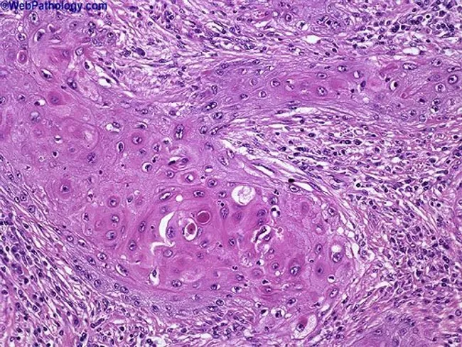
This histopathology image depicts a cutaneous squamous cell carcinoma on routine hematoxylin and eosin stained section. The biopsy shows nests of malignant squamous cells with keratinization and abundant keratin pearls, indicating squamous differentiation. The tumor exhibits moderate differentiation, evidenced by intercellular bridges, eosinophilic cytoplasm, nuclear pleomorphism, hyperchromasia, and mitotic activity. Invasion into the surrounding dermal stroma is evident, with desmoplastic-type fibrous response in some areas. The epidermis overlying the lesion shows dysplastic changes, and keratin pearl formation within tumor nests is a hallmark of keratinizing SCC. The architecture includes irregular cords and nests of polygonal cells, occasional central keratinization, and keratinized pearls of varying sizes. Clinically, these features suggest a malignant cutaneous neoplasm with potential for local invasion; staging would consider depth of invasion and perineural or lymphovascular involvement. This image is relevant for diagnostic pathology, histology education, and tumor biology research, illustrating characteristic squamous differentiation, keratin production, and invasion patterns. Differential considerations include well-differentiated keratinizing SCC versus moderately-to-poorly differentiated forms, verrucous carcinoma, and basal cell carcinoma with squamous features. The visual cues—keratin pearls, keratinization, and cohesive epidermal-derived tumor islands—assist in confirming diagnosis and guiding surgical management. Correlation with clinical data will inform prognosis and adjuvant therapy decisions and multidisciplinary care planning.
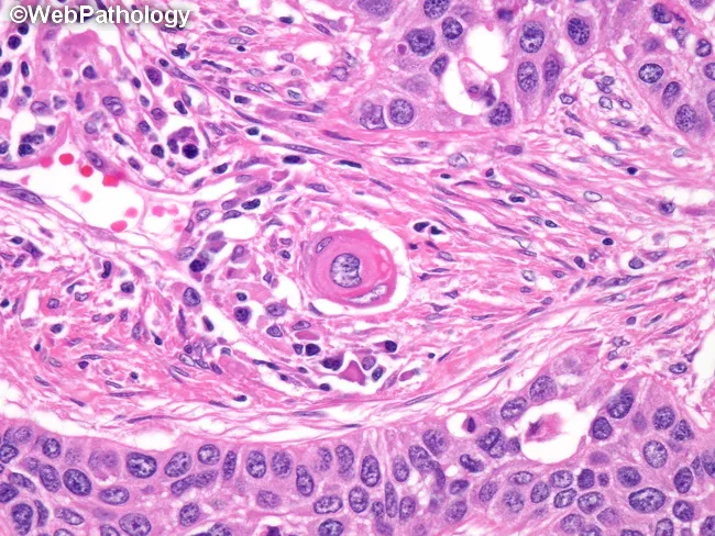
This is a brightfield histopathology image of an invasive, well-differentiated squamous cell carcinoma in epidermal/mucosal squamous epithelium. The H&E stained section demonstrates malignant squamous cells in nests and cords penetrating the dermis with desmoplastic stromal reaction. Characteristic features include abundant eosinophilic cytoplasm, keratinization in the form of keratin pearls, and pronounced intercellular bridges between adjacent tumor cells. Some tumor clusters show concentric keratinization around a central keratin pearl; intracellular mucin may be present in scattered malignant cells, though not a dominant feature in well-differentiated tumors. The cells exhibit minimal pleomorphism and relatively preserved nuclear-to-cytoplasmic ratio compared with poorly differentiated SCC. The presence of keratin pearls and intercellular bridges supports squamous differentiation and a diagnosis of squamous cell carcinoma. In non-keratinizing or poorly differentiated variants, immunohistochemistry for squamous markers (p40, p63, CK5/6) and absence of TTF-1 would aid differential; mucin positivity would favor adenosquamous or mucinous differentiation. Clinically, this finding is relevant for tumor staging and treatment planning, including surgical excision with clear margins and consideration of adjuvant therapy depending on invasion depth and perineural or lymphovascular invasion. This image is suitable for educational reference and image-based pathology teaching, and for validating histologic criteria of keratinizing SCC.
Histopathology of Oral Squamous Cell Carcinoma (SCC)
Overview
Grading System (Broders' Classification)
| Grade | Name | Key Features |
|---|---|---|
| Grade I | Well differentiated | Closely resembles normal squamous epithelium; abundant keratin pearl formation; intercellular bridges prominent |
| Grade II | Moderately differentiated | Intermediate features; fewer keratin pearls; more nuclear atypia; more mitoses |
| Grade III | Poorly differentiated | Little resemblance to squamous epithelium; rare/absent keratin pearls; marked atypia; many atypical mitoses |
| Grade IV | Undifferentiated (Anaplastic) | No squamous differentiation; extreme pleomorphism; bizarre cells |
Grade I: Well-Differentiated SCC (Most Common in Oral Cavity)
Low-Power (Scanning) Appearance:
- Irregular nests, islands, and cords of squamous cells infiltrating the connective tissue
- Irregular "stellate" or "tongue-like" projections of tumour downward through the basement membrane
- Desmoplastic stromal reaction - dense fibrous connective tissue response around tumour nests
High-Power Features:
- Abundant eosinophilic cytoplasm - reflecting squamous differentiation
- Intercellular bridges (prickles) - the hallmark of squamous differentiation; seen as small spiny projections between cells under high magnification
- Keratin pearl (squamous pearl / epithelial pearl) - PATHOGNOMONIC feature
- Concentric, whorled lamellae of keratin in the centre of tumour nests
- Appear as pale pink concentric rings on H&E
- Represent abortive attempts at keratinization within the tumour nest
- Individual cell keratinization (dyskeratosis) - premature keratinization of single cells; appear as bright pink cells with pyknotic nucleus
- Nuclear pleomorphism - variation in nuclear size and shape
- Hyperchromatic nuclei - darkly staining, coarsely granular chromatin
- Prominent nucleoli
- Increased N:C ratio - less prominent in well-differentiated tumours
- Present but relatively few in well-differentiated SCC
- Some atypical mitoses (tripolar, ring-shaped)
- Breached - the key feature distinguishing invasive SCC from CIS
- Confirmed with PAS stain (shows disrupted basement membrane)
- Desmoplasia - reactive fibroblast proliferation producing dense collagen around tumour islands
- Chronic inflammatory infiltrate at tumour-host interface:
- Lymphocytes (TCD4+, TCD8+), plasma cells, macrophages
- A dense lymphocytic infiltrate is a sign of host immune response and is associated with better prognosis
Grade II: Moderately Differentiated SCC
- Intermediate features between well and poorly differentiated
- Fewer keratin pearls - may be absent in some areas; focal individual cell keratinization
- More nuclear pleomorphism - nuclei vary greatly in size and shape
- More mitotic figures, including atypical forms
- Intercellular bridges less distinct but identifiable
- Tumour nests less well-defined; irregular margins
- Moderate desmoplasia and inflammatory reaction
Grade III: Poorly Differentiated SCC
- Tumour cells show minimal squamous differentiation
- No keratin pearls; keratinization absent or confined to scattered individual cells
- Intercellular bridges absent or barely detectable
- Extreme nuclear pleomorphism - giant nuclei, multinucleated tumour giant cells
- Numerous atypical mitoses - tripolar, quadripolar, ring-form mitoses
- High N:C ratio - cells resemble basal cells
- Tumour cells arranged in sheets rather than discrete nests
- Spindle cell change may be present (sarcomatoid/spindle cell carcinoma variant)
- Heavy desmoplasia and inflammatory reaction
- Lymphovascular invasion and perineural invasion more common
General Histopathological Features Common to All Grades
1. Surface Epithelium Changes
- Adjacent surface epithelium may show dysplasia or CIS (field cancerization)
- Surface may show ulceration - loss of epithelial covering
- Hyperkeratosis or parakeratosis in areas adjacent to the tumour
2. Tumour Architecture
- Invasive irregular islands and cords of squamous cells extending into the lamina propria, submucosa, and deeper tissues
- Irregular, jagged, infiltrating margins - not encapsulated
- "Spray of cells" pattern at the advancing front in poorly differentiated tumours
3. Cytological Features
| Feature | Description |
|---|---|
| Cell size | Large, polygonal cells with defined cell borders |
| Cytoplasm | Abundant, eosinophilic (pink) |
| Nucleus | Large, hyperchromatic, irregular |
| Nucleolus | Prominent, often multiple |
| N:C ratio | Increased (more in high grades) |
| Mitoses | Present; atypical in higher grades |
| Intercellular bridges | Prominent in Grade I; absent in Grade III |
| Keratin pearls | Prominent in Grade I; absent in Grade III |
| Individual cell keratinization | Present in all grades |
4. Basement Membrane
- Disrupted / breached - mandatory criterion for invasion
- PAS stain confirms disruption
- Absence of myoepithelial layer (unlike adenoid cystic carcinoma)
5. Stromal Response (Tumour Microenvironment)
- Desmoplasia - reactive dense fibrous tissue encircling tumour nests
- Chronic inflammatory infiltrate - lymphocytes, plasma cells, eosinophils, macrophages
- Dense lymphocytic infiltrate = better prognosis (immune response)
- Sparse infiltrate = worse prognosis
- Vascular changes - dilated capillaries, angiogenesis
- Perineural invasion - tumour cells tracking along nerve sheaths (bad prognostic sign)
- Lymphovascular invasion - tumour emboli in lymphatics/blood vessels (predicts metastasis)
6. Depth of Invasion
- Shallow invasion: into lamina propria only
- Deep invasion: into muscle, bone, periosteum
- Depth of invasion > 4 mm is associated with significantly higher risk of lymph node metastasis
Histological Image
Special Stains and IHC
| Stain / Marker | Use in SCC |
|---|---|
| PAS stain | Demonstrates disrupted basement membrane |
| Reticulin | Confirms stromal invasion pattern |
| CK5/6, CK14 (Cytokeratins) | Positive - confirms squamous differentiation |
| p63 / p40 | Squamous cell marker; strongly positive |
| p16 | Surrogate marker for HPV; positive in HPV-associated oropharyngeal SCC |
| Ki-67 | Proliferation index; elevated, especially in high-grade SCC |
| p53 | Overexpressed in tobacco/alcohol-related SCC |
| E-cadherin | Reduced/lost in invasive SCC |
Variants of SCC - Histological Subtypes
| Variant | Histological Feature |
|---|---|
| Verrucous carcinoma | Exophytic, broad pushing margin, minimal atypia, abundant keratin, NO destructive invasion |
| Spindle cell (sarcomatoid) SCC | Spindle-shaped tumour cells; IHC positive for CK confirms epithelial origin |
| Basaloid SCC | Peripheral palisading of basaloid cells; comedo-type necrosis |
| Adenosquamous carcinoma | Mixed squamous and glandular (mucin-producing) differentiation |
| Papillary SCC | Exophytic papillary architecture; less invasive pattern |
| Acantholytic (adenoid) SCC | Pseudo-glandular spaces from acantholysis of tumour cells |
Summary of Key Histopathological Points (Shafer's)
| Feature | Significance |
|---|---|
| Keratin pearl | Pathognomonic of well-differentiated SCC |
| Intercellular bridges | Proves squamous differentiation |
| Breached basement membrane | Distinguishes invasive SCC from CIS |
| Desmoplasia | Stromal reaction to invasion |
| Atypical mitoses | Marker of malignancy |
| Perineural invasion | Poor prognosis; predicts recurrence |
| Lymphocytic infiltrate | Host immune response; better prognosis when dense |
| Depth > 4 mm | High risk of nodal metastasis |
"The histological hallmark of well-differentiated oral SCC is the KERATIN PEARL - a concentric laminated mass of keratin within the centre of a tumour nest - representing abortive keratinization by malignant cells. Its presence, together with intercellular bridges and breaching of the basement membrane, confirms the diagnosis. The key distinction from CIS is the disruption of the basement membrane."
Epithelial dysplasia
oral epithelial dysplasia grading mild moderate severe histopathology
A multi-modal comparison chart displaying clinical photographs (column A), Optical Coherence Tomography (OCT) scans (column B), and histopathology slides (column C) for a spectrum of oral cavity conditions. The rows categorize findings into: non-dysplastic lesions (Lichen planus, Pyogenic granuloma, Hyperkeratosis), progressive stages of dysplasia (mild, moderate, and severe), Oral Squamous Cell Carcinoma (OSCC), and normal buccal mucosa. Clinical images show various presentations including white lacy patches, reddish nodules, and ulcerated masses. OCT cross-sections demonstrate progressive loss of tissue stratification and increased epithelial thickness or signal disruption associated with malignant transformation. Histopathology (Hematoxylin and Eosin stain, 100x magnification) provides the definitive diagnosis, showing structural changes such as epithelial thickening, cellular atypia, and basement membrane disruption in severe dysplasia and OSCC. This diagnostic panel illustrates the correlation between non-invasive imaging modalities and gold-standard biopsy results in the detection and grading of oral premalignant and malignant lesions.
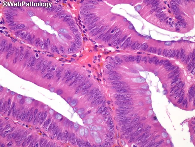
Histopathology-grade colonic adenoma demonstrates low-grade dysplasia characterized by mildly enlarged, elongated, hyperchromatic nuclei that maintain polarity and uniform cytomorphology. Nuclear crowding and pseudostratification are observed, while nucleoli remain inconspicuous. Goblet cell differentiation is reduced, contributing to a more compact mucosal epithelium. Architecturally, the mucosal glands in the polyp are crowded and branched, forming tubular glands with increased gland-to-stroma ratio. The surface epithelium shows longitudinal orientation and slight loss of mucinous content, corresponding to decreased goblet cell density. Despite these changes, cytologic atypia is limited, and invasion is not evident. The image represents a formalin-fixed, paraffin-embedded colonic mucosa section stained with Hematoxylin and Eosin, highlighting small, dysplastic crypts rising above adjacent non-neoplastic mucosa. This histology corresponds to a low-grade dysplasia category (mild-to-moderate) as part of colonic adenoma grading, with the alternative high-grade category including severe dysplasia and carcinoma in situ. Clinically, recognizing low-grade vs high-grade dysplasia influences surveillance intervals, polypectomy strategy, and cancer risk stratification. In a diagnostic workup, these features guide decisions about endoscopic removal, histologic follow-up, and correlation with endoscopic appearance to rule out invasive carcinoma. This description supports precise labeling of lesion grade, informs management planning, and facilitates standardized reporting in multidisciplinary tumor boards and pathology education globally.
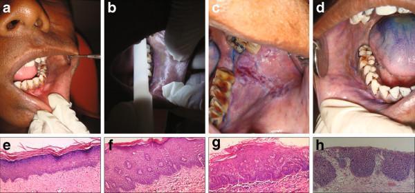
This composite educational graphic details clinical and histopathological diagnostic screening protocols for oral dysplastic lesions. Panels A-D demonstrate clinical examination techniques: (A) conventional visual inspection under incandescent light showing a suspicious oral mucosa lesion; (B) chemiluminescent illumination (430-580nm) where positive lesions appear blue-white/acetowhite; (C) application of toluidine blue showing dark royal blue retention; and (D) a combined diagnostic protocol. Panels E-H present H&E stained photomicrographs (10x magnification) representing the histopathological grading scale for epithelial dysplasia: (E) No dysplasia, showing regular epithelial architecture and lack of atypia; (F) Mild dysplasia, with cellular atypia and architectural changes limited to the lower third of the epithelium and superficial keratosis; (G) Moderate dysplasia, where architectural changes extend into the middle third of the epithelial layer; and (H) Severe dysplasia, characterized by full-thickness architectural disturbances and marked cellular atypia. This visual resource illustrates the correlation between clinical screening adjunctive tools and gold-standard histopathological classification in oral oncology.
Epithelial Dysplasia - Oral Pathology (10 Marks)
Definition
- WHO definition: "A lesion of altered epithelium with cellular atypism and loss of normal maturation, which does not fulfil criteria for carcinoma in situ."
- Synonyms: Intraepithelial neoplasia (IEN), oral intraepithelial neoplasia (OIN), precancerous epithelial change
- Dysplasia is a histopathological diagnosis - it cannot be made clinically
Significance
- Epithelial dysplasia is the most important prognostic indicator in oral PMDs (leukoplakia, erythroplakia, OSMF)
- Presence and grade of dysplasia directly determines risk of malignant transformation
- Dysplasia is the primary reason all oral PMD biopsies are mandatory
- ~15% of oral premalignant lesions with dysplasia progress to SCC over a mean interval of 5 years
Histological Features of Epithelial Dysplasia
A. Architectural (Structural) Changes
- Irregular epithelial stratification - loss of the orderly progression from basal → spinous → granular → keratin layers
- Loss of polarity of basal cells - basal cells no longer oriented perpendicular to the basement membrane; cells show chaotic orientation
- Drop-shaped rete ridges - bulbous, club-shaped or teardrop-shaped rete pegs instead of the normal slender finger-like projections
- Increased number of mitotic figures - abnormal distribution; mitoses found in suprabasal layers (normally only in basal layer)
- Abnormal superficial mitoses - mitotic figures in the upper half of the epithelium (should never be seen in normal epithelium)
- Premature keratinization in individual cells below the surface (dyskeratosis)
- Keratin pearls within the rete pegs - concentric laminated whorls of keratin within the spinous layer
- Irregular distribution of keratinisation
B. Cytological (Cellular) Changes
- Nuclear enlargement - nuclei disproportionately large relative to cell size
- Increased nuclear-cytoplasmic (N:C) ratio - nucleus occupies most of the cell
- Nuclear hyperchromatism - darkly staining nuclei due to increased DNA content; coarsely granular chromatin
- Nuclear pleomorphism - variation in size and shape of nuclei within the same lesion
- Prominent and enlarged nucleoli (often multiple)
- Abnormal (atypical) mitotic figures - tripolar, ring-shaped, asymmetric mitoses (usually only in higher grades)
- Increased number and size of nucleoli
- Cellular pleomorphism - variation in cell size and shape
- Dyskeratosis - premature individual cell keratinization (cells in lower layers showing abundant eosinophilic cytoplasm)
- Loss of intercellular cohesion - loosening of contact between epithelial cells
- Basal cell hyperplasia - increased number of basal cells; 2-3 layers of basal cells instead of one
WHO Criteria for Diagnosing Oral Epithelial Dysplasia
- Irregular epithelial stratification
- Loss of polarity of basal cells
- Drop-shaped rete ridges
- Increased mitotic figures
- Abnormal superficial mitoses
- Premature keratinization in single cells (dyskeratosis)
- Keratin pearls within rete pegs
- Abnormal variation in nuclear size (anisokaryosis)
- Abnormal variation in nuclear shape (nuclear pleomorphism)
- Abnormal variation in cell size (anisocytosis)
- Abnormal variation in cell shape (cellular pleomorphism)
- Increased N:C ratio
- Increased number and size of nucleoli
- Atypical mitotic figures
- Hyperchromasia
Grading of Epithelial Dysplasia
WHO Three-Tier Grading System (Classic / Shafer's)
| Grade | Thickness Affected | Features |
|---|---|---|
| Mild Dysplasia | Lower 1/3 (basal and parabasal layer only) | Minimal architectural and cytological changes; normal maturation in upper 2/3 |
| Moderate Dysplasia | Lower 2/3 of epithelium | Moderate atypia; changes extend to middle third; some abnormal mitoses |
| Severe Dysplasia | >2/3 of epithelial thickness | Marked atypia throughout most of epithelium; many atypical mitoses; nearly full-thickness involvement |
| Carcinoma in Situ | Full thickness | All cellular features of malignancy throughout entire epithelium; basement membrane intact |
Histological Image
Binary (Ljubljana / WHO 2017) System
| Grade | Equivalent to |
|---|---|
| Low-grade dysplasia (LGD) | Mild + Moderate dysplasia |
| High-grade dysplasia (HGD) | Severe dysplasia + CIS |
Grading Comparison Table
| Feature | Mild | Moderate | Severe | CIS |
|---|---|---|---|---|
| Thickness involved | Lower 1/3 | Lower 2/3 | >2/3 | Full |
| Nuclear atypia | Minimal | Moderate | Marked | Extreme |
| Atypical mitoses | Absent | Rare | Present | Many |
| Basement membrane | Intact | Intact | Intact | Intact |
| Progression to SCC (5yr) | 1-2% | 3-5% | 5-15% | ~18% |
Special Points in Dysplasia Assessment
1. Basement Membrane
- Always intact in all grades of dysplasia including CIS
- Breaching = invasive SCC
- Confirmed with PAS or reticulin stain
2. Subepithelial Inflammatory Infiltrate
- Chronic inflammatory cells (lymphocytes, plasma cells) frequently present beneath dysplastic epithelium
- Represents host immune surveillance
- Dense infiltrate may indicate higher grade lesion
3. Surface Keratinization
- Hyperorthokeratosis or hyperparakeratosis commonly overlies dysplastic epithelium
- The keratinized surface is the basis for the white (leukoplakic) clinical appearance
4. Limitations of Grading
- High inter-observer variability - same biopsy may be graded differently by different pathologists
- Distinction between mild and moderate is particularly subjective
- Only representative sampling of the most suspicious area gives meaningful results
Malignant Transformation Risk by Grade
| Dysplasia Grade | 3-Year Risk of Invasive SCC |
|---|---|
| None (no dysplasia) | ~3.7% |
| Mild/Moderate | ~10.1% |
| Severe/CIS | ~18.1% |
Relationship to Oral Potentially Malignant Disorders
| PMD | Frequency of Dysplasia |
|---|---|
| Leukoplakia | ~5-25% of biopsies show dysplasia |
| Erythroplakia | ~90% show severe dysplasia/CIS |
| OSMF | 46% mild, 52% moderate, 2% severe dysplasia |
| Oral lichen planus (erosive) | 0.5-5% |
| Actinic cheilitis | Significant proportion |
Investigations
- Incisional biopsy - mandatory; gold standard
- Take from the most suspicious area (most erythematous, indurated)
- Multiple biopsies for large lesions
- Toluidine blue staining - vital stain; retained by dysplastic cells (guides biopsy site)
- Autofluorescence (VELscope) - loss of fluorescence in dysplastic areas
- Narrow-band imaging (NBI) - highlights abnormal vascularity
- Brush biopsy (OralCDx) - cytological screening adjunct
- Immunohistochemistry:
- Ki-67 - elevated proliferative index correlates with grade
- p53 - overexpression indicates mutation
- p16 - evaluates HPV association
- Cyclin D1 - overexpressed in higher grades
- LOH (Loss of Heterozygosity) analysis - chromosomes 3p, 9p, 17p loss predicts progression
Treatment Based on Grade
| Grade | Management |
|---|---|
| Mild dysplasia | Habit cessation; close surveillance every 3-6 months; repeat biopsy if change |
| Moderate dysplasia | Excision preferred; risk factor elimination; close follow-up |
| Severe dysplasia / CIS | Surgical excision with margins; CO2 laser; mandatory follow-up |
- Cessation of tobacco and alcohol - mandatory at all grades; mild dysplasia may regress
- Surgical excision (scalpel or CO2 laser) for moderate-severe grades
- Photodynamic therapy (PDT) - for extensive/multifocal lesions
- Long-term surveillance - mandatory regardless of treatment; field cancerization means entire at-risk mucosa must be monitored
- Patient education and counselling
Summary Table
| Feature | Description |
|---|---|
| Definition | Premalignant epithelial change; cellular atypia + loss of maturation |
| Diagnosis | Histopathological only (biopsy) |
| Grading | Mild / Moderate / Severe / CIS (WHO 3-tier); Low/High grade (WHO 2017 binary) |
| Key architectural feature | Drop-shaped rete ridges; abnormal superficial mitoses |
| Key cytological feature | Nuclear hyperchromatism; increased N:C ratio; atypical mitoses |
| Basement membrane | Always intact (breached = invasive SCC) |
| Highest risk lesion | Erythroplakia (90% CIS/severe dysplasia) |
| Transformation risk | 3.7% (none) → 18.1% (severe/CIS) over 3 years |
| Treatment | Excision + habit cessation + surveillance |
- Dysplasia is a histopathological diagnosis - clinical appearance alone is unreliable; biopsy is always mandatory
- The two essential criteria are: (a) cellular atypia and (b) loss of normal maturation/stratification
- The drop-shaped rete ridge and abnormal superficial mitoses are the most reliable architectural markers
- Grading is notoriously subjective - the same slide may be called mild by one pathologist and moderate by another; clinical correlation is essential
- Severe dysplasia and CIS are clinically managed the same way - both require excision
- Erythroplakia is the oral lesion most likely to harbour severe dysplasia or CIS (~90%)
- The basement membrane is intact in ALL grades of dysplasia including CIS - its breach defines invasive SCC
Squamous cell carcinoma
oral squamous cell carcinoma tongue ulcer indurated border clinical
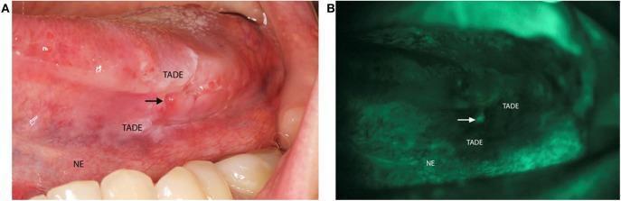
This dual-panel clinical photograph demonstrates oral squamous cell carcinoma screening using conventional white light and VELscope® tissue autofluorescence imaging. Panel A is a clinical photograph of the left lateral border of the tongue showing an indurated, erythematous ulcerated lesion (marked by an arrow) consistent with biopsy-proven squamous cell carcinoma. The surrounding area is labeled as Tumor Adjacent Dysplastic Epithelium (TADE), which appears clinically suspicious, while the more distal tissue is labeled as Normal Epithelium (NE). Panel B illustrates the same anatomical region under VELscope® examination. The malignant ulcer and the surrounding TADE exhibit significant fluorescence visualization loss (appearing as dark, non-fluorescent patches), whereas the NE maintains normal pale green autofluorescence. This comparison highlights the clinical utility of autofluorescence imaging in delineating surgical margins and identifying dysplastic changes that may not be fully apparent under standard illumination. The image serves as an educational tool for oral oncology, pathology, and diagnostic screening techniques.
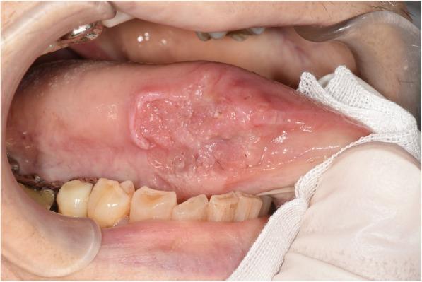
This clinical photograph displays a primary lesion on the right lateral border of the tongue in an adult patient. The lesion is a large, irregularly shaped ulcer with raised, indurated, and rolled-out edges, characteristic of oral squamous cell carcinoma. The central portion of the lesion exhibits a heterogeneous surface with areas of granulation, erosion, and mucosal thickening. The surrounding tongue tissue appears edematous. Visible anatomical context includes the lower dentition, which shows signs of wear and attrition, and the oral vestibule retracted by a dental speculum. A gloved hand and surgical gauze are visible at the right of the frame, indicating a clinical examination or preoperative setting. The lesion is located approximately midway between the tongue tip and the posterior lateral border, illustrating a classic presentation for intraoral malignancy. This image is an educational resource for dental and medical students to identify suspicious oral mucosal changes and understand the clinical morphology of tongue ulcers.
Oral Squamous Cell Carcinoma (OSCC) - Oral Pathology (10 Marks)
Definition
- 95% of all oral cancers are SCC; the remainder are largely adenocarcinomas of salivary gland origin
- HNSCC (Head and Neck SCC) is the 6th most common neoplasm worldwide
- ~650,000 new cases globally per year; ~50,000 in the USA annually
- (Robbins, Cotran & Kumar Pathologic Basis of Disease)
Epidemiology
- Age: Most common in 5th-7th decade (40-70 years); rising incidence in younger patients (<40 yrs) especially tongue SCC
- Sex: Males > Females (historically 2:1; ratio narrowing with changing habits)
- Geography: High incidence in India, Southeast Asia (betel nut), and increasingly in Western countries (HPV-related)
- In India, oral cancer is the most common cancer in men and accounts for ~30% of all cancers
Etiology and Risk Factors
A. Carcinogen-Associated (Classic / HPV-Negative) - Oral Cavity
- Tobacco - single most important risk factor
- Smoked: cigarettes, bidis, cigars, pipes, chutta (reverse smoking)
- Smokeless: chewing tobacco, snuff, gutka, mawa
- Carcinogens: polycyclic aromatic hydrocarbons, nitrosamines, benzopyrene
- Dose-response relationship - risk increases with quantity and duration
- Alcohol - acts synergistically with tobacco (multiplicative risk, not additive)
- Acetaldehyde (carcinogenic metabolite)
- Increases mucosal permeability to carcinogens
- Heavy drinkers have 6-fold increased risk
- Areca nut / Betel quid (paan) - major risk in South and Southeast Asia
- Chronic mucosal irritation - ill-fitting dentures, sharp cusps, chronic trauma
- Nutritional deficiencies - iron (Plummer-Vinson syndrome), vitamins A, C, E
- Syphilis (historical) - chronic atrophic glossitis
- Actinic radiation - solar UV light for lip SCC (pipe smokers + sun exposure)
- Immunosuppression - HIV, organ transplant recipients
- Field cancerization - pre-existing premalignant lesions
B. HPV-Associated - Oropharynx (Tonsil, Base of Tongue)
- HPV types 16 and 18 - account for ~90% of oropharyngeal SCC
- Transmitted via orogenital sexual contact
- HPV E6 oncoprotein degrades p53; E7 oncoprotein inactivates Rb
- HPV-associated SCC: younger patients, better prognosis, overexpresses p16
- Increasing dramatically in Western countries - now outnumbers tobacco-related oral SCC in USA/Europe
C. Premalignant Lesions (Direct Precursors)
| Lesion | Transformation Rate |
|---|---|
| Leukoplakia | 5-25% |
| Erythroplakia | >50% (91% already dysplasia/CIS at biopsy) |
| OSMF | ~7.6% over 17 years |
| Oral lichen planus (erosive) | 0.5-5% |
| Actinic cheilitis | Significant |
Pathogenesis (Molecular)
- Mutational signature from carcinogen exposure
- Key mutations: TP53 (most common), CDKN2A (p16), PIK3CA, RAS
- Loss of function: NOTCH1, FAT1 (cadherin family tumour suppressor)
- Progression: Normal → Hyperplasia → Dysplasia → CIS → Invasive SCC
- HPV-16 E6 → degrades p53 (bypasses TP53 mutation)
- HPV-16 E7 → inactivates Rb (bypasses CDKN2A mutation)
- Hence HPV-positive tumours have fewer somatic mutations
- Overexpresses p16 (as a compensatory response to Rb inactivation)
- Better clinical outcomes despite nodal metastasis at presentation
- Years of carcinogen exposure creates a genetically altered mucosal field
- Multiple independent tumour clones arise simultaneously
- Explains 3-7% per year rate of second primary tumours
- ~35% chance of developing a new primary within 5 years of first tumour
Clinical Features
Sites of Predilection
- Ventral and lateral tongue - most common site overall
- Floor of mouth - "coffin of the mouth" (high-risk zone)
- Lower lip - especially pipe smokers, outdoor workers
- Soft palate and fauces
- Gingiva / alveolar mucosa
- Buccal mucosa - especially in betel nut/tobacco chewers (India)
- Tonsillar crypts, base of tongue, posterior pharyngeal wall, soft palate
Gross Morphology / Clinical Appearances
- Raised, firm, pearly white plaque or irregular mucosal thickening
- Roughened, verrucous, or granular surface
- May be preceded by / superimposed on leukoplakia or erythroplakia
- Often asymptomatic in early stages
| Feature | Description |
|---|---|
| Ulceration | Central necrotic ulcer with irregular base; gray/yellow slough |
| Rolled/everted border | Raised, thickened, indurated margins - classic "rolled border" |
| Induration | Hard, firm consistency on palpation - ALWAYS suspicious |
| Fixation | Fixed to underlying structures (muscle, bone) in advanced disease |
| Colour | Red, white, or mixed (red+white most suspicious) |
| Surface | Irregular, granular, verrucous, or ulcerated |
| Bleeding | Spontaneous or on palpation; bleeds easily |
Symptoms (in order of progression):
- Painless ulcer or lump - early; classically painless until advanced
- Persistent sore that does not heal (>3 weeks = investigate)
- Pain - develops as tumour invades nerves; may radiate to ear (otalgia)
- Trismus - involvement of pterygoid muscles (buccal/tongue SCC)
- Dysphagia - difficulty swallowing (floor of mouth, tongue)
- Dysarthria - altered speech (tongue involvement)
- Halitosis - fetid odour from necrotic tumour
- Bleeding from the lesion
- Numbness / paraesthesia - nerve invasion (mental nerve, inferior alveolar nerve)
- Neck lump - cervical lymphadenopathy (regional metastasis)
Clinical Image
Histopathological Features
- Grade I (Well differentiated): Keratin pearls, intercellular bridges, moderate pleomorphism, basement membrane breached
- Grade II (Moderately differentiated): Fewer pearls, more atypia, more mitoses
- Grade III (Poorly differentiated): No pearls, sheets of cells, extreme atypia, many atypical mitoses
- HPV-associated SCC: Non-keratinizing; basaloid cells in nests within lymphocytic stroma; p16 strongly positive
- Desmoplasia: Reactive fibrous stroma around invading nests
- Perineural invasion and lymphovascular invasion - poor prognostic features
- Depth of invasion >4 mm - significantly increased nodal metastasis risk
- Grade I: >75% differentiated
- Grade II: 50-75% differentiated
- Grade III: 25-50% differentiated
- Grade IV: <25% differentiated
Spread and Metastasis
Local Spread:
- Infiltrates along tissue planes, muscles, periosteum, bone
- Perineural spread (along nerves) - explains distant pain and numbness
Regional (Lymphatic):
- Cervical lymph nodes - most common route
- Level I (submental, submandibular) → Level II (upper jugular) → Level III/IV/V
- Floor of mouth / tongue → bilateral nodal spread (midline lesions)
- Ipsilateral nodes first, then contralateral
Distant Metastasis:
- Mediastinal lymph nodes → Lungs → Liver → Bones
- Distant mets often present at time of discovery of primary
Investigations
1. Biopsy
- Incisional biopsy from most suspicious area - gold standard
- Avoid necrotic centre; take from periphery (advancing edge)
- Multiple biopsies if large lesion
2. Imaging
| Investigation | Purpose |
|---|---|
| MRI | Soft tissue extent, depth of invasion, perineural spread, base of tongue |
| CT (contrast) | Neck nodal assessment, bone erosion (mandible, maxilla) |
| OPG / CBCT | Mandibular/maxillary bone involvement |
| PET-CT | Distant metastasis detection; staging; post-treatment surveillance |
| Chest X-ray / CT chest | Pulmonary metastasis |
| USG neck | Node assessment; USG-guided FNAC of nodes |
3. Laboratory
- CBC, LFT, RFT, coagulation - pre-surgical workup
- Serum calcium - if bone metastasis suspected
4. Auxiliary Aids for Screening
- Toluidine blue vital staining
- VELscope (autofluorescence) - loss of fluorescence in dysplastic/malignant areas
- NBI (Narrow band imaging) - abnormal vasculature
- Brush biopsy (OralCDx) - cytological screening
TNM Staging (AJCC 8th Edition)
| T | Primary Tumour |
|---|---|
| T1 | ≤ 2 cm; depth of invasion (DOI) ≤ 5 mm |
| T2 | ≤ 2 cm with DOI > 5 mm; or 2-4 cm with DOI ≤ 10 mm |
| T3 | > 4 cm; or any size with DOI > 10 mm |
| T4a | Moderately advanced: invades cortical bone, inferior alveolar nerve, floor of mouth, skin |
| T4b | Very advanced: masticator space, pterygoid plates, skull base, encases carotid artery |
| N | Regional Nodes |
|---|---|
| N0 | No regional node metastasis |
| N1 | Single ipsilateral node ≤ 3 cm |
| N2a | Single ipsilateral node 3-6 cm |
| N2b | Multiple ipsilateral nodes ≤ 6 cm |
| N2c | Bilateral/contralateral nodes ≤ 6 cm |
| N3 | Node > 6 cm |
| M | Distant Metastasis |
|---|---|
| M0 | None |
| M1 | Present |
Treatment
Principles: Surgery is primary; radiation and chemotherapy are adjuncts
A. Surgery
- Wide local excision with adequate margins (minimum 1-1.5 cm clear margin)
- Confirm with frozen section intraoperatively
- Composite resection (for bone involvement) - segmental mandibulectomy / maxillectomy
- Neck dissection for nodal disease:
- Elective (staging/prophylactic) for N0 neck if DOI > 4 mm
- Therapeutic neck dissection for N+ disease
- Types: Radical, Modified radical, Selective (levels I-III or I-IV)
- Reconstruction:
- Primary closure (small defects)
- Local flaps: nasolabial, buccal fat pad, tongue flap
- Regional flaps: pectoralis major myocutaneous flap (PMMF)
- Free flaps: radial forearm free flap (RFFF), fibula free flap (for bone reconstruction)
B. Radiotherapy
- Post-operative (adjuvant) radiation - for high-risk features:
- Positive/close margins, perineural/lymphovascular invasion, extranodal extension, N2/N3 disease
- Definitive radiation - for patients unfit for surgery or organ-preservation intent
- Dose: typically 60-66 Gy in 30-33 fractions
- IMRT (Intensity Modulated Radiation Therapy) - minimises parotid gland damage (reduces xerostomia)
C. Chemotherapy
- Cisplatin - most widely used; sensitises tumour to radiation
- Concurrent chemoradiation (CRT) - standard for locally advanced or unresectable disease
- Induction chemotherapy (TPF: Taxane + Platinum + 5-FU) - for very advanced disease to reduce tumour bulk before definitive therapy
- Cetuximab (anti-EGFR monoclonal antibody) - for platinum-ineligible patients
- Checkpoint inhibitors (PD-1 inhibitors): Pembrolizumab, Nivolumab - approved for recurrent/metastatic HNSCC
Prognosis
| Stage | 5-Year Survival |
|---|---|
| Stage I (T1N0M0) | ~80-85% |
| Stage II | ~70-75% |
| Stage III | ~50-60% |
| Stage IV | ~20-30% |
| Overall (all stages) | ~50% |
- Good prognosis: Early stage, well-differentiated, negative nodes, HPV-positive (oropharynx), clear surgical margins, dense lymphocytic infiltrate
- Poor prognosis: Advanced stage, perineural invasion, lymphovascular invasion, positive nodes (especially extranodal extension), positive/close margins, poorly differentiated, new second primary tumours
Summary Table
| Feature | OSCC |
|---|---|
| Frequency | 95% of oral malignancies |
| Most common site | Lateral/ventral tongue, floor of mouth |
| Most common risk factor | Tobacco + alcohol (synergistic) |
| HPV association | Types 16, 18 - especially oropharynx |
| Precursor lesions | Leukoplakia, erythroplakia, dysplasia |
| Classic clinical sign | Indurated ulcer with rolled/everted borders |
| Histology hallmark | Keratin pearls + intercellular bridges + stromal invasion |
| Grading | Broders' I-IV (well → undifferentiated) |
| Spread | Local → cervical nodes → lungs, liver, bone |
| Treatment | Surgery + neck dissection ± adjuvant CRT |
| 5-year survival | ~50% overall |
- The "high-risk triad" of sites in oral cavity: ventral/lateral tongue + floor of mouth + soft palate - any persistent lesion here must be biopsied
- Painless indurated ulcer that does not heal in 3 weeks = OSCC until proven otherwise
- Field cancerization explains why 3-7% of patients develop new primary tumours annually - the entire at-risk mucosa must be surveilled
- HPV-associated oropharyngeal SCC behaves differently from classic tobacco-related oral SCC - better prognosis, younger patients, small primary with bulky nodes, p16 positive
- Depth of invasion (not just size) is the most important determinant of nodal metastasis and is now incorporated in the AJCC 8th edition T-staging
- Early detection is the most critical factor - Stage I/II has ~80% 5-year survival vs 20% for Stage IV
Basal cell carcinoma
basal cell carcinoma nodular pearly border telangiectasia face nose
This clinical photograph displays a nodular basal cell carcinoma located on the right side of a patient's nose. The lesion is a well-circumscribed, oval-shaped, pinkish-flesh-colored nodule approximately 1.5 cm in diameter, circled in purple marker for clinical identification. Key diagnostic features include a distinct pearly, translucent surface and classic rolled borders. A central focal ulceration with hemorrhagic crusting is evident, indicating a 'rodent ulcer' presentation. Arborescent telangiectasias (fine, dilated blood vessels) are visible on the surface of the nodule and extending onto the adjacent skin of the cheek. The surrounding skin shows signs of chronic actinic damage, including solar lentigines. This image serves as a classic educational example of the most common subtype of basal cell carcinoma, highlighting the importance of recognizing texture, vascularity, and border morphology in dermatological diagnosis.
Clinical skin photography of a solitary nodular basal cell carcinoma on the forehead. Modality: clinical photography; frontal close-up view with natural color balance to depict surface features. The lesion is a dome-shaped, pearly papule with an elevated translucent border and fine arborizing telangiectasias. Central depression or ulceration is present, consistent with a nodular (rodent ulcer) subtype. Location is sun-exposed facial skin, typical for basal cell carcinoma; surrounding skin may show mild erythema and faint indistinct margins. Impression: classic BCC morphology with characteristic nesting of basaloid cells on histology, palisading peripheral arrangement, and retraction artifacts. Clinically, these findings carry high local invasion potential but low metastatic risk; management commonly involves complete surgical excision, Mohs micrographic surgery, or standard excision with clear margins, sometimes imiquimod or curettage depending on subtype and resources. Differential diagnoses include pigmented BCC variants, keratoacanthoma, sebaceous hyperplasia, or trichoblastic tumors; however, pearly edge and telangiectasia strongly favor BCC. The image is educational for dermatology trainees, medical students, and clinicians assessing cutaneous tumors; it illustrates key features of sun-damaged skin neoplasms and reinforces the importance of biopsy for confirmation. Revisit if lesion changes or enlarges, or if there is pain or bleeding; document growth behavior and plan definitive treatment.
Basal Cell Carcinoma (BCC) - Oral Pathology / Dermatology (10 Marks)
Definition
- Most common human malignancy - >3 million new cases per year in the USA
- Accounts for ~75% of all non-melanoma skin cancers (NMSC) and ~25% of all cancers in the USA
- Incidence increasing worldwide by 3-10% per year
- Also called "rodent ulcer" (advanced ulcerated nodular type) - a historical term referring to its locally destructive, "gnawing" nature
- BCC is a malignant neoplasm of follicular germinative differentiation (Fitzpatrick's)
- (Fitzpatrick's Dermatology, p. 1915)
Epidemiology
- Age: Most common in patients >60 years; increasingly seen in those <50 years (especially women <40)
- Sex: Males > Females (slightly)
- Race: Predominantly in fair-skinned (Fitzpatrick types I-II) individuals; very rare in dark skin (melanin provides photoprotection)
- Geography: Higher incidence in regions with intense sunlight (Australia, Southern USA)
- Incidence: ~19 times more common in whites than blacks
- Lifetime risk in whites: ~1 in 5 (increases to 1 in 3 for those >65 years in southern latitudes)
- Organ transplant recipients: 5-10 times greater risk
Etiology and Risk Factors
Primary - Ultraviolet Radiation (UVR):
- UVB (290-320 nm) - primary carcinogenic wavelength
- Causes DNA damage → mutations in tumour suppressor genes
- Cumulative lifetime UV exposure is the most important risk factor
- Both chronic sun exposure and intermittent intense sun exposure (sunburns) contribute
Risk Factors:
- UV radiation (sunlight, tanning beds, phototherapy)
- Fair skin, light eyes, light hair (Fitzpatrick type I-II)
- Northern European ancestry
- Inability to tan / tendency to burn
- Ionising radiation - prior radiation therapy (latency of years to decades)
- Chronic arsenic exposure (arsenical keratoses precede BCC)
- Immunosuppression - organ transplant recipients, HIV
- Genetic syndromes:
- Gorlin-Goltz syndrome (Basal Cell Naevus Syndrome / BCNS) - multiple BCCs, jaw keratocysts, calcified falx cerebri; PTCH1 germline mutation
- Xeroderma Pigmentosum - defective DNA repair
- Bazex syndrome (follicular atrophoderma + BCCs)
- Rombo syndrome
- Chronic mucosal trauma / scars (rare)
- Tobacco - minor association
Pathogenesis - Hedgehog Signalling Pathway (KEY)
Normal Hedgehog Pathway:
- PTCH1 (Patched-1) receptor normally inhibits Smoothened (SMO)
- When Sonic Hedgehog (SHH) ligand binds PTCH1 → PTCH1 inhibition lifted → SMO becomes active → downstream transcription factors activated → cell proliferation
In BCC - Aberrant Activation:
- PTCH1 mutation (loss of function) - 73% of sporadic BCCs - removes brake on SMO → constitutive pathway activation
- SMO mutation (gain of function) - 20% of sporadic BCCs
- SUFU mutation (loss of function) - 8%
- TP53 mutations - found in 61% (UV-induced)
- Result: uncontrolled basal cell proliferation even without SHH ligand
Most Common Mutated Genes in BCC (Fitzpatrick's):
| Gene | Role | Prevalence |
|---|---|---|
| PTCH1 | Tumour suppressor (HH pathway) | 73% |
| TP53 | Tumour suppressor | 61% |
| SMO | Oncogene (HH pathway) | 20% |
| SUFU | Tumour suppressor (HH pathway) | 8% |
Clinical Features
Common Sites:
- Head and neck (80%) - especially nose, nasolabial folds, periocular region, ears, forehead, scalp
- "H-zone" (high-risk zone) of the face: nose, eyelids, eyebrows, temples, lips, ear, periauricular skin - higher risk of subclinical extension
- Trunk and extremities (superficial BCC)
- Oral cavity BCC: Rare (oral mucosa has no hair follicles); mainly lip/perioral skin
Subtypes and Clinical Appearances:
1. Nodular BCC (Most Common ~60-70%)
- Translucent (pearly), dome-shaped papule or nodule
- Rolled/raised borders - classic "rolled border"
- Telangiectasias (arborizing blood vessels) overlying the lesion
- Central depression or ulceration with bleeding
- Advanced lesion = "Rodent ulcer" - central ulcer with raised pearly rolled borders
- Slow growing, usually < 2 cm at diagnosis
2. Superficial BCC (Second Most Common ~15-30%)
- Flat, well-demarcated erythematous patch or plaque
- Scaly surface with thin, thread-like rolled borders
- Most common on trunk and extremities
- May be confused with eczema, psoriasis, or Bowen's disease
3. Morpheaform / Sclerosing / Infiltrating BCC (Least Common, ~5%)
- Flat, pale, scar-like, indurated plaque
- Poorly defined margins (resembles a morphea / scleroderma patch)
- Most aggressive subtype; highest subclinical extension
- Often missed or misdiagnosed as a scar
- Requires wider excision margins
4. Pigmented BCC
- Subtype of nodular BCC with increased melanin
- Hyperpigmented, translucent papule - may resemble melanoma
- Melanocytes interspersed between tumour cells
5. Basosquamous (Metatypical) BCC
- Features of both BCC and SCC
- More aggressive; slightly higher metastatic potential
General Clinical Features:
- Usually asymptomatic - discovered incidentally or noticed as non-healing lesion
- Does not heal - patient may report "recurrent sore" or "spot that bleeds"
- May bleed with minor trauma
- Painless in most cases
- Metastasis is extremely rare (<0.1%) but possible in very advanced, neglected, or basosquamous type
Clinical Image
Histopathological Features (KEY)
1. Tumour Cell Characteristics:
- Basaloid cells - cells closely resembling the basal cells of the epidermis
- Large, oval, hyperchromatic nuclei - dark-staining, scanty cytoplasm
- High N:C ratio
- Scant pale cytoplasm - "naked nuclei" appearance
- Indistinct cell borders
- Mitotic figures present but not numerous
- Minimal cytological atypia (much less than SCC)
- No intercellular bridges (unlike SCC)
- No keratin pearls (unlike well-differentiated SCC)
2. Architectural Features:
- Nests, lobules, and islands of basaloid cells within the dermis
- Peripheral palisading - the most important architectural feature:
- Cells at the periphery of tumour nests are arranged in a single orderly layer, standing in a picket-fence pattern (nuclei aligned parallel to each other, perpendicular to the basement membrane)
- Retraction artefact (stromal clefting) - artificial separation between tumour nests and surrounding stroma (due to mucinous stroma and processing artefact)
- This is a key diagnostic feature distinguishing BCC from trichoblastoma
- Mucinous stroma (myxoid ground substance) surrounding tumour nests
- Connection to overlying epidermis or hair follicles often seen
3. Histological Subtypes:
| Subtype | Histological Feature |
|---|---|
| Nodular | Large nodules of basaloid cells; peripheral palisading; stromal retraction |
| Micronodular | Multiple small nodules < 15 μm; aggressive |
| Superficial | Small buds of basaloid cells budding downward from basal layer; limited to upper dermis |
| Morpheaform/Sclerosing | Thin elongated strands of basaloid cells in dense fibrous (sclerotic) stroma; no retraction; poorly defined margin |
| Infiltrating | Irregular spiky islands; aggressive stromal infiltration |
| Pigmented | Melanin granules within tumour cells and melanophages in stroma |
| Basosquamous | Areas of both BCC and SCC differentiation |
4. Stroma:
- Mucinous (myxoid) stroma - alcian blue positive
- Retraction artefact between tumour nests and stroma
- Fibroblastic stromal reaction
- Chronic inflammatory infiltrate (lymphocytes, plasma cells)
5. Differentiating BCC from Trichoblastoma:
| Feature | BCC | Trichoblastoma |
|---|---|---|
| Cytological atypia | Present | Minimal |
| Mitotic rate | Higher | Low |
| Necrosis | May be present | Absent |
| Stromal mucinosis | Yes | No |
| Clefting | Between nests and stroma | Within stroma |
| CK20+ Merkel cells | Absent | Present |
Investigations
- Skin biopsy - gold standard
- Shave biopsy - most common; usually adequate for diagnosis
- Punch biopsy - for flat morpheaform BCC or recurrent BCC in scar
- Dermoscopy - arborizing telangiectasias, blue-grey ovoid nests, leaf-like areas, spoke-wheel areas
- Reflectance confocal microscopy (RCM) - non-invasive in vivo tool
- Imaging (MRI/CT) - for locally advanced BCC with suspected deep invasion or perineural spread
- Histopathological examination - subtype determination guides treatment
Treatment
A. Surgical Treatments (Preferred)
| Method | Indication / Details |
|---|---|
| Standard surgical excision | Nodular BCC on low-risk sites; 3-5 mm margins (4 mm standard) |
| Mohs Micrographic Surgery (MMS) | Gold standard for high-risk BCC; complete margin examination; cure rate ~99% primary; ~92% recurrent BCC |
| Electrodesiccation and curettage (ED&C) | Small, low-risk, superficial or nodular BCC; not for H-zone |
| Cryosurgery | Small, low-risk lesions; cure rate 95-99%; poor cosmesis |
- Location in H-zone (nose, eyelids, ears, lips)
- Diameter > 2 cm
- Poorly defined clinical margins
- Aggressive histological subtypes (morpheaform, infiltrating, micronodular)
- Recurrent BCC
- Perineural invasion
B. Non-Surgical Treatments
| Agent | Use |
|---|---|
| Imiquimod 5% cream (immune modulator) | Superficial BCC; 5x/week for 6 weeks; 80-90% clearance |
| 5-Fluorouracil (5-FU) cream | Superficial BCC; twice daily up to 12 weeks; 90% histologic cure |
| Photodynamic therapy (PDT) | Superficial and thin nodular BCC |
| Radiotherapy | Elderly/unfit patients; inoperable cases; adjuvant for perineural invasion |
C. Targeted Therapy (Advanced/Metastatic BCC)
| Drug | Mechanism | Indication |
|---|---|---|
| Vismodegib (GDC-0449) | SMO inhibitor - blocks Hedgehog pathway | Locally advanced or metastatic BCC |
| Sonidegib (LDE225) | SMO inhibitor | Locally advanced BCC |
| Cemiplimab (anti-PD-1) | Checkpoint inhibitor | Advanced BCC not responding to Hedgehog inhibitors |
Prognosis
- Excellent prognosis with appropriate treatment
- Metastasis rate: < 0.1% (extremely rare)
- Recurrence rate after Mohs: 1-4% for primary BCC; 4-8% for recurrent BCC
- Recurrence rate after standard excision: ~5-10%
- Patients have ~30% higher risk of developing another BCC within the first 5 years
- Gorlin syndrome patients develop hundreds of BCCs over their lifetime
- Morpheaform / infiltrating subtypes have worse local prognosis due to subclinical extension
Summary Table
| Feature | BCC |
|---|---|
| Most common | Human malignancy overall |
| Origin | Basal layer of epidermis / hair follicle |
| Primary cause | UV radiation |
| Key pathway | Hedgehog (PTCH1/SMO) |
| Most common site | Head and neck (nose, periocular) |
| Most common subtype | Nodular (~60-70%) |
| Hallmark clinical sign | Pearly papule with rolled borders + telangiectasias |
| Advanced type | Rodent ulcer (central ulceration) |
| Histology hallmark | Peripheral palisading + stromal retraction |
| Metastasis | Extremely rare (<0.1%) |
| Treatment of choice | Surgical excision / Mohs surgery |
| Targeted therapy | Vismodegib (SMO inhibitor) |
- BCC does NOT typically occur within the oral mucosa - the oral cavity lacks hair follicles. When seen in the oral/perioral area, it involves the lip skin, perioral facial skin, and occasionally the jaw following tumour extension
- The histological hallmark of BCC is peripheral palisading of basaloid cells + stromal retraction artefact - these two features together are pathognomonic
- BCC and SCC must be distinguished: BCC has no keratin pearls, no intercellular bridges, and peripheral palisading; SCC has keratin pearls and intercellular bridges
- All BCCs share one molecular event - Hedgehog signalling pathway activation (PTCH1/SMO mutations)
- Morpheaform BCC is the most dangerous subtype due to ill-defined margins and extensive subclinical spread
- Gorlin-Goltz syndrome = multiple BCCs + odontogenic keratocysts (jaw) + skeletal anomalies - PTCH1 germline mutation (relevant to oral pathology!)
Veruccous ca
verrucous carcinoma oral cavity cauliflower exophytic white lesion
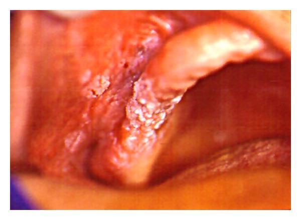
This clinical intraoral photograph displays a prominent, exophytic white lesion with a classic verrucous (wart-like) texture. The lesion is primarily localized to the molar region of the right maxillary alveolar ridge. It exhibits a distinctive cauliflower-like or papillary surface morphology and extends beyond the gingival tissues to involve the adjacent buccal vestibule and the mucosal lining of the inner cheek. The overall appearance is characterized by thick, hyperkeratotic white plaques that are confluent and well-demarcated from the surrounding erythematous oral mucosa. This visual presentation is highly characteristic of verrucous carcinoma, a low-grade variant of squamous cell carcinoma. The image serves as an educational example of oral oncology, highlighting the clinical features of proliferative epithelial lesions in the oral cavity for dental and medical students.
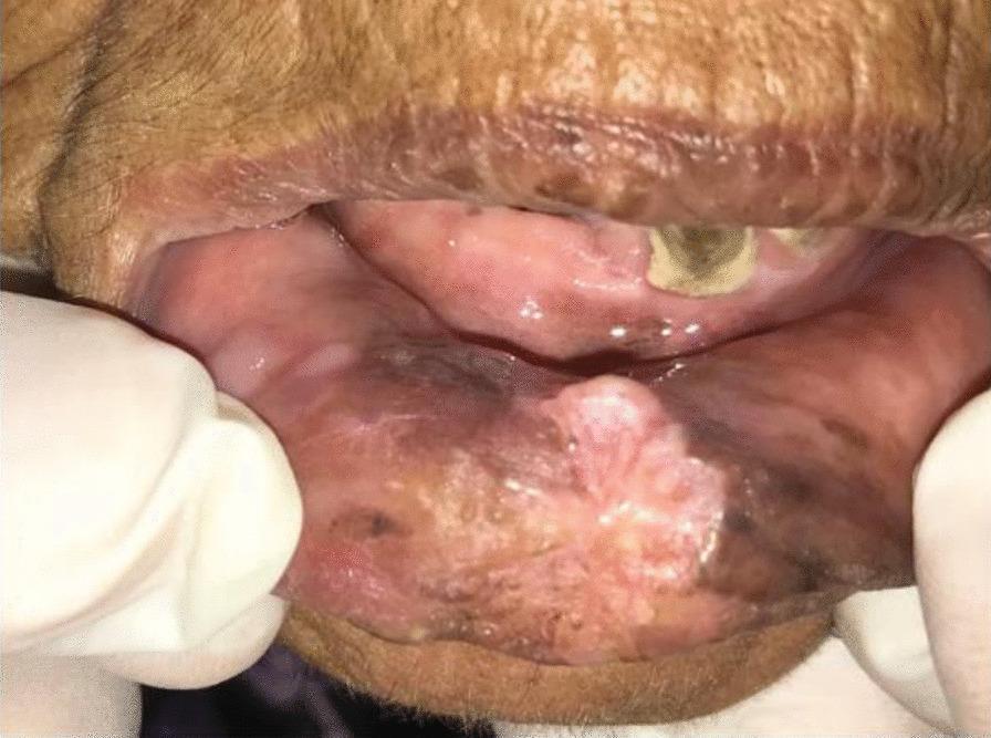
Clinical photograph of the oral cavity and lower lip in a 70-year-old patient, demonstrating an exophytic, verrucous lesion consistent with Oral Squamous Cell Carcinoma (OSCC). The lesion on the lower lip is characterized by a mixed red-white (erythroplakic and leukoplakic) appearance with an irregular, cauliflower-like texture. Significant dark hyperpigmentation and discoloration are visible in the surrounding labial mucosa. Intraoral examination reveals severe dental staining, particularly on the upper dentition, and mucosal changes on the upper gingiva including white patches and areas of erythematous inflammation. The overall presentation is characteristic of long-term tobacco or khat use, illustrating the progression from chronic mucosal irritation to verrucous carcinoma. This image serves as an educational example for identifying premalignant and malignant oral lesions during clinical screening.
Verrucous Carcinoma - Oral Pathology (10 Marks)
Definition
- First described by Ackermann (1948) as a distinct entity - hence also called "Ackermann's tumour"
- Synonymous with "Oral Florid Papillomatosis" when occurring in the oral cavity (term introduced by Kraus and Perezmesa, 1966)
- It is an intermediate-grade malignancy - more aggressive than a papilloma but far less so than conventional SCC
- Key histological dictum: "Looks benign, behaves malignant"
Subtypes / Sites (Three Classic Types)
| Subtype | Site |
|---|---|
| Oral Florid Papillomatosis | Oral mucosa - most relevant to oral pathology |
| Epithelioma Cuniculatum (Carcinoma Cuniculatum) | Plantar surface of foot; "rabbit burrow"-like sinuses |
| Giant Condyloma Acuminatum (Buschke-Löwenstein Tumour) | Genitoanal region |
Epidemiology
- Rare - accounts for ~2-3% of all oral malignancies
- Age: Predominantly elderly patients; most common in 5th-7th decades
- Sex: Males > Females (associated with smokeless tobacco use)
- Particularly prevalent in South and Southeast Asia (gutka, paan chewing)
- In Western countries - linked to snuff and chewing tobacco use
Etiology and Risk Factors
- Smokeless tobacco (chewing tobacco, snuff, gutka, mawa) - strongest association in oral VC
- Tobacco is placed in the buccal/labial sulcus - explains predilection for buccal mucosa
- Reverse smoking (chutta) - associated with palatal VC
- Areca nut / Betel quid - especially in South Asia
- HPV (Human Papillomavirus) - controversial association
- Low-risk types (HPV 6, 11) - found in some cases, especially Buschke-Löwenstein tumour
- High-risk types (HPV 16, 18) - occasionally implicated
- Role in oral VC is debated - HPV not always found; may not be etiologically essential
- Chronic mucosal irritation - ill-fitting dentures, sharp teeth, chronic trauma
- Poor oral hygiene
Clinical Features
Sites of Predilection (Oral Cavity):
- Buccal mucosa - most common (classically in tobacco chewers)
- Alveolar mucosa / Gingiva
- Hard palate
- Lip mucosa (lower lip)
- Tongue (less common)
- Floor of mouth (least common)
Gross / Clinical Appearance:
| Feature | Description |
|---|---|
| Shape | Large, exophytic, cauliflower-like (verrucous/papillary) mass |
| Surface | Thick, white, hyperkeratotic projections; warty, papillomatous texture |
| Colour | White (from heavy keratinization); may have red areas |
| Borders | Well-defined, broad-based; not infiltrating |
| Consistency | Soft initially; may become firm with invasion |
| Clefts/Crypts | Deep keratin-filled clefts between papillary projections - pathognomonic |
| Growth | Slow-growing - over months to years |
| Ulceration | Usually absent in early stages; may occur late |
| Induration | Minimal initially; increases as lesion enlarges |
| Bleeding | Uncommon until late |
| Size | Can become very large - sometimes enormous ("florid papillomatosis") |
Symptoms:
- Usually asymptomatic for prolonged periods
- Patient may notice a growing "wart" or "white patch" in the mouth
- No pain in early stages
- Difficulty chewing, speaking, swallowing as lesion enlarges
- Foul odour from keratin-filled clefts (halitosis)
- Gradual expansion with local bone destruction in advanced cases (alveolar bone, palate)
Clinical Image
Histopathological Features (KEY)
Low-Power (Scanning) Appearance:
- Broadly based, bulbous, blunt rete ridges pushing down into the connective tissue
- Undulating (verrucous) surface with broad papillary projections
- Keratin-filled crypts between papillary projections (endophytic invaginations)
- Pushing (compressive) margin - broad front advancing into stroma, without finger-like infiltration
- No spiky, irregular infiltrating islands (unlike conventional SCC)
Epithelial Features:
- Well-differentiated stratified squamous epithelium - highly orderly maturation
- Prominent hyperorthokeratosis or hyperparakeratosis
- Acanthosis - greatly thickened epithelium
- Minimal or absent nuclear atypia - cells look deceptively normal (key feature)
- Minimal mitotic activity - few mitoses, and only in basal/parabasal layers (no atypical mitoses)
- Cytoplasm often apple-pink and glassy in appearance
- Prominent granular cell layer (hypergranulosis)
- No keratin pearls within the stroma (unlike conventional SCC) - keratin confined to surface and crypts
- No intercellular bridges disruption
- Broad "elephant foot" rete ridges - bulbous, pushing downward as a blunt front
Stroma:
- Dense chronic inflammatory infiltrate at the tumour-stroma interface - prominent lymphocytes and plasma cells
- Peritumoral inflammation is characteristic and prominent
- No desmoplasia (unlike conventional SCC)
- Stroma compressed by pushing margin rather than invaded
Critical Distinguishing Feature:
"The squamous epithelium is well differentiated, and cytologic atypia is minimal. The tumor border is smooth and pushing, rather than spiky and infiltrative." - Andrews' Diseases of the Skin
Hybridisation with SCC:
- Hybrid verrucous carcinoma - contains foci of conventional SCC within an otherwise typical VC
- This hybrid form can metastasise - must be specifically reported
- Regular VC does not metastasise
Biopsy Challenge
- Superficial biopsies are inadequate - they only show hyperkeratosis and acanthosis with minimal atypia, which looks benign
- The diagnostic pushing margin and deep architecture can only be seen on deep incisional biopsy
- Many patients undergo multiple biopsies before diagnosis is confirmed
- Histopathologist must have clinical context - a large exophytic white lesion with minimal histological atypia should raise suspicion for VC
Differential Diagnosis
| Condition | Distinguishing Feature |
|---|---|
| Oral squamous papilloma | Small, pedunculated, koilocytes, fibrovascular cores, HPV clearly present; benign |
| Verruca vulgaris (common wart) | Small; viral inclusions (keratohyaline granules); HPV 2/4; koilocytes |
| Condyloma acuminatum | Multiple; genital involvement; HPV 6/11; koilocytes; sexually transmitted |
| Proliferative verrucous leukoplakia (PVL) | Multiple sites; slowly progresses to VC or SCC; no single dominant mass |
| Conventional SCC (well-differentiated) | Infiltrating margin; cytological atypia; atypical mitoses; keratin pearls in stroma |
| Inflammatory papillary hyperplasia | Under ill-fitting denture; many small nodules; no pushing margin |
| Oral florid papillomatosis | Essentially a synonym for oral VC; same entity |
Investigations
- Deep incisional biopsy - must include the base/advancing margin
- Multiple biopsies often required
- Superficial biopsy is inadequate - diagnosis missed
- Histopathological examination - gold standard
- MRI - to assess depth of invasion, bone involvement
- CT scan - bone erosion, extent of lesion
- OPG / CBCT - alveolar bone destruction
- HPV typing (PCR/ISH) - research purposes; does not change immediate management
Treatment
Treatment of Choice: Surgical Excision
- Wide local excision with adequate margins - primary treatment
- Confirm clear margins with frozen section / histopathology
- Conservative excision where possible (slow-growing, locally destructive only)
- Mohs Micrographic Surgery (MMS) - for cutaneous/perioral VC; maximises tissue conservation
- Neck dissection - NOT indicated for pure VC (does not metastasise)
- Required for hybrid VC with SCC component
Radiotherapy - Controversial:
- Traditionally avoided due to concern of "anaplastic transformation" (dedifferentiation to poorly differentiated SCC after RT)
- This risk was previously thought to be high but reviews have concluded these cases were likely hybrid tumours misdiagnosed as pure VC initially
- RT is now accepted as an alternative for:
- Extensive lesions where only treatment otherwise would be total laryngectomy
- Patients unfit for surgery
- Oral VC where surgery would cause severe morbidity
- Less radiosensitive than conventional SCC - higher doses required
- (Cummings Otolaryngology)
Other Modalities:
- CO2 laser excision - effective for accessible lesions; good healing
- Chemotherapy (bleomycin, cisplatin, 5-FU, methotrexate) - variable results
- Intralesional interferon-alfa - some reports of response
- Imiquimod (for small cutaneous VC)
- PDT - photodynamic therapy; limited data
Prognosis
- Excellent overall prognosis - 5-year survival approaching 90% (Cummings)
- Pure VC does not metastasise - no nodal spread, no distant metastasis
- Surgical salvage after recurrence is universally successful (Cummings)
- Hybrid VC (with SCC foci) - behaves like conventional SCC; may metastasise; treated as conventional SCC
- Recurrence possible after incomplete excision
- Multiple recurrences possible if habit is not discontinued
Summary Table
| Feature | Verrucous Carcinoma |
|---|---|
| Definition | Well-differentiated low-grade SCC variant |
| Described by | Ackermann (1948) |
| Oral synonym | Oral Florid Papillomatosis |
| Primary cause | Smokeless tobacco (chewing tobacco) |
| Most common oral site | Buccal mucosa |
| Appearance | White, cauliflower-like, exophytic |
| Histology hallmark | Pushing margin + minimal atypia + keratin-filled crypts |
| Atypia | Minimal/absent |
| Infiltrating margin | ABSENT (pushing margin only) |
| Metastasis | Absent (pure VC); possible in hybrid VC |
| Treatment | Wide surgical excision |
| Radiotherapy | Controversial; generally avoided |
| Prognosis | Excellent (~90% 5-year survival) |
- Verrucous carcinoma is described as "looks benign microscopically, behaves malignant clinically" - minimal cytological atypia despite being a carcinoma
- The key histological features are: broad pushing/blunt margin + minimal atypia + hyperkeratosis + keratin-filled crypts - absence of infiltrating spiky islands is the key distinction from conventional SCC
- Deep biopsy is mandatory - superficial biopsies miss the diagnosis; multiple biopsies are often needed before confirming VC
- Oral Florid Papillomatosis = VC of the oral cavity; strongly linked to smokeless tobacco in South Asian populations
- Radiotherapy is avoided - risk of anaplastic transformation (dedifferentiation); surgery is the treatment of choice
- Pure VC never metastasises - neck dissection is not required; this distinguishes it from conventional SCC
Keratoacanthoma
keratoacanthoma crateriform nodule central keratin plug face
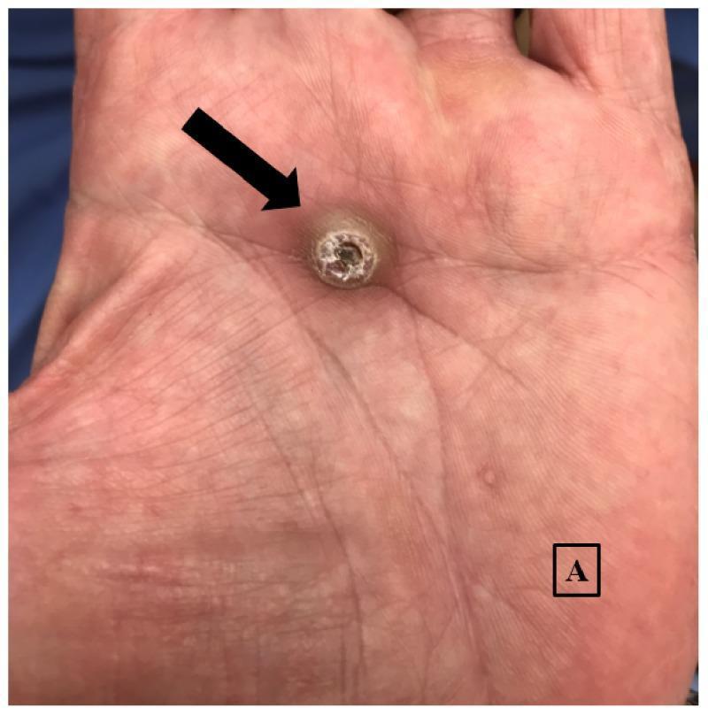
This clinical photograph displays a solitary, well-circumscribed lesion on the central aspect of a human palm. The lesion is a firm, round, dome-shaped nodule measuring approximately 1.5 x 1.2 cm, characterized by a classic crateriform morphology. A prominent central hyperkeratotic keratin plug is visible within the ulceration, which is a hallmark feature of keratoacanthoma, a variant of squamous cell carcinoma. The surrounding palmar skin shows normal dermatoglyphic lines with mild erythema. A black arrow points directly to the primary nodule, and a smaller, less distinct papule is marked with the label 'A' in the distal palmar region for clinical reference. This image serves as a textbook example of a palmar keratoacanthoma, illustrating the importance of recognizing specific morphologic patterns in dermatologic diagnosis.
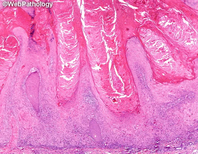
Light microscopy of a skin biopsy stained with Hematoxylin and Eosin reveals crateriform epidermal proliferation consisting of well-differentiated keratinizing squamous epithelium with eosinophilic ground-glass cytoplasm. Central keratinization forms a keratin plug, surrounded by an abrupt epithelial crater. Microabscesses with neutrophils are commonly present, and a mixed inflammatory infiltrate including eosinophils may accompany deeper tissue involvement. The tumor typically displays pushing margins with little true infiltration into adjacent dermis; however, rare cases show invasion into underlying skeletal muscle, and infrequent perineural or vascular invasion. Deeper aspects may show cellular maturation toward the surface, while the basement membrane remains relatively intact in noninvasive areas. When involution occurs, the keratin plug is lost, the epithelial proliferation diminishes markedly, and the lesion floor flattens. Chronic inflammation and fibrosis with a foreign body giant cell reaction to released keratin are characteristic of the involutional phase. Nuclear atypia is typically absent, though some instances may exhibit mild pleomorphism, necrosis, or mitotic activity restricted to basal layers. The overall pattern - crateriform architecture, abrupt keratinization, and a brisk inflammatory milieu with keratin debris - helps distinguish this entity from conventional squamous cell carcinoma. Clinically, keratoacanthoma often presents as a rapidly growing skin nodule and may spontaneously regress, informing management decisions and differential diagnoses.
Keratoacanthoma (KA) - Oral Pathology (10 Marks)
Definition
- Nosological status is controversial:
- Traditionally classified as a benign, self-limiting pseudomalignancy or pseudocarcinoma
- Currently widely regarded as a highly differentiated variant of SCC - "SCC with KA-type features"
- WHO (2018) classification now includes KA under SCC
- Historical term: "Molluscum sebaceum" (Jonathan Hutchinson, 1889)
- Term "keratoacanthoma" coined by Freudenthal (1936)
- Key clinical adage: "Rapid growth in, rapid growth out" - rapid evolution and spontaneous involution
Epidemiology
- Common benign/low-grade tumour; accounts for approximately ~25% of all KA/SCC diagnoses
- Age: Predominantly elderly patients (>50 years); rare in young patients
- Sex: Males > Females (2-3:1) - likely due to more occupational/recreational sun exposure
- Race: More common in fair-skinned (Fitzpatrick type I-II) individuals; rare in dark skin
- Oral cavity KA: Rare; < 1% of all KAs; intraoral location is an unusual subsite
Etiology and Risk Factors
- Ultraviolet radiation (sunlight) - primary cause for cutaneous KA; UVB most important
- Chemical carcinogens - tar, pitch, mineral oils; occupational exposures
- Tobacco - associated with oral and lip KA
- HPV infection - some cases, especially multiple KA variants; not universally identified
- Trauma / skin injury - KA may arise at sites of trauma, tattoos, surgical scars
- Immunosuppression - organ transplant recipients have much higher incidence
- BRAF inhibitors (vemurafenib, dabrafenib) - drug-induced multiple KAs; important side effect
- Chemical exposure (arsenic, industrial carcinogens)
- Genetic syndromes:
- Muir-Torre syndrome (autosomal dominant) - multiple KAs + sebaceous tumours + internal malignancies (colorectal, GU); MSH2/MLH1 mismatch repair gene mutations
- Ferguson-Smith syndrome - multiple self-regressing KAs; autosomal dominant; TGFBR1 mutation
- Grzybowski syndrome - multiple eruptive generalised KAs; rare
Clinical Features
Sites:
- Head and neck (predominant) - especially face (nose, cheeks, forehead, ears)
- Sun-exposed surfaces of extremities - dorsum of hands, forearms
- Lips - especially lower lip
- Oral mucosa - unusual; buccal mucosa, gingiva, palate, tongue (rare)
- Periungual / subungual - associated with underlying bone destruction
- Trunk - less common
Phases of Growth (Classic):
| Phase | Duration | Description |
|---|---|---|
| Proliferative (Growth) phase | 4-8 weeks | Rapid enlargement from a papule to a dome-shaped nodule; may double in size within weeks |
| Mature (Stationary) phase | 4-8 weeks | Lesion reaches maximum size; central keratin crater fully developed |
| Involuting (Regression) phase | 2-6 months | Spontaneous regression; central keratin plug expelled; leaves atrophic scar |
| Total duration | ~6 months | Most lesions completely self-resolve |
Gross Appearance (Fully Developed / Mature KA):
| Feature | Description |
|---|---|
| Shape | Dome-shaped, symmetric, firm nodule |
| Surface | Smooth dome with central keratotic "crater" or "plug" |
| Central crater | Filled with horny keratin - the hallmark |
| Borders | Rolled borders - smooth, raised, overhanging the crater |
| Colour | Flesh-coloured to slightly pink; keratotic crater is yellowish-white |
| Size | Usually 1-2 cm; giant KA can reach 5-10 cm |
| Consistency | Firm (more sclerotic and fibrous than BCC on curettage) |
| Symmetry | Symmetric at scanning magnification - important histological clue |
| Number | Usually solitary (most common); multiple in special syndromes |
Symptoms:
- Usually asymptomatic - noticed as rapidly growing skin lump
- May have mild tenderness or pruritus
- Subungual KA - painful; associated with bony destruction
- No spontaneous bleeding in uncomplicated cases
Clinical Image
Histopathological Features (KEY)
Architecture (Low-Power / Scanning Magnification):
- Symmetric, dome-shaped, crater-like architecture - the most important low-power feature
- "KA relies mainly on its silhouette as assessed at scanning magnification" (Fitzpatrick's)
- Flask-shaped / crateriform configuration - a central keratin-filled invagination (crater) surrounded by a collar of proliferating epithelium
- Central large keratinous mass - massive keratin plug occupying the central crater
- Overhanging "lipping" of epithelial shoulders - the adjacent epidermis extends over the crater margins like a lip
- Symmetric profile - both walls of the crater are symmetric (unlike irregular SCC)
Epithelial Features (High-Power):
- Well-differentiated squamous epithelium - highly orderly maturation
- Cells with abundant glassy (ground-glass) eosinophilic cytoplasm - hallmark cytological feature
- "Apple-pink" or "pale glassy" cytoplasm - characteristic
- Small, bland nuclei with little pleomorphism (in classic KA)
- Prominent granular cell layer (hypergranulosis)
- Hyperorthokeratosis - surface and crater keratinization
- Minimal nuclear atypia in typical cases
- Mitotic figures present - can be numerous (making distinction from SCC difficult)
- No atypical mitoses (in typical KA; atypical mitoses = SCC)
- Intraepithelial microabscesses - collections of neutrophils and eosinophils within the epithelium - characteristic feature
- Dyskeratosis - scattered individually keratinizing cells
Advancing Margins:
- Pushing (expansile) margin - base of lesion pushes into dermis as a broad front
- Some tongues of epithelium may extend irregularly at the base, making SCC distinction difficult
- Deep invasive tongues reaching fat/muscle = suspicious for SCC
Stroma:
- Dense chronic inflammatory infiltrate - lymphocytes, plasma cells, eosinophils, neutrophils
- Granulomatous reaction with foreign body giant cells (especially in involuting phase)
- No desmoplasia (unlike SCC typically)
- Fibrosis prominent in regressing lesions
Three Histological Phases:
| Phase | Microscopy |
|---|---|
| Early | Markedly hyperplastic epithelium; central keratotic plug not fully formed |
| Fully developed | Large central keratin core; well-differentiated squamous proliferation; glassy cells; inflammatory infiltrate |
| Involuting | Hypoplastic epithelium; no hyperplasia or atypia; crater flattening; fibrosis; foreign body reaction to expelled keratin |
Histopathological Image
KA vs SCC - The Critical Distinction
| Feature | Keratoacanthoma | SCC |
|---|---|---|
| Growth | Rapid (weeks) → spontaneous regression | Progressive, no regression |
| Symmetry | Symmetric crater | Asymmetric, irregular |
| Architecture | Crateriform | Variable; no crater |
| Cytoplasm | Glassy, eosinophilic (ground-glass) | Variable; less glassy |
| Atypia | Minimal | Variable; may be marked |
| Atypical mitoses | Absent | May be present |
| Margins | Pushing/broad | Infiltrating/spiky |
| Microabscesses | Characteristic | Less common |
| Invasive depth | Limited | Variable; may be deep |
| Inflammation | Dense, peritumoral | Variable |
| Spontaneous regression | YES - characteristic | No |
| Clinical history | Essential for diagnosis | - |
Variants of KA
| Variant | Features |
|---|---|
| Solitary KA | Most common; classic presentation on sun-exposed skin |
| Giant KA | >3 cm; persistent; may not regress |
| KA centrifugum marginatum | Centrifugally expanding; central healing while periphery grows; can reach several cm |
| Multiple self-regressing (Ferguson-Smith) | Autosomal dominant; adolescence; self-resolving |
| Eruptive KA (Grzybowski) | Multiple (hundreds); generalised; non-regressing; pruritic; rare |
| Subungual KA | Under nail; associated with underlying bone destruction |
| Intraoral KA | Rare; buccal mucosa, gingiva, lip; clinically and histologically identical |
| KA in Muir-Torre syndrome | KA + sebaceous neoplasms + visceral malignancy; mismatch repair defect |
Differential Diagnosis
| Condition | Distinguishing Feature |
|---|---|
| SCC | Progressive growth; no spontaneous regression; marked atypia; infiltrating margin; atypical mitoses |
| BCC (nodular) | Pearly border + telangiectasias; no central keratin crater; peripheral palisading on histology |
| Verruca vulgaris (wart) | HPV; koilocytes; digitate projections; no crater |
| Cutaneous horn | Hard keratin projection; no dome-shaped base |
| Molluscum contagiosum | Viral inclusions; multiple lesions; younger patients |
| Infundibular cyst | No central crater; cyst wall; no inflammatory infiltrate |
| Sebaceous hyperplasia | Yellow, lobulated; no crater; middle-aged to elderly |
Investigations
- Clinical diagnosis - characteristic rapid growth + crateriform morphology often sufficient
- Excisional biopsy - recommended; provides entire lesion for diagnosis
- Incisional biopsy inadequate - misses overall silhouette
- Shave biopsy misses the base (where SCC features may be present)
- Histopathological examination - mandatory
- Must examine entire lesion for accurate diagnosis
- Clinical history of rapid growth must be communicated to pathologist
- Dermoscopy - central keratin mass, white circles, hairpin vessels
- Imaging (MRI/CT) - for giant KA or subungual KA with suspected bone involvement
- Molecular testing - for Muir-Torre syndrome if multiple KAs (MSH2/MLH1 immunostaining)
Treatment
Approach:
| Method | Details |
|---|---|
| Surgical excision | Treatment of choice; 3-5 mm margins; provides complete tissue for histology |
| Deep saucer-type shave excision | Acceptable for small lesions |
| Electrodesiccation and curettage (ED&C × 3) | For small lesions on low-risk sites |
| Cryotherapy | For small lesions only; modest cure rate |
| Mohs Micrographic Surgery | For large, recurrent, or high-risk site KA |
| Radiotherapy | For inoperable/large lesions; elderly patients unfit for surgery |
| Intralesional therapies | Methotrexate, 5-FU, interferon-alfa; for large/giant/multiple KA |
| Systemic retinoids | Acitretin; for multiple KA (Ferguson-Smith, Grzybowski) |
| Observation | Only if confident diagnosis, small lesion, patient preference; with close monitoring |
Prognosis
- Excellent for solitary typical KA - complete resolution with treatment
- Spontaneous regression occurs in most untreated solitary KAs over 6 months, leaving an atrophic scar
- Recurrence after excision is possible
- Giant KA may not regress and can cause significant local destruction
- Subungual KA may destroy underlying phalanx
- KA in Muir-Torre syndrome - internal malignancy surveillance required
Summary Table
| Feature | Keratoacanthoma |
|---|---|
| Nature | Benign / well-differentiated SCC variant (controversial) |
| Origin | Hair follicle infundibulum |
| Growth | Rapid - weeks to mature |
| Spontaneous regression | Yes - 6 months |
| Classic appearance | Dome-shaped nodule + central keratin crater |
| Most common site | Head and face (sun-exposed) |
| Histology hallmark | Crateriform architecture + glassy eosinophilic cytoplasm + pushing margin |
| Microabscesses | Characteristic (neutrophils + eosinophils) |
| Difficult to distinguish from | SCC |
| Treatment | Surgical excision |
| Prognosis | Excellent |
| Key syndrome | Muir-Torre syndrome |
- KA is the "great masquerader" - clinically and histologically mimics SCC; always treat as SCC until proven otherwise
- The three-phase clinical course (rapid growth → stasis → spontaneous regression) is the most important distinguishing clinical feature from SCC
- Histological diagnosis requires the entire lesion - the symmetric crateriform silhouette at scanning magnification is the key; superficial or small biopsies are inadequate
- "Glassy (ground-glass) eosinophilic cytoplasm" of tumour cells is the key cytological hallmark on high-power view
- Microabscesses (neutrophils and eosinophils) within the epithelium are characteristic of KA
- Clinical history of rapid growth must be communicated to the pathologist - without it, pathologist may report "cannot rule out SCC" (Pfenninger's)
- Muir-Torre syndrome = multiple KAs + sebaceous tumours → screen for colorectal/GU malignancy (relevant to oral medicine)
Tnm staging
TNM Staging of Oral Cancer - Oral Pathology (10 Marks)
Introduction
- T = Primary Tumour (size and local extent)
- N = Regional lymph Node involvement
- M = Distant Metastasis
Key Principle of AJCC 8th Edition:
Depth of Invasion (DOI) ≠ Tumour thickness
- DOI is measured from the "horizon" (level of adjacent normal mucosal basement membrane) to the deepest point of tumour invasion - not from the surface
- Exophytic tumours may be thick but have shallow DOI
- Endophytic/ulcerated tumours may have greater DOI than their surface dimensions suggest
Subsites Included in "Oral Cavity" Staging
- Oral tongue (anterior 2/3)
- Buccal mucosa
- Hard palate
- Upper alveolar ridge (maxilla)
- Lower alveolar ridge (mandible)
- Retromolar trigone
- Floor of mouth
- Lip (vermilion border)
T Staging - Primary Tumour (Oral Cavity)
| T Category | Description |
|---|---|
| TX | Primary tumour cannot be assessed |
| Tis | Carcinoma in situ |
| T1 | Tumour ≤ 2 cm AND Depth of Invasion (DOI) ≤ 5 mm |
| T2 | Tumour ≤ 2 cm with DOI > 5 mm; OR Tumour > 2 cm and ≤ 4 cm with DOI ≤ 10 mm |
| T3 | Tumour > 4 cm; OR any tumour with DOI > 10 mm |
| T4a | Moderately advanced local disease: Invades adjacent structures - cortical bone of mandible/maxilla, inferior alveolar nerve, floor of mouth, skin of face, extrinsic muscles of tongue (genioglossus, hyoglossus, palatoglossus, styloglossus), maxillary sinus |
| T4b | Very advanced local disease: Tumour invades masticator space, pterygoid plates, skull base; OR encases the internal carotid artery |
Important Notes on T Staging:
- Superficial bone erosion (e.g., of tooth socket by gingival tumour) is not sufficient for T4a classification
- T4b is unresectable - encasement of carotid or skull base invasion makes surgical resection technically not feasible
- DOI inclusion in AJCC 8th edition was based on evidence that DOI >4-5 mm predicts significantly higher nodal metastasis risk
N Staging - Regional Lymph Nodes
Clinical N Staging (cN) - Oral Cavity, Oropharynx (HPV-), Hypopharynx
| N Category | Description |
|---|---|
| NX | Regional lymph nodes cannot be assessed |
| N0 | No regional lymph node metastasis |
| N1 | Single ipsilateral node ≤ 3 cm, ENE(-) |
| N2a | Single ipsilateral node > 3 cm but ≤ 6 cm, ENE(-) |
| N2b | Multiple ipsilateral nodes, none > 6 cm, ENE(-) |
| N2c | Bilateral or contralateral nodes, none > 6 cm, ENE(-) |
| N3a | Any node > 6 cm, ENE(-) |
| N3b | Any node with ENE(+) (extranodal extension on clinical/imaging assessment); OR single ipsilateral node > 3 cm with ENE(+) |
Pathologic N Staging (pN) - Key Additions in AJCC 8th Ed:
| pN Category | Description |
|---|---|
| pN1 | Single ipsilateral node ≤ 3 cm, ENE(-) |
| pN2a | Single ipsilateral/contralateral node ≤ 3 cm with ENE(+); OR single ipsilateral node 3-6 cm without ENE |
| pN2b | Multiple ipsilateral nodes, none > 6 cm, ENE(-) |
| pN2c | Bilateral/contralateral nodes, none > 6 cm, ENE(-) |
| pN3a | Node > 6 cm, ENE(-) |
| pN3b | Single ipsilateral node > 3 cm with ENE(+); OR multiple ipsilateral/contralateral/bilateral nodes with any ENE |
ENE = Extranodal Extension (AJCC 8th Edition NEW addition):
- Tumour extending beyond the capsule of the lymph node into surrounding soft tissue
- Clinically: matted nodes, skin invasion, nerve involvement
- ENE(+) automatically upgrades to N3b regardless of node size
- Strong independent predictor of poor prognosis
M Staging - Distant Metastasis
| M Category | Description |
|---|---|
| MX | Distant metastasis cannot be assessed (workup not completed) |
| M0 | No distant metastasis |
| M1 | Distant metastasis present |
Common sites of distant metastasis in oral SCC:
Overall Stage Grouping (Oral Cavity)
| Stage | TNM Combination |
|---|---|
| Stage I | T1 N0 M0 |
| Stage II | T2 N0 M0 |
| Stage III | T3 N0 M0; OR T1/T2/T3 N1 M0 |
| Stage IVA | T4a N0/N1 M0; OR T1-T4a N2 M0 |
| Stage IVB | T4b any N M0; OR any T N3 M0 |
| Stage IVC | Any T, any N, M1 |
TNM Stage Grouping Diagram:
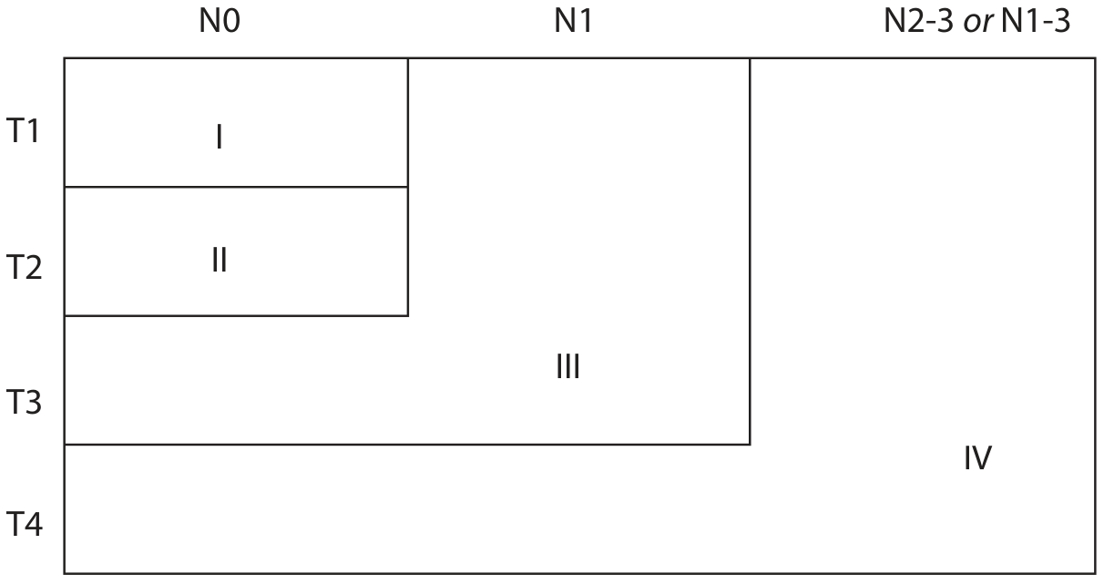
Prefix and Suffix Modifiers
| Modifier | Meaning |
|---|---|
| c prefix (cTNM) | Clinical staging - based on examination, imaging |
| p prefix (pTNM) | Pathological staging - based on histopathology after surgical resection |
| y prefix (yTNM) | Post-treatment staging (after neoadjuvant chemo/RT) |
| r prefix (rTNM) | Restaging at recurrence |
| a prefix (aTNM) | Staging at autopsy |
| R suffix | Residual tumour after treatment: R0 = no residual; R1 = microscopic; R2 = macroscopic |
HPV-Associated Oropharyngeal SCC - Separate Staging
Clinical N (HPV+ Oropharynx):
| N | Description |
|---|---|
| N0 | No nodes |
| N1 | One or more ipsilateral nodes, none > 6 cm |
| N2 | Contralateral or bilateral nodes, none > 6 cm |
| N3 | Any node > 6 cm |
Pathologic N (HPV+ Oropharynx):
| pN | Description |
|---|---|
| pN0 | No nodes |
| pN1 | Metastasis in ≤ 4 lymph nodes |
| pN2 | Metastasis in > 4 lymph nodes |
Key Changes in AJCC 8th Edition vs 7th Edition
| Feature | 7th Edition (2010) | 8th Edition (2017) |
|---|---|---|
| T staging | Based on size alone | Size + DOI combined |
| T1 | ≤ 2 cm | ≤ 2 cm AND DOI ≤ 5 mm |
| N staging | Size-based only | Size + ENE (Extranodal Extension) added |
| N3b | Not defined | ENE(+) = N3b (any node with ENE) |
| HPV oropharynx | Same as oral cavity | Separate staging system |
| Stage IV | Less granular | IVA, IVB, IVC more precisely defined |
Significance of Depth of Invasion (DOI)
| DOI | Clinical Significance |
|---|---|
| ≤ 2 mm | Very low nodal metastasis risk (~2%) |
| 2-4 mm | Low risk |
| > 4-5 mm | Significant nodal risk - threshold for elective neck dissection |
| > 10 mm | T3 regardless of surface dimensions |
Five-Year Survival by Stage
| Stage | 5-Year Survival (Oral SCC) |
|---|---|
| Stage I | ~80-85% |
| Stage II | ~70-75% |
| Stage III | ~50-60% |
| Stage IVA | ~30-40% |
| Stage IVB | ~15-20% |
| Stage IVC | <10% |
Summary Table
| Category | Key Criteria | Threshold |
|---|---|---|
| T1 | ≤ 2 cm AND DOI ≤ 5 mm | Size + DOI |
| T2 | ≤ 4 cm AND DOI ≤ 10 mm | Size + DOI |
| T3 | > 4 cm OR DOI > 10 mm | Size OR DOI |
| T4a | Cortical bone/IAN/floor/skin/tongue muscles | Structure invasion |
| T4b | Masticator space/pterygoid/skull base/carotid | Unresectable |
| N1 | Single ipsilateral ≤ 3 cm, ENE(-) | - |
| N2a | Single ipsilateral 3-6 cm, ENE(-) | - |
| N2b | Multiple ipsilateral ≤ 6 cm, ENE(-) | - |
| N2c | Bilateral/contralateral ≤ 6 cm, ENE(-) | - |
| N3a | Any node > 6 cm, ENE(-) | - |
| N3b | Any ENE(+) | ENE |
| M0 | No distant spread | - |
| M1 | Distant metastasis | - |
- AJCC 8th Edition introduced DOI into T staging - a major change from the 7th edition which relied on size alone. DOI is the most powerful predictor of nodal spread in oral SCC
- DOI ≠ Tumour thickness - DOI is measured from the mucosal "horizon" to the deepest point of invasion
- ENE (extranodal extension) was introduced in the 8th edition and immediately upgrades any node to N3b - reflects the severe prognostic impact of capsular breach
- T4b = unresectable - masticator space, pterygoid plates, skull base, carotid artery encasement
- Stage IVC = any distant metastasis (M1) - palliative intent
- HPV+ oropharyngeal SCC has its own separate staging (better prognosis - pN1 = ≤4 nodes, pN2 = >4 nodes)
- DOI > 4-5 mm = threshold for elective neck dissection of the clinically N0 neck
oral lichen planus
oral lichen planus reticular Wickham striae buccal mucosa white lacy
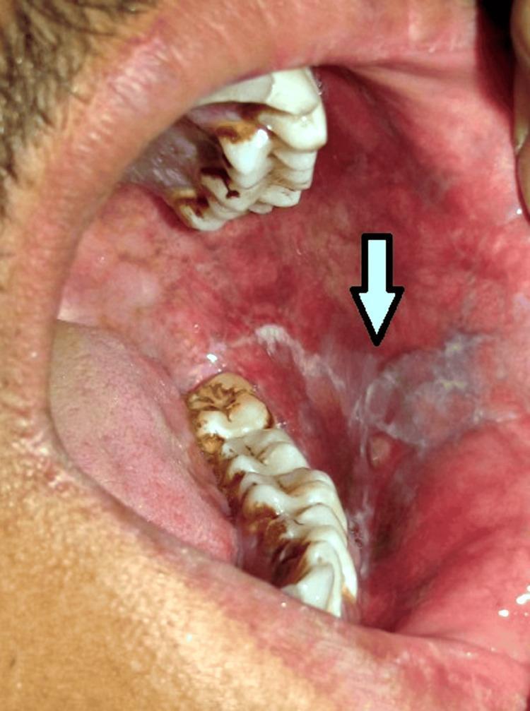
This clinical photograph shows an intraoral view of the left buccal mucosa, illustrating characteristic signs of oral lichen planus. The primary finding is the presence of reticular white striae, also known as Wickham striae, which form a distinct lacy or net-like pattern across the mucosal surface. These lesions are situated adjacent to the mandibular molar region. The surrounding background mucosa exhibits significant erythema, suggesting an inflammatory component consistent with the erosive or erythematous subtype of the condition. A white arrow points toward the concentrated area of reticular striae. The image serves as a classic diagnostic reference for medical and dental students to identify oral mucosal auto-inflammatory diseases. Key educational concepts include recognizing non-scrappable white patches, distinguishing reticular patterns from other leukoplakic lesions, and understanding the clinical presentation of lichen planus within the oral cavity.
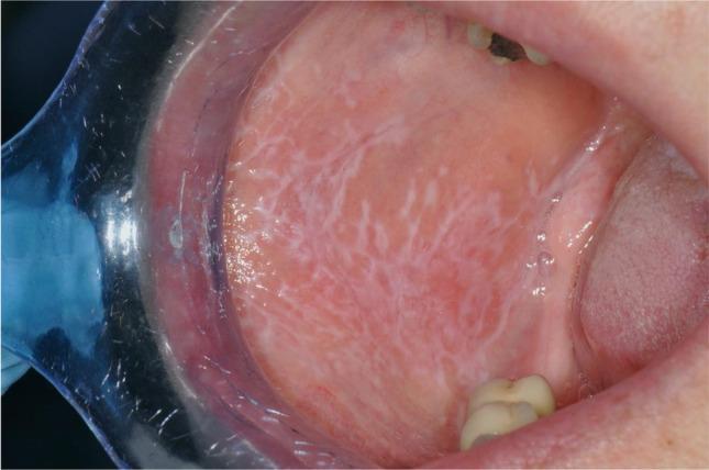
This clinical intraoral photograph demonstrates reticular oral lichen planus (OLP) on the buccal mucosa, categorized as a Thongprasom score 1. The primary visual finding is a network of delicate, white, lacy thread-like lines known as Wickham striae. These striae exhibit a classic arborescent or branching morphology against a background of slightly erythematous but otherwise intact mucosa. The lesion is well-distributed across the mucosal surface without evidence of ulceration, erosion, or significant atrophy, which is characteristic of the asymptomatic reticular subtype. Anatomical landmarks visible include the retracted labial commissure (using a clear plastic retractor), the maxillary and mandibular teeth, and a portion of the tongue. The image serves as a high-quality educational example of the classic diagnostic presentation of reticular OLP in oral medicine and dermatology, highlighting the distinction between white reticulated patterns and the more severe erosive forms of the disease.
Oral Lichen Planus (OLP) - Oral Pathology (10 Marks)
Definition
- First described by Wilson (1869) as a cutaneous disease
- Named from the Greek/Latin: "lichen" = tree moss (resembling the appearance); "planus" = flat
- Frequency: 0.2% to 3% of the general population
- WHO classified OLP as an OPMD in 2005
Epidemiology
- Age: Middle-aged adults; most common in 4th-6th decades
- Sex: Women > Men (up to 75% female in some series); oral LP in women begins ~10 years later than in men (age 57 vs. 47)
- No racial predilection
- Oral involvement occurs in ~50-75% of cutaneous LP patients; oral LP may exist without skin involvement
Etiology and Pathogenesis
General Etiology:
Triggering/Associated Factors:
- Idiopathic (most cases) - no identifiable trigger
- Drugs → Lichenoid drug reactions (similar but not identical to true LP):
- NSAIDs (aspirin, ibuprofen)
- Antihypertensives: beta-blockers, ACE inhibitors, thiazides
- Antimalarials: hydroxychloroquine, chloroquine
- Gold salts
- Oral hypoglycemics (tolbutamide, chlorpropamide)
- Dental materials → Lichenoid contact reactions:
- Amalgam restorations (mercury)
- Gold, composite resins
- Systemic diseases:
- Hepatitis C virus (HCV) - strong association (especially in Italy, Japan)
- Primary biliary cirrhosis
- Primary sclerosing cholangitis
- Sjögren's syndrome
- Lupus erythematosus
- Stress and anxiety - psychological factors may trigger/exacerbate
- Graft-versus-host disease (GVHD) - produces lichenoid reactions
- Genetic predisposition - familial cases reported
Pathogenesis (Immunological Mechanism):
Antigenic stimulus (unknown / exogenous antigen)
        ↓
Antigen processing by resident dendritic cells in epithelium
        ↓
Antigen presentation via MHC class I → CD8+ cytotoxic T cells
Antigen presentation via MHC class II → CD4+ helper T cells
        ↓
T-cell recruitment: CD8+ (majority) + CD4+ cells
Both express α-1 integrin molecules
        ↓
Elaboration of cytokines: RANTES, TNF-α, IL-1β, IFN-γ
Activation of matrix metalloproteinases (MMPs)
Mast cell degranulation
        ↓
Lymphocytes migrate through basement membrane into epithelium
        ↓
CD8+ cytotoxic T cells → Keratinocyte APOPTOSIS → Civatte bodies
        ↓
Basement membrane damage → Fibrinogen deposition
        ↓
Band-like lymphocytic infiltrate at epithelial-connective tissue junction
Clinical Features
Sites of Predilection (in order):
- Buccal mucosa - most common; bilateral, symmetric
- Tongue (dorsum and lateral borders)
- Gingiva (especially atrophic/erosive type)
- Palate (hard and soft)
- Lips (vermilion)
- Floor of mouth (less common)
Cardinal Clinical Characteristics:
- Bilateral and symmetric distribution - most important distinguishing feature from leukoplakia
- Multiple lesions present simultaneously
- Chronic, relapsing, remitting course
- Cannot be wiped off (unlike candidiasis)
Types / Clinical Variants (Andreason's Classification):
1. Reticular OLP (Most Common, ~60%)
- White, lacy, interlacing striae - Wickham's striae - the pathognomonic feature
- Fine white lines in a net-like, arborizing, or annular pattern
- Background mucosa: normal to slightly erythematous
- Asymptomatic in most cases
- Does not require treatment
- Striae may be preceded by white keratotic papules that fuse to form the network
2. Erosive / Ulcerative OLP (Most Significant Clinically)
- Central, painful ulceration with superficial erosions
- Covered by pseudomembrane or fibrinous plaque
- Periphery shows characteristic white striae - important diagnostic clue
- Most symptomatic form - burning, pain, exacerbated by spicy food
- Most common form to show malignant transformation
- Most common form causing patients to seek treatment
3. Atrophic / Erythematous OLP
- Thinned, reddened mucosa with faint/absent striae
- Desquamative gingivitis - most common form on gingiva
- Loss of normal pink, stippled appearance of gingiva
- Glossy, edematous, erythematous, bleeds easily
- Symptomatic; interferes with oral hygiene
4. Plaque-Type OLP
- Smooth, homogeneous white patch - resembles leukoplakia
- Slightly elevated, minimally fissured
- May develop from resolving erosive LP
- Can be confused with leukoplakia - bilaterality and associated striae help
5. Papular OLP
- Multiple small, discrete, white papules
- Often represent early stage before striae formation
6. Bullous OLP (Rare)
- Transient blisters/bullae (few mm to > 1 cm)
- Located on posterior/inferior buccal mucosa
- Rupture → painful ulcers
- Usually associated with other LP types simultaneously
Clinical Image
Histopathological Features (KEY)
Epithelial Changes:
- Hyperkeratosis or hyperparakeratosis at the surface
- Acanthosis (thickened spinous layer) in reticular type
- Atrophy of the spinous layer in erosive/atrophic type - fewer cell layers
- "Sawtooth" rete ridges - the rete pegs assume a pointed, saw-tooth or zigzag pattern - characteristic
- Liquefaction (vacuolar) degeneration of basal cells - basal cells show intracellular vacuolation, disrupted outline, eventually lost
- Basal cell disruption - loss of the normal columnar/cuboidal appearance of basal cells
Connective Tissue Changes (KEY):
- Dense, band-like subepithelial lymphocytic infiltrate - THE most important histological feature
- Predominantly T lymphocytes (CD4+ and CD8+, mostly CD8+)
- Infiltrate hugs the epithelial-connective tissue interface in a "band-like" pattern
- Confined to the superficial lamina propria - does not extend deep
- Civatte bodies (Colloid bodies / Hyaline bodies) - highly characteristic:
- Ovoid, anucleate, eosinophilic, hyaline droplets scattered at the epithelial-connective tissue junction
- Represent apoptotic keratinocytes that have been pushed from the epithelium by the lymphocytic infiltrate
- Non-specific but highly suggestive when combined with other features
- Lymphocyte exocytosis - lymphocytes migrating into the lower epithelium
- Dilated capillaries in lamina propria
Summary of Histological Hallmarks:
"Sawtooth rete ridges + band-like lymphocytic infiltrate at BMZ + vacuolar degeneration of basal cells + Civatte bodies"
Direct Immunofluorescence (DIF) - KEY Investigation
| Finding | Description |
|---|---|
| Linear fibrinogen deposition along the basement membrane | PATHOGNOMONIC - present in 90-100% of OLP cases |
| IgM deposits in Civatte bodies | Characteristic but not specific |
| Shaggy fibrin deposits | Present at dermo-epidermal junction |
- DIF pattern: "Fibrinogen - linear/shaggy deposition at BMZ"
- Distinguishes OLP from pemphigus (IgG/C3 intercellular), pemphigoid (IgG/C3 linear BMZ), lupus (IgG/IgM/C3 - "lupus band test")
Diagnostic Criteria (Modified WHO Criteria for OLP)
- Bilateral, symmetric lesions
- White reticular/papular/plaque type; OR erythematous/erosive/bullous type with peripheral white striae
- Cannot be attributed to other disease or drug reaction
- Thickened ortho/parakeratotic epithelium
- Sawtooth rete ridges
- Liquefaction degeneration of basal layer
- Band-like subepithelial lymphocytic infiltrate
- Absence of epithelial dysplasia
Differential Diagnosis
| Condition | Distinguishing Feature |
|---|---|
| Leukoplakia | Unilateral; no bilateral striae; smoking/tobacco history |
| Lichenoid drug reaction | Drug history; unilateral; resolves on drug withdrawal |
| Oral hairy leukoplakia | Lateral tongue; EBV-associated; HIV patients; cannot be wiped off; vertical white folds |
| Candidiasis (atrophic) | Responds to antifungals; Candida on smear/culture |
| Lupus erythematosus | Central atrophy + radiating striae (butterfly pattern); ANA positive; DIF shows IgG/IgM/C3 |
| GVHD (lichenoid form) | History of bone marrow transplant |
| Pemphigus vulgaris | Nikolsky positive; DIF shows IgG/C3 intercellular |
| Mucous membrane pemphigoid | Subepithelial bulla; scarring; DIF shows IgG/C3 linear BMZ |
| Erythema multiforme | Acute onset; crusted lips; "target lesions" on skin; younger |
Association With Systemic Disease
- Hepatitis C virus (HCV) - especially in Italy and Japan
- Primary biliary cirrhosis
- Primary sclerosing cholangitis
- Sjögren's syndrome
- Lupus erythematosus
- Diabetes mellitus (Grinspan syndrome: OLP + DM + hypertension)
Malignant Transformation
- Controversial but accepted as a legitimate risk
- Overall transformation rate: < 1% to ~5% (WHO recognises OLP as OPMD)
- Erosive / atrophic forms carry significantly higher risk than reticular type
- The issue of "lichenoid dysplasia" vs. true OLP with dysplasia complicates assessment
- Lesions with associated epithelial dysplasia carry much higher risk
- Long-term follow-up is mandatory
- (Cummings Otolaryngology, p. 1948-1950)
Investigations
- Biopsy + H&E histopathology - gold standard
- Direct Immunofluorescence (DIF) - fibrinogen at BMZ; distinguishes from pemphigus/pemphigoid
- Patch testing - for lichenoid contact reactions (dental amalgam)
- Liver function tests / Hepatitis C serology - screen for HCV
- Blood glucose - Grinspan syndrome
- ANA, anti-dsDNA - rule out lupus
Treatment
A. General Measures (All Types):
- Reassurance - chronic but manageable; not contagious
- Eliminate triggering factors - stop offending drugs; replace amalgam restorations if lichenoid reaction suspected
- Good oral hygiene - essential, especially in atrophic/gingival LP
- Asymptomatic reticular LP does not require treatment - periodic monitoring only
B. Topical Treatment (First Line):
| Agent | Use |
|---|---|
| Topical corticosteroids (triamcinolone acetonide paste, clobetasol gel, fluocinonide) | First line; moderate-to-ultrapotent agents preferred; symptomatic erosive/atrophic LP |
| Topical tacrolimus (0.1% ointment) | Calcineurin inhibitor; effective without steroid side effects; for refractory cases |
| Topical pimecrolimus (1% cream) | Similar to tacrolimus; calcineurin inhibitor |
| Topical cyclosporine rinse | Where steroids contraindicated |
| Intralesional triamcinolone (10-40 mg/mL injection) | Focal/recalcitrant lesions |
C. Systemic Treatment (Severe/Extensive Disease):
| Agent | Details |
|---|---|
| Systemic corticosteroids (prednisolone) | Short-term for severe flares; bridge to topical maintenance |
| Hydroxychloroquine | Anti-malarial; adjunct for chronic/refractory OLP |
| Azathioprine | Immunosuppressive; for recalcitrant disease |
| Systemic retinoids (acitretin) | For plaque-type or extensive OLP |
| Mycophenolate mofetil | Immunosuppressive adjunct |
| Dapsone | Second-line; erosive LP |
D. Monitoring:
- Regular follow-up every 3-6 months
- Biopsy of any changing, indurated, or persistently erosive area
- Annual biopsy of high-risk (erosive) areas
Summary Table
| Feature | Oral Lichen Planus |
|---|---|
| Nature | Chronic inflammatory autoimmune |
| Prevalence | 0.2-3% population |
| Sex | Females > Males |
| Pathogenesis | CD8+ T-cell mediated keratinocyte apoptosis |
| Most common site | Bilateral buccal mucosa |
| Most common type | Reticular (white lacy Wickham striae) |
| Most symptomatic | Erosive type |
| Histology hallmark | Band-like lymphocytic infiltrate + sawtooth rete + Civatte bodies |
| Pathognomonic cell | Civatte body |
| DIF finding | Linear fibrinogen at BMZ (90-100%) |
| Malignant potential | <1-5%; erosive type highest |
| Treatment | Topical steroids / tacrolimus + monitoring |
- OLP is bilateral and symmetric - this single feature differentiates it from most other white lesions including leukoplakia
- Wickham's striae - white lacy, interlacing lines - are pathognomonic of the reticular type
- Histologically: "Band-like lymphocytic infiltrate at the BMZ + sawtooth rete ridges + vacuolar basal cell degeneration + Civatte bodies" - the four pillars
- Civatte bodies = apoptotic keratinocytes pushed into the connective tissue by lymphocytes - highly suggestive
- DIF: linear fibrinogen at BMZ = most important diagnostic test; positive in 90-100%
- Erosive OLP = most dangerous subtype clinically; most symptomatic; highest malignant transformation risk
- Asymptomatic reticular LP needs monitoring, not treatment; symptomatic forms need topical steroids/tacrolimus
Genereate me a pdf consisting all the above lesions without missing any important points
mkdir -p /tmp/workspace/oral-pathology-notes && ls /tmp/workspace/
Genereate me a pdf consisting all the above lesions without missing any important points
mkdir -p /tmp/workspace/oral-pathology-notes
~/oral-pathology-notes/generate_pdf.py
#!/usr/bin/env python3
"""Comprehensive Oral Pathology Notes PDF Generator"""
from reportlab.lib.pagesizes import A4
from reportlab.lib.styles import getSampleStyleSheet, ParagraphStyle
from reportlab.lib.units import cm
from reportlab.lib import colors
from reportlab.platypus import (
SimpleDocTemplate, Paragraph, Spacer, Table, TableStyle,
PageBreak, HRFlowable, KeepTogether
)
from reportlab.lib.enums import TA_LEFT, TA_CENTER, TA_JUSTIFY
from reportlab.platypus import ListFlowable, ListItem
# ── Output path ──────────────────────────────────────────────────────────────
OUTPUT = "/tmp/workspace/oral-pathology-notes/Oral_Pathology_Notes.pdf"
# ── Document setup ───────────────────────────────────────────────────────────
doc = SimpleDocTemplate(
OUTPUT,
pagesize=A4,
leftMargin=2*cm, rightMargin=2*cm,
topMargin=2.2*cm, bottomMargin=2.2*cm,
title="Oral Pathology – Comprehensive Exam Notes",
author="Orris AI",
)
# ── Colour palette ───────────────────────────────────────────────────────────
NAVY = colors.HexColor("#1a3a5c")
TEAL = colors.HexColor("#0d7377")
GOLD = colors.HexColor("#b8860b")
LIGHT_BG = colors.HexColor("#eaf4fb")
TABLE_HDR = colors.HexColor("#1a3a5c")
TABLE_ALT = colors.HexColor("#f0f7ff")
RED_WARN = colors.HexColor("#8b0000")
WHITE = colors.white
BLACK = colors.black
GREY_LINE = colors.HexColor("#cccccc")
# ── Styles ───────────────────────────────────────────────────────────────────
base = getSampleStyleSheet()
def S(name, parent="Normal", **kw):
return ParagraphStyle(name, parent=base[parent], **kw)
cover_title = S("CoverTitle", "Title", fontSize=28, textColor=NAVY, spaceAfter=10, alignment=TA_CENTER, fontName="Helvetica-Bold")
cover_sub = S("CoverSub", "Normal", fontSize=14, textColor=TEAL, spaceAfter=6, alignment=TA_CENTER, fontName="Helvetica-Oblique")
cover_info = S("CoverInfo", "Normal", fontSize=10, textColor=colors.grey, alignment=TA_CENTER)
chapter_style = S("Chapter", "Heading1", fontSize=16, textColor=WHITE, spaceAfter=4, spaceBefore=6, fontName="Helvetica-Bold", backColor=NAVY, leftIndent=-0.3*cm, rightIndent=-0.3*cm, leading=22)
h2 = S("H2", "Heading2", fontSize=12, textColor=NAVY, spaceAfter=3, spaceBefore=8, fontName="Helvetica-Bold")
h3 = S("H3", "Heading3", fontSize=10.5, textColor=TEAL, spaceAfter=2, spaceBefore=5, fontName="Helvetica-Bold")
h4 = S("H4", "Heading4", fontSize=10, textColor=GOLD, spaceAfter=2, spaceBefore=4, fontName="Helvetica-BoldOblique")
body = S("Body", "Normal", fontSize=9.5, leading=14, spaceAfter=3, alignment=TA_JUSTIFY)
bullet_style = S("Bullet", "Normal", fontSize=9.5, leading=13, leftIndent=14, firstLineIndent=-10, spaceAfter=2)
bullet2 = S("Bullet2", "Normal", fontSize=9, leading=12, leftIndent=24, firstLineIndent=-10, spaceAfter=1.5)
box_style = S("Box", "Normal", fontSize=9.5, leading=13, backColor=LIGHT_BG, leftIndent=8, rightIndent=8, spaceBefore=4, spaceAfter=4, borderPadding=6)
key_box = S("KeyBox", "Normal", fontSize=9.5, leading=13, backColor=colors.HexColor("#fff8e1"), leftIndent=8, rightIndent=8, spaceBefore=4, spaceAfter=4)
warn_box = S("WarnBox", "Normal", fontSize=9.5, leading=13, textColor=RED_WARN, backColor=colors.HexColor("#fff0f0"), leftIndent=8, rightIndent=8, spaceBefore=4, spaceAfter=4, fontName="Helvetica-Bold")
def bp(text, style=bullet_style): return Paragraph(f"• {text}", style)
def bp2(text): return Paragraph(f" • {text}", bullet2)
def h(text, style=h2): return Paragraph(text, style)
def p(text, style=body): return Paragraph(text, style)
def sp(n=4): return Spacer(1, n)
def hr(): return HRFlowable(width="100%", thickness=0.5, color=GREY_LINE, spaceAfter=4, spaceBefore=2)
# ── Table helper ─────────────────────────────────────────────────────────────
def make_table(data, col_widths=None):
t = Table(data, colWidths=col_widths, repeatRows=1)
n = len(data)
style_cmds = [
('BACKGROUND', (0,0), (-1,0), TABLE_HDR),
('TEXTCOLOR', (0,0), (-1,0), WHITE),
('FONTNAME', (0,0), (-1,0), 'Helvetica-Bold'),
('FONTSIZE', (0,0), (-1,0), 9),
('ALIGN', (0,0), (-1,-1), 'LEFT'),
('VALIGN', (0,0), (-1,-1), 'TOP'),
('FONTSIZE', (0,1), (-1,-1), 8.5),
('FONTNAME', (0,1), (-1,-1), 'Helvetica'),
('ROWBACKGROUNDS', (0,1), (-1,-1), [WHITE, TABLE_ALT]),
('GRID', (0,0), (-1,-1), 0.4, GREY_LINE),
('TOPPADDING', (0,0), (-1,-1), 4),
('BOTTOMPADDING',(0,0),(-1,-1), 4),
('LEFTPADDING', (0,0), (-1,-1), 5),
('RIGHTPADDING',(0,0), (-1,-1), 5),
('WORDWRAP', (0,0), (-1,-1), True),
]
t.setStyle(TableStyle(style_cmds))
return t
def chapter(num, title):
return [
sp(8),
Paragraph(f" CHAPTER {num} | {title}", chapter_style),
sp(4),
]
# ═══════════════════════════════════════════════════════════════════════════════
# BUILD CONTENT
# ═══════════════════════════════════════════════════════════════════════════════
story = []
# ─── COVER PAGE ────────────────────────────────────────────────────────────
story += [
Spacer(1, 3.5*cm),
Paragraph("ORAL PATHOLOGY", cover_title),
Paragraph("Comprehensive Exam Notes – Shafer's Style", cover_sub),
Spacer(1, 0.4*cm),
HRFlowable(width="70%", thickness=2, color=TEAL, spaceAfter=10, spaceBefore=4),
Spacer(1, 0.4*cm),
Paragraph("Covering 11 High-Yield Topics", cover_sub),
Spacer(1, 1.5*cm),
Paragraph(
"1. Erythroplakia | 2. Oral Submucous Fibrosis | 3. Carcinoma In Situ<br/>"
"4. Oral Squamous Papilloma | 5. Histopathology of SCC | 6. Epithelial Dysplasia<br/>"
"7. Oral SCC | 8. Basal Cell Carcinoma | 9. Verrucous Carcinoma<br/>"
"10. Keratoacanthoma | 11. Oral Lichen Planus",
cover_info),
Spacer(1, 1.5*cm),
Paragraph("Compiled from: Shafer's Oral Pathology · Cummings Otolaryngology · Fitzpatrick's Dermatology<br/>"
"Andrews' Diseases of the Skin · K.J. Lee's Essential Otolaryngology · Robbins Pathology",
cover_info),
Spacer(1, 1*cm),
Paragraph("Prepared by Orris AI | July 2026", cover_info),
PageBreak(),
]
# ═══════════════════════════════════════════════════════════════════════════════
# CHAPTER 1 – ERYTHROPLAKIA
# ═══════════════════════════════════════════════════════════════════════════════
story += chapter(1, "ERYTHROPLAKIA")
story += [
h("Definition"),
p("Erythroplakia is defined by the <b>WHO (1978)</b> as: <i>\"A fiery red patch that cannot be characterised clinically or pathologically as any other definable disease.\"</i> It is a diagnosis of exclusion and is considered the <b>most dangerous oral potentially malignant disorder (OPMD)</b> due to its very high rate of malignant transformation (>50%)."),
sp(),
h("Epidemiology"),
bp("Prevalence: <b>0.02–0.83%</b> of the general population"),
bp("Sex: <b>Males > Females</b>; predominantly middle-aged to elderly"),
bp("High incidence in <b>chutta smokers</b> (reverse smoking) in South India"),
bp("Less common than leukoplakia but far more sinister"),
sp(),
h("Etiology"),
bp("<b>Tobacco</b> (smoked and smokeless) – primary etiological factor"),
bp("<b>Alcohol</b> – synergistic with tobacco"),
bp("<b>Areca nut / betel quid</b>"),
bp("<b>HPV</b> (types 16, 18) – subset of cases"),
bp("<b>p53 gene mutation</b> – molecular basis; loss of tumour suppressor function"),
bp("Chronic <b>iron deficiency</b>, nutritional deficiencies"),
sp(),
h("Clinical Features"),
bp("<b>Fiery red, velvety, well-demarcated</b> patch or plaque"),
bp("Soft, smooth, sometimes <b>granular</b> or lobulated surface"),
bp("Cannot be wiped off"),
bp("Margins: abrupt, well-defined"),
bp("<b>Asymptomatic</b> in early stages; burning/pain if erosive"),
h3("Sites of Predilection:"),
bp2("Floor of mouth (most common)"),
bp2("Soft palate / palatal complex"),
bp2("Buccal mucosa"),
bp2("Ventral/lateral tongue"),
h3("Variants (Shafer & Waldron):"),
bp("<b>Homogeneous erythroplakia</b> – uniformly red, velvety"),
bp("<b>Erythroplakia interspersed with leukoplakia</b> – mixed red-white"),
bp("<b>Granular/speckled erythroplakia</b> – stippled red surface"),
sp(),
h("Histopathology"),
p("<b>Shafer & Waldron (1975): In 58 patients – 91% showed significant pathology:</b>"),
make_table(
[["Histopathological Finding", "Percentage"],
["Invasive squamous cell carcinoma", "51%"],
["Carcinoma in situ (CIS)", "40%"],
["Mild-to-moderate dysplasia", "9%"],
["Normal / Hyperkeratosis alone", "<1%"]],
col_widths=[10*cm, 5*cm]
),
sp(6),
bp("Thin, atrophic epithelium – reduced keratin layer explains the red colour (submucosa visible through)"),
bp("Severe epithelial dysplasia or carcinoma in situ in most cases"),
bp("Marked nuclear pleomorphism, hyperchromatism, atypical mitoses"),
bp("Dense chronic inflammatory infiltrate in lamina propria"),
sp(),
h("Differential Diagnosis"),
make_table(
[["Condition", "Distinguishing Feature"],
["Atrophic candidiasis", "Responds to antifungals; Candida on smear"],
["Erythematous lichen planus", "Bilateral; white striae; band-like lymphocytes on biopsy"],
["Lupus erythematosus", "Butterfly; ANA+; DIF shows lupus band"],
["Atrophic oral submucous fibrosis", "Fibrous bands; restricted mouth opening"],
["Hemangioma/vascular lesion", "Blanches on pressure; no dysplasia"]],
col_widths=[7*cm, 8*cm]
),
sp(6),
h("Malignant Transformation"),
Paragraph("<b>KEY FACT: >50% of erythroplakia shows invasive carcinoma or CIS at biopsy</b>", warn_box),
bp("Transformation rate: <b>14–50 times higher</b> than leukoplakia"),
bp("Highest risk of all OPMDs"),
sp(),
h("Investigations"),
bp("Incisional / punch <b>biopsy</b> – mandatory for all lesions"),
bp("<b>Toluidine blue</b> vital staining – marks areas of high dysplasia"),
bp("<b>Chemiluminescence / VELscope</b> – adjunct screening"),
bp("<b>p53 immunohistochemistry</b>, Ki-67 proliferation index"),
sp(),
h("Treatment"),
bp("<b>Surgical excision</b> with clear margins – treatment of choice"),
bp("<b>CO₂ laser excision</b> – for extensive lesions"),
bp("Cryotherapy – limited role"),
bp("<b>Long-term follow-up every 3–6 months</b> – high recurrence and second primary risk"),
bp("3–7% annual risk of new primary (field cancerization)"),
PageBreak(),
]
# ═══════════════════════════════════════════════════════════════════════════════
# CHAPTER 2 – ORAL SUBMUCOUS FIBROSIS
# ═══════════════════════════════════════════════════════════════════════════════
story += chapter(2, "ORAL SUBMUCOUS FIBROSIS (OSMF)")
story += [
h("Definition"),
p("OSMF is defined by <b>Pindborg and Sirsat (1966)</b> as: <i>\"An insidious chronic disease affecting any part of the oral cavity and sometimes the pharynx, although occasionally preceded by and/or associated with vesicle formation; always associated with juxta-epithelial inflammatory reaction followed by a fibro-elastic change of the lamina propria with epithelial atrophy, leading to stiffness of oral mucosa and causing trismus and inability to eat.\"</i>"),
sp(),
h("Epidemiology"),
bp("Prevalence: <b>0.2–0.5% in India</b>; higher in pan/areca chewers"),
bp("Predominantly South and Southeast Asian populations"),
bp("Age: 20–40 years (young adults)"),
bp("Sex: Equal; some series show male predominance"),
bp("~5 million affected in India"),
sp(),
h("Etiology"),
bp("<b>Areca nut (betel nut)</b> – PRIMARY and most important factor; arecoline is the active alkaloid"),
bp("<b>Betel quid / pan masala / gutka</b> – chewing habit"),
bp("Tobacco – contributory"),
bp("Nutritional deficiencies (iron, vitamins B complex, C)"),
bp("Genetic predisposition (HLA-DR3, HLA-DR7 associations)"),
bp("Autoimmune component – elevated ANA in some patients"),
sp(),
h("Pathogenesis"),
Paragraph(
"<b>Arecoline (alkaloid in areca nut)</b><br/>"
"↓<br/>"
"Stimulates fibroblasts → ↑ <b>TGF-β1</b> secretion<br/>"
"↓<br/>"
"↑ <b>Collagen synthesis</b> + ↓ <b>Matrix Metalloproteinase (MMP)</b> activity<br/>"
"↓<br/>"
"<b>Collagen accumulation → Submucosal fibrosis</b><br/>"
"↓<br/>"
"Epithelial atrophy + Reduced vascularity<br/>"
"↓<br/>"
"<b>Trismus + Pallor + Leathery mucosa</b>",
box_style),
sp(4),
h("Classifications"),
h3("A. Pindborg Histological Classification (1989):"),
make_table(
[["Stage", "Histological Features"],
["Very early", "Fine fibrils in subepithelial region; oedematous stroma; minimal inflammation"],
["Early", "Fine fibrous bands; mild juxta-epithelial inflammation"],
["Moderately advanced", "Moderately thick fibrous bands; moderate inflammation; epithelial atrophy begins"],
["Advanced", "Very thick, dense hyalinized bands; epithelial atrophy; minimal inflammation"]],
col_widths=[4*cm, 11*cm]
),
sp(6),
h3("B. Khanna & Andrade Classification (1995) – Clinical (Based on Interincisal Distance):"),
make_table(
[["Group", "Interincisal Distance (IID)", "Features"],
["Group I (Very early)", ">35 mm", "Burning sensation, no trismus"],
["Group II (Early)", "26–35 mm", "Soft fibrous bands; mild trismus"],
["Group III (Moderate)", "15–25 mm", "Firm fibrous bands; moderate trismus"],
["Group IVA (Severe)", "<15 mm", "Trismus + leathery mucosa; pan masala addiction"],
["Group IVB (Severe+)", "<15 mm", "All of IVA + SCC / other severe changes"]],
col_widths=[3.5*cm, 5*cm, 6.5*cm]
),
sp(6),
h("Clinical Features"),
h3("Symptoms (Chronological Progression):"),
bp("<b>Stage 1 – Stomatitis:</b> Burning sensation, vesicle formation, ulceration"),
bp("<b>Stage 2 – Fibrosis:</b> Fibrous bands appear; restricted mouth opening begins"),
bp("<b>Stage 3 – Sequelae:</b> Trismus, difficulty eating/swallowing; hearing loss (Eustachian tube)"),
h3("Signs:"),
bp("<b>Fibrous bands</b> – palpable, vertical white bands on buccal mucosa, palate, lips"),
bp("<b>Marble-white / blanched mucosa</b> – loss of normal stippled pink appearance"),
bp("<b>Trismus</b> – cardinal sign; progressive restriction of mouth opening"),
bp("<b>Atrophic tongue</b> – loss of filiform papillae; depapillation"),
bp("<b>Leathery mucosa</b> – stiff, inelastic feel on palpation"),
bp("<b>Restricted tongue movement</b>"),
bp("<b>Shrunken, fibrotic uvula</b>"),
bp("Petechiae and pallor of mucosa"),
sp(),
h("Histopathology"),
bp("<b>Epithelium:</b> Atrophic, flat rete ridges; hyperkeratosis; dysplasia in advanced stages"),
bp("<b>Juxta-epithelial zone:</b> Dense hyalinization of collagen; fibro-elastic change"),
bp("<b>Lamina propria:</b> Thick, homogeneous, eosinophilic collagen bundles (hyalinized)"),
bp("<b>Chronic inflammatory infiltrate</b> – lymphocytes, plasma cells; decreases as fibrosis advances"),
bp("<b>Reduced vascularity</b> – explains pallor"),
bp("<b>Muscle degeneration</b> in advanced cases"),
sp(),
h("Differential Diagnosis"),
make_table(
[["Condition", "Distinguishing Feature"],
["Systemic sclerosis (scleroderma)", "Systemic features; anti-Scl-70/ANA+; skin tightening"],
["Cicatricial pemphigoid", "Subepithelial bullae; DIF: IgG/C3 linear BMZ"],
["Temporal mandibular joint ankylosis", "X-ray shows joint pathology; no mucosal change"],
["Chronic submucosal fibrosis from trauma", "Localised; no bilateral bands"]],
col_widths=[6*cm, 9*cm]
),
sp(6),
h("Malignant Transformation"),
Paragraph("<b>Overall transformation rate: 7.6% (Pindborg)</b>", warn_box),
bp("Risk increases with duration of habit and severity of fibrosis"),
bp("Dysplastic epithelium + reduced immune surveillance = carcinogenic milieu"),
bp("Biopsy of any suspicious area is mandatory"),
sp(),
h("Treatment"),
h3("Medical (Conservative):"),
make_table(
[["Agent", "Mechanism / Use"],
["Intralesional corticosteroids (triamcinolone 40 mg/mL)", "Anti-inflammatory; first-line for early-moderate"],
["Hyaluronidase (1500 IU/mL intralesional)", "Breaks down hyaluronic acid in fibrous tissue"],
["Pentoxifylline (400 mg TDS oral)", "Improves microcirculation; anti-fibrotic (↓TGF-β)"],
["Lycopene (8 mg BD)", "Antioxidant; evidence for symptom relief"],
["Vitamins A, B, C, E", "Antioxidants; mucosal health"],
["Physiotherapy / mouth exercises", "Maintain mouth opening; prevent further restriction"]],
col_widths=[7*cm, 8*cm]
),
sp(6),
h3("Surgical (Severe Cases – Group IVA/IVB):"),
bp("<b>Fibrotomy</b> – surgical release of fibrous bands"),
bp("<b>Nasolabial flap reconstruction</b> – most commonly used; provides well-vascularised tissue"),
bp("<b>Buccal fat pad (BFP) flap</b> – for posterior defects"),
bp("<b>Split-thickness skin graft</b> – for large defects"),
bp("Stop areca/tobacco habit – <b>mandatory</b>"),
PageBreak(),
]
# ═══════════════════════════════════════════════════════════════════════════════
# CHAPTER 3 – CARCINOMA IN SITU
# ═══════════════════════════════════════════════════════════════════════════════
story += chapter(3, "CARCINOMA IN SITU (CIS)")
story += [
h("Definition"),
p("Carcinoma in Situ (CIS) is defined as a lesion in which <b>the full thickness of the epithelium shows the cellular features of carcinoma (malignant cytology) with no evidence of invasion through the basement membrane</b>. It represents the most severe end of the epithelial dysplasia spectrum and is synonymous with <b>severe dysplasia (Grade III)</b> in many classification schemes."),
sp(),
h("Position in the Dysplasia Spectrum"),
make_table(
[["Grade", "Extent of Atypia", "Basement Membrane"],
["Mild dysplasia", "Lower 1/3 of epithelium", "Intact"],
["Moderate dysplasia", "Lower 2/3 of epithelium", "Intact"],
["Severe dysplasia", ">2/3 of epithelium", "Intact"],
["Carcinoma in Situ (CIS)", "Full thickness of epithelium", "INTACT"],
["Invasive SCC", "Full thickness + invasion", "BREACHED"]],
col_widths=[5*cm, 6*cm, 4*cm]
),
sp(6),
Paragraph("<b>KEY:</b> CIS is distinguished from invasive SCC solely by the INTACT basement membrane (confirmed by PAS stain or reticulin stain)", key_box),
sp(4),
h("Histopathological Features"),
h3("Architectural Features:"),
bp("Loss of normal stratification / maturation throughout full thickness"),
bp("Disorganised arrangement of epithelial cells"),
bp("Saw-tooth or irregular rete ridges"),
bp("Surface keratinocytes show malignant features"),
h3("Cytological Features:"),
bp("<b>Nuclear hyperchromatism</b> – enlarged, hyperchromatic nuclei"),
bp("<b>Increased nuclear:cytoplasmic (N:C) ratio</b>"),
bp("<b>Atypical mitoses</b> – tripolar, star-shaped, abnormal spindles; present at all levels including surface"),
bp("<b>Cellular pleomorphism</b> – marked variation in cell size and shape"),
bp("<b>Loss of cellular polarity</b> – random orientation"),
bp("<b>Abnormal keratinisation</b> – individual cell keratinisation (dyskeratosis)"),
bp("<b>Prominent nucleoli</b>"),
h3("Special Stains to Confirm Intact BM:"),
bp("<b>PAS stain</b> – highlights intact basement membrane as a continuous magenta/pink line"),
bp("<b>Reticulin stain</b> – confirms intact reticulin fibre network at BMZ"),
bp("<b>Laminin / Type IV collagen IHC</b> – continuous linear staining confirms no invasion"),
sp(),
h("Malignant Transformation"),
Paragraph("<b>~18.1% of CIS progresses to invasive SCC within 3 years (Shafer)</b>", warn_box),
bp("Untreated CIS will eventually progress to invasive carcinoma"),
bp("Early detection and treatment are therefore essential"),
sp(),
h("Treatment"),
bp("<b>Surgical excision</b> with 5–10 mm clear margins – treatment of choice"),
bp("<b>CO₂ laser excision</b> – for extensive/multifocal lesions"),
bp("Radiotherapy – controversial; not preferred due to mucositis/xerostomia side effects and risk of osteoradionecrosis"),
bp("Regular follow-up; repeat biopsy of suspicious areas"),
PageBreak(),
]
# ═══════════════════════════════════════════════════════════════════════════════
# CHAPTER 4 – ORAL SQUAMOUS PAPILLOMA
# ═══════════════════════════════════════════════════════════════════════════════
story += chapter(4, "ORAL SQUAMOUS PAPILLOMA")
story += [
h("Definition"),
p("Oral squamous papilloma is a <b>benign epithelial neoplasm</b> caused by <b>Human Papillomavirus (HPV types 6 and 11)</b>, characterised by a pedunculated or sessile exophytic growth with a cauliflower-like or warty surface, lined by stratified squamous epithelium with fibrovascular cores."),
sp(),
h("Epidemiology"),
bp("Most common benign oral epithelial neoplasm"),
bp("Any age; peak in 3rd–4th decades"),
bp("No sex predilection"),
bp("Caused by <b>HPV 6 and HPV 11</b> (low-risk genotypes – not oncogenic)"),
sp(),
h("Sites of Predilection"),
bp("<b>Tongue</b> (most common) – especially dorsum and lateral borders"),
bp("<b>Soft palate</b>"),
bp("Hard palate, uvula"),
bp("Labial/buccal mucosa, gingiva"),
sp(),
h("Clinical Features"),
bp("<b>Exophytic, pedunculated</b> or sessile growth"),
bp("Surface: <b>cauliflower-like / papillary / verrucous / wart-like</b>"),
bp("Colour: <b>white to pink</b> (white if keratinised; pink/normal if not)"),
bp("Size: usually <b>< 1 cm</b>"),
bp("<b>Asymptomatic</b>; soft on palpation"),
bp("Single lesion (usually); multiple lesions suggest florid papillomatosis or immunosuppression"),
sp(),
h("Histopathology"),
bp("<b>Multiple finger-like papillary projections / fronds</b> of stratified squamous epithelium"),
bp("Each frond contains a <b>central fibrovascular core</b> – characteristic"),
bp("<b>Koilocytes</b> – pathognomonic cells: superficial keratinocytes with <b>perinuclear cytoplasmic clearing (halo)</b> and pyknotic/raisin-like nucleus; represent HPV-infected cells"),
bp("<b>Hyperkeratosis / hyperparakeratosis</b> on surface"),
bp("<b>Acanthosis</b>"),
bp("<b>No dysplasia</b> – distinguishes from verrucous carcinoma and SCC"),
bp("Mild chronic inflammation in fibrovascular cores"),
Paragraph("<b>Pathognomonic Cell = KOILOCYTE</b> (perinuclear halo + pyknotic nucleus = HPV cytopathic effect)", warn_box),
sp(),
h("Differential Diagnosis"),
make_table(
[["Condition", "Distinguishing Feature"],
["Condyloma acuminatum", "Broader base; HPV 6/11; sexually transmitted; broader papillary fronds"],
["Verruca vulgaris (common wart)", "HPV 2/4; more common on skin; koilocytes present; parakeratotic columns"],
["Verrucous carcinoma", "Larger; blunt pushing margins; minimal atypia; no discrete peduncle"],
["Fibroma", "Smooth surface; dense fibrous stroma; no papillary fronds"],
["Focal epithelial hyperplasia (Heck's disease)", "HPV 13/32; flat/sessile; multiple lesions; children"]],
col_widths=[5.5*cm, 9.5*cm]
),
sp(6),
h("Treatment"),
bp("<b>Surgical excision to the base</b> including a margin of normal mucosa – treatment of choice"),
bp("<b>CO₂ laser ablation</b> – for multiple/recurrent lesions"),
bp("Recurrence rare if completely excised"),
bp("<b>No malignant transformation</b> – HPV 6/11 are low-risk types"),
PageBreak(),
]
# ═══════════════════════════════════════════════════════════════════════════════
# CHAPTER 5 – HISTOPATHOLOGY OF SCC
# ═══════════════════════════════════════════════════════════════════════════════
story += chapter(5, "HISTOPATHOLOGY OF SQUAMOUS CELL CARCINOMA")
story += [
h("Overview"),
p("Oral SCC is histologically characterised by <b>malignant proliferation of epithelial cells</b> derived from the stratified squamous epithelium, showing <b>invasion through the basement membrane</b> into the connective tissue. The tumour cells attempt to replicate the features of normal squamous epithelium but in a disorganised, atypical manner."),
sp(),
h("Key Histopathological Features of Invasive SCC"),
bp("<b>Basement membrane BREACH</b> – sine qua non of invasive carcinoma"),
bp("<b>Islands, nests, cords, strands</b> of malignant squamous cells invading connective tissue"),
bp("<b>Keratin pearls (epithelial pearls)</b> – concentrically arranged layers of keratin; present in well-differentiated SCC"),
bp("<b>Intercellular bridges (desmosomes)</b> – visible between tumour cells; confirms squamous differentiation"),
bp("<b>Desmoplasia</b> – reactive fibrous stroma surrounding tumour islands; hallmark of invasive carcinoma"),
bp("Nuclear pleomorphism, hyperchromatism, prominent nucleoli"),
bp("<b>Atypical mitoses</b> at all levels"),
bp("<b>Perineural invasion</b> – tumour cells wrapping around nerves; poor prognostic sign"),
bp("<b>Lymphovascular invasion</b> – tumour emboli in lymphatics/blood vessels; poor prognostic sign"),
sp(),
h("Broders' Grading of SCC (1920)"),
p("A.C. Broders graded oral SCC based on the <b>proportion of differentiated (keratinising) to undifferentiated cells</b>:"),
make_table(
[["Grade", "Name", "Differentiated Cells", "Features"],
["Grade I", "Well differentiated", ">75%", "Abundant keratin pearls; prominent intercellular bridges; minimal atypia; resembles normal squamous epithelium closely"],
["Grade II", "Moderately differentiated", "50–75%", "Occasional keratin pearls; moderate nuclear pleomorphism; desmoplasia"],
["Grade III", "Poorly differentiated", "25–50%", "Few/no keratin pearls; marked pleomorphism; many atypical mitoses; desmoplasia prominent"],
["Grade IV", "Anaplastic/Undifferentiated", "<25%", "No keratin; no intercellular bridges; spindle cells; giant cells; highly aggressive"]],
col_widths=[2.5*cm, 4*cm, 3.5*cm, 5*cm]
),
sp(6),
h("Special Stains and Immunohistochemistry"),
make_table(
[["Stain/Marker", "Result in SCC", "Purpose"],
["CK5/6 (Cytokeratin)", "Positive", "Confirms squamous differentiation"],
["p63", "Positive (nuclear)", "Squamous cell marker; basal cell marker"],
["p16 (INK4a)", "Positive (block positivity)", "Surrogate marker for HPV high-risk (oropharyngeal SCC)"],
["PAS stain", "Highlights BM breach", "Confirms invasion"],
["Ki-67", "High index", "Proliferation marker; correlates with grade"],
["p53", "Overexpression", "Mutant p53; aggressive tumour behaviour"]],
col_widths=[4*cm, 3.5*cm, 7.5*cm]
),
sp(6),
h("Histological Variants of Oral SCC"),
make_table(
[["Variant", "Key Histological Feature", "Clinical Note"],
["Verrucous carcinoma", "Pushing/blunt margins; minimal atypia; no infiltrating margin", "Low-grade; rarely metastasises"],
["Spindle cell SCC (sarcomatoid)", "Spindle-shaped tumour cells; may lack squamous differentiation", "Aggressive; post-radiation field"],
["Basaloid SCC", "Peripheral palisading; comedo-like necrosis; hyaline material", "High-grade; HPV association"],
["Adenosquamous carcinoma", "SCC + adenocarcinoma components", "Rare; aggressive"],
["Papillary SCC", "Exophytic papillary fronds; invasion at base", "Favourable prognosis"]],
col_widths=[4*cm, 5.5*cm, 5.5*cm]
),
sp(6),
PageBreak(),
]
# ═══════════════════════════════════════════════════════════════════════════════
# CHAPTER 6 – EPITHELIAL DYSPLASIA
# ═══════════════════════════════════════════════════════════════════════════════
story += chapter(6, "EPITHELIAL DYSPLASIA")
story += [
h("Definition"),
p("Epithelial dysplasia is defined as a <b>spectrum of architectural and cytological changes in the stratified squamous epithelium</b> resulting from accumulation of genetic alterations, indicating an increased risk of malignant transformation. The basement membrane remains <b>intact</b> in all grades of dysplasia."),
sp(),
h("WHO Criteria for Dysplasia (2005)"),
h3("A. Architectural Changes (7):"),
bp("1. Irregular epithelial stratification"),
bp("2. Loss of polarity of basal cells"),
bp("3. Drop-shaped rete ridges"),
bp("4. Increased number of mitotic figures"),
bp("5. Abnormally superficial mitoses"),
bp("6. Premature keratinisation in individual cells (dyskeratosis)"),
bp("7. Keratin pearls within rete ridges"),
h3("B. Cytological Changes (6+):"),
bp("1. Abnormal variation in nuclear size (anisonucleosis)"),
bp("2. Abnormal variation in nuclear shape (nuclear pleomorphism)"),
bp("3. Abnormal variation in cell size (anisocytosis)"),
bp("4. Abnormal variation in cell shape (cellular pleomorphism)"),
bp("5. Increased nuclear:cytoplasmic (N:C) ratio"),
bp("6. Atypical mitotic figures"),
bp("7. Increased number and size of nucleoli"),
bp("8. Nuclear hyperchromatism"),
sp(),
h("Grading (Three-Tier WHO System)"),
make_table(
[["Grade", "Extent of Epithelial Involvement", "Malignant Transformation Rate"],
["Mild dysplasia", "Lower 1/3 (basal & parabasal layers only)", "~3.7%"],
["Moderate dysplasia", "Lower 2/3", "~10–15%"],
["Severe dysplasia", ">2/3 but not full thickness", "~15%"],
["Carcinoma in Situ (CIS)", "Full thickness; BM intact", "~18.1% (Shafer)"]],
col_widths=[4*cm, 7*cm, 4*cm]
),
sp(6),
h("Binary System (WHO 2017 / Ljubljana Classification)"),
make_table(
[["Category", "Corresponds to 3-tier", "Rationale"],
["Low-Grade Dysplasia (LGD)", "Mild + Moderate dysplasia", "Less reliable grading reproducibility for mild vs moderate"],
["High-Grade Dysplasia (HGD)", "Severe dysplasia + CIS", "Both warrant active intervention"]],
col_widths=[5*cm, 5*cm, 5*cm]
),
sp(6),
h("Investigations"),
bp("<b>Incisional biopsy</b> with H&E – gold standard"),
bp("<b>Toluidine blue vital staining</b> – highlights dysplastic/malignant areas (nuclei stain blue)"),
bp("<b>Lugol's iodine</b> – normal glycogen-rich epithelium stains brown; dysplastic areas = unstained"),
bp("<b>Ki-67 immunohistochemistry</b> – proliferation marker; ↑ in dysplasia"),
bp("<b>p53 immunohistochemistry</b> – overexpression suggests mutation; ↑ risk"),
bp("<b>Loss of heterozygosity (LOH)</b> at 3p, 9p, 17p – molecular risk stratification"),
bp("<b>AgNOR staining</b> – nucleolar organiser regions; ↑ in dysplasia"),
sp(),
h("Management by Grade"),
make_table(
[["Grade", "Management"],
["Mild dysplasia", "Eliminate risk factors; review every 3–6 months; rebiopsy if change"],
["Moderate dysplasia", "Biopsy confirmation; eliminate risk factors; consider excision; 3–6 monthly review"],
["Severe dysplasia / CIS", "Surgical excision with clear margins; CO₂ laser; close follow-up"]],
col_widths=[5*cm, 10*cm]
),
sp(6),
PageBreak(),
]
# ═══════════════════════════════════════════════════════════════════════════════
# CHAPTER 7 – ORAL SQUAMOUS CELL CARCINOMA
# ═══════════════════════════════════════════════════════════════════════════════
story += chapter(7, "ORAL SQUAMOUS CELL CARCINOMA (OSCC)")
story += [
h("Definition"),
p("Oral squamous cell carcinoma (OSCC) is a <b>malignant neoplasm of squamous epithelial cells</b> arising from the oral mucosa, characterised by <b>invasion through the basement membrane</b> into the underlying connective tissue. It accounts for <b>~95%</b> of all oral cavity malignancies."),
sp(),
h("Epidemiology"),
bp("6th most common cancer worldwide"),
bp("~95% of oral cancers; most common oral malignancy"),
bp("Incidence: <b>~300,000 new cases/year worldwide</b>"),
bp("India accounts for ~30% of global oral cancer burden"),
bp("Age: predominantly >40 years; sex: <b>Males > Females (2:1)</b>"),
bp("High incidence in South/Southeast Asia (tobacco/areca chewing habits)"),
sp(),
h("Etiology"),
h3("A. Carcinogen-Induced Pathway (HPV-negative):"),
bp("<b>Tobacco</b> – smoked (cigarettes, beedis, chutta) and smokeless (khaini, snuff) – most important etiological factor"),
bp("<b>Alcohol</b> – synergistic with tobacco; 15-fold risk increase when combined"),
bp("<b>Areca nut</b> (betel quid) – particularly South Asian populations"),
bp("UV radiation (lip SCC)"),
bp("Chronic trauma (sharp teeth, ill-fitting dentures)"),
bp("Iron deficiency / Plummer-Vinson syndrome"),
bp("Syphilis (historically); immunosuppression"),
h3("B. HPV-Associated Pathway:"),
bp("<b>HPV 16 and 18</b> (high-risk types) – primarily oropharyngeal SCC"),
bp("E6 oncoprotein degrades <b>p53</b>; E7 oncoprotein inactivates <b>Rb</b>"),
bp("HPV+ oral SCC: younger patients, non-smokers/drinkers, better prognosis"),
sp(),
h("Field Cancerization (Slaughter, 1953)"),
Paragraph(
"<b>Field Cancerization</b> (Slaughter et al., 1953): The entire oral mucosal field is exposed to the same carcinogenic stimulus, resulting in multiple independent foci of premalignant change throughout the mucosa. This explains:<br/>"
"- Multiple synchronous lesions<br/>"
"- <b>3–7% annual risk of second primary oral cancer</b><br/>"
"- Recurrence at margins despite clear resection",
box_style),
sp(4),
h("Sites of Predilection"),
bp("<b>Ventral and lateral tongue</b> – most common intraoral site"),
bp("<b>Floor of mouth</b> – highly dangerous; proximity to mandible and submandibular nodes"),
bp("<b>Lower lip</b> – most common oral SCC overall (includes lips); UV-related"),
bp("Soft palate, retromolar trigone, buccal mucosa"),
bp("Least common: hard palate, dorsum of tongue"),
sp(),
h("Clinical Features"),
h3("Early:"),
bp("Asymptomatic white or red patch"),
bp("Painless indurated ulcer"),
bp("Mucosal thickening or roughness"),
h3("Established / Classic:"),
bp("<b>Indurated ulcer with everted (rolled) margins</b> – most characteristic presentation"),
bp("Fixed to underlying structures – <b>induration/fixation</b> indicates invasion"),
bp("Pain – often referred; earache via auriculotemporal nerve (tongue SCC)"),
bp("Trismus – invasion of masticator muscles or pterygoids"),
bp("<b>Cervical lymphadenopathy</b> – firm, non-tender, matted nodes"),
bp("Neck nodes: first echelon = Level I (submandibular) and Level II (jugulodigastric)"),
bp("Ankyloglossia (tongue fixation)"),
bp("Weight loss, dysphagia (advanced)"),
sp(),
h("Histopathology (Broders' Grading) – See Chapter 5"),
sp(2),
h("HPV+ vs HPV- Oral/Oropharyngeal SCC"),
make_table(
[["Feature", "HPV-Positive", "HPV-Negative"],
["Age", "Younger (40–55 years)", "Older (>60 years)"],
["Risk factors", "Multiple sexual partners, oral sex", "Tobacco + alcohol"],
["Site", "Oropharynx (tonsil, BOT)", "Oral cavity"],
["Histology", "Basaloid, non-keratinising", "Keratinising, Broders' gradable"],
["p16 IHC", "Diffuse block positive", "Negative or focal"],
["Prognosis", "Better (80% 5-yr survival)", "Worse (40–60% 5-yr survival)"],
["Staging", "Separate AJCC 8th ed. system", "Standard TNM"]],
col_widths=[4*cm, 5.5*cm, 5.5*cm]
),
sp(6),
h("Spread"),
bp("<b>Local invasion:</b> Bone (mandible/maxilla), muscles, floor of mouth, skin"),
bp("<b>Lymphatic:</b> Cervical lymph nodes → Level I → II → III → IV → V (in order)"),
bp("<b>Haematogenous:</b> Lungs (most common) → Liver → Bone (rare for early stage)"),
sp(),
h("TNM Staging (AJCC 8th Edition, 2017) – Summary"),
make_table(
[["T Category", "Criteria"],
["T1", "≤2 cm AND DOI ≤5 mm"],
["T2", "≤2 cm with DOI >5 mm; OR >2–4 cm with DOI ≤10 mm"],
["T3", ">4 cm OR DOI >10 mm"],
["T4a", "Invades cortical bone, IAN, floor of mouth, facial skin, extrinsic tongue muscles"],
["T4b", "Masticator space, pterygoid plates, skull base, internal carotid artery (UNRESECTABLE)"]],
col_widths=[2.5*cm, 12.5*cm]
),
sp(4),
Paragraph("<b>KEY CHANGE (AJCC 8th Ed.):</b> Depth of Invasion (DOI) incorporated into T staging. DOI ≠ tumour thickness – measured from mucosal 'horizon' to deepest point of invasion.", key_box),
sp(4),
h3("Stage Grouping:"),
make_table(
[["Stage", "TNM"],
["Stage I", "T1 N0 M0"],
["Stage II", "T2 N0 M0"],
["Stage III", "T3 N0 M0; OR T1-T3 N1 M0"],
["Stage IVA", "T4a N0/N1 M0; OR T1-T4a N2 M0"],
["Stage IVB", "T4b any N M0; OR any T N3 M0"],
["Stage IVC", "Any T, any N, M1"]],
col_widths=[3*cm, 12*cm]
),
sp(6),
h("Treatment"),
h3("Surgery:"),
bp("Wide local excision with <b>1–1.5 cm clear margins</b>"),
bp("<b>Neck dissection</b> – elective (N0 with DOI >4 mm) or therapeutic (N+)"),
bp("Types: Radical neck dissection (RND) / Modified RND / Selective neck dissection"),
bp("Reconstruction: Primary closure / local flaps / free flap (radial forearm, fibula)"),
h3("Adjuvant Therapy:"),
bp("<b>Radiotherapy (RT)</b> – postoperative for close/positive margins, perineural invasion, ≥2 nodes"),
bp("<b>Concurrent chemoradiotherapy (CRT)</b> – cisplatin-based; for extranodal extension (ENE)"),
bp("<b>Cetuximab</b> – anti-EGFR monoclonal antibody; alternative to cisplatin"),
h("Prognosis (5-Year Survival by Stage)"),
make_table(
[["Stage", "5-Year Survival"],
["Stage I", "~80–85%"],
["Stage II", "~70–75%"],
["Stage III", "~50–60%"],
["Stage IVA", "~30–40%"],
["Stage IVB/C", "<20%"]],
col_widths=[5*cm, 10*cm]
),
sp(6),
PageBreak(),
]
# ═══════════════════════════════════════════════════════════════════════════════
# CHAPTER 8 – BASAL CELL CARCINOMA
# ═══════════════════════════════════════════════════════════════════════════════
story += chapter(8, "BASAL CELL CARCINOMA (BCC)")
story += [
h("Definition"),
p("Basal cell carcinoma (BCC) is a <b>locally invasive malignant neoplasm</b> arising from <b>follicular germinative epithelium (basal cells of the epidermis and hair follicles)</b>. It is the <b>most common human malignancy</b> (>3 million new cases/year in the USA). BCC rarely metastasises but is locally destructive."),
sp(),
h("Epidemiology"),
bp("Most common skin cancer; most common malignancy in humans"),
bp(">3 million cases/year in USA"),
bp("Age: usually >50 years; incidence rising due to UV exposure and ageing populations"),
bp("Sex: Males > Females"),
bp("Highest risk: Fair skin (Fitzpatrick type I/II), red/blonde hair, blue eyes"),
sp(),
h("Etiology and Molecular Pathogenesis"),
h3("Hedgehog (Hh) Signalling Pathway – Central Mechanism:"),
Paragraph(
"Normal: PTCH1 (Patched-1) inhibits SMO (Smoothened) → no transcription of GLI target genes<br/><br/>"
"In BCC: PTCH1 mutated/lost → SMO constitutively active → GLI1/2 transcription factors activated → tumour growth<br/><br/>"
"Mutations: <b>PTCH1 (73%)</b>, <b>SMO (20%)</b>, <b>TP53 (61%)</b>",
box_style),
sp(4),
bp("UV radiation – primary trigger; causes C→T transitions at dipyrimidine sites in PTCH1/TP53"),
bp("Ionising radiation"),
bp("Arsenic exposure (chronic)"),
bp("Immunosuppression"),
sp(),
h("Sites of Predilection"),
bp("<b>H-zone of the face</b> – highest risk areas: periorbital, perinasal, perioral, preauricular"),
bp("Nose (most common specific site ~25%), periocular region, cheeks"),
bp("<b>Sun-exposed areas</b>: head and neck (80%), trunk, extremities"),
bp("BCC is RARE inside the oral cavity (oral mucosa lacks hair follicles; UV not primary trigger inside)"),
sp(),
h("Clinical Subtypes"),
make_table(
[["Subtype", "Clinical Features", "Histological Correlate"],
["Nodular BCC (most common ~60%)", "Pearly, translucent, dome-shaped nodule; telangiectasias; rolled border; central ulceration = 'rodent ulcer'", "Large rounded nests; peripheral palisading; stromal retraction"],
["Superficial BCC", "Flat, erythematous, scaly plaque; well-defined; trunk common", "Buds from epidermis; thin peripheral palisading"],
["Morphoeic/Sclerosing BCC (aggressive)", "Scar-like, indurated, ill-defined; no raised border", "Thin strands in dense fibrous stroma; poor palisading"],
["Pigmented BCC", "Nodular + melanin pigment; brown/black", "Melanin in tumour cells and stroma"],
["Fibroepithelioma of Pinkus", "Pedunculated, smooth; back common", "Anastomosing cords in fibrous stroma"]],
col_widths=[3.5*cm, 5.5*cm, 6*cm]
),
sp(6),
h("Histopathology – KEY"),
bp("Tumour cells resemble <b>basal cells</b> – small, uniform, basaloid, hyperchromatic nuclei, scant cytoplasm"),
bp("<b>Peripheral palisading</b> – outermost cell layer arranged perpendicularly; like a 'picket fence' – PATHOGNOMONIC"),
bp("<b>Stromal retraction artefact (clefting)</b> – separation between tumour nests and surrounding stroma – CHARACTERISTIC"),
bp("<b>NO keratin pearls</b> – distinguishes from SCC"),
bp("<b>NO intercellular bridges</b> – distinguishes from SCC"),
bp("Mucin in the stroma"),
bp("Apoptosis within tumour nests"),
Paragraph("<b>KEY HISTOLOGY = Peripheral palisading + Stromal retraction clefting + NO keratin pearls</b>", warn_box),
sp(),
h("Gorlin-Goltz Syndrome (Nevoid BCC Syndrome)"),
Paragraph(
"Autosomal dominant; <b>germline PTCH1 mutation</b><br/>"
"Triad: <b>Multiple BCCs</b> (100s) + <b>Odontogenic keratocysts (jaw keratocysts)</b> + <b>Skeletal anomalies</b><br/>"
"Also: Calcification of falx cerebri, medulloblastoma, bifid ribs, frontal bossing",
key_box),
sp(4),
h("Treatment"),
bp("<b>Surgical excision</b> – standard; 3–5 mm margins for low-risk; 5–10 mm for high-risk"),
bp("<b>Mohs micrographic surgery</b> – highest cure rate (99%); for high-risk/recurrent/facial BCC"),
bp("Curettage and electrodesiccation – small, low-risk lesions"),
bp("<b>Imiquimod 5% cream</b> – topical immune modulator; superficial BCC"),
bp("<b>Photodynamic therapy (PDT)</b> – superficial BCC"),
bp("<b>Vismodegib (Hedgehog pathway inhibitor)</b> – SMO inhibitor; for advanced/metastatic BCC unresectable"),
bp("Radiotherapy – elderly patients not suitable for surgery"),
PageBreak(),
]
# ═══════════════════════════════════════════════════════════════════════════════
# CHAPTER 9 – VERRUCOUS CARCINOMA
# ═══════════════════════════════════════════════════════════════════════════════
story += chapter(9, "VERRUCOUS CARCINOMA (VC)")
story += [
h("Definition"),
p("Verrucous carcinoma is a <b>low-grade, well-differentiated variant of squamous cell carcinoma</b> described by <b>Ackermann (1948)</b> as <i>\"oral florid papillomatosis\"</i>. It is characterised by an <b>exophytic, warty, slowly growing, locally invasive mass</b> that pushes rather than infiltrates adjacent tissues and very rarely metastasises."),
sp(),
h("Three Subtypes (Ackermann's Classification)"),
bp("<b>Oral florid papillomatosis</b> – oral mucosa"),
bp("<b>Epithelioma cuniculatum</b> – sole of the foot"),
bp("<b>Buschke-Löwenstein tumour</b> – anogenital region"),
sp(),
h("Etiology"),
bp("<b>Smokeless tobacco</b> (snuff, chewing tobacco) – primary aetiological factor"),
bp("Oral snuff dipping – strong association"),
bp("<b>HPV (especially HPV 16)</b> – controversial but noted in subset"),
bp("Betel quid / areca nut"),
bp("Alcohol – synergistic factor"),
sp(),
h("Clinical Features"),
bp("<b>Buccal mucosa</b> – most common site; also gingiva/alveolus, tongue, palate"),
bp("White, <b>cauliflower-like / papillary / warty</b> exophytic mass"),
bp("Broad sessile base – does not have a narrow pedicle"),
bp("<b>Slow-growing</b>; may be present for years"),
bp("<b>Locally aggressive</b> – invades bone (mandible), hard palate"),
bp("Pain is variable; often painless until advanced"),
bp("Lymph nodes: usually <b>NOT involved</b> (unless hybrid VC)"),
sp(),
h("Histopathology – KEY FEATURES"),
bp("<b>Exophytic papillary surface</b> – broad, bulbous, frond-like papillae"),
bp("<b>Pushing / blunt (club-like) margin</b> – broad front of invasion compressing adjacent tissue; NOT infiltrating"),
bp("<b>Minimal cytological atypia</b> – nuclear pleomorphism is slight; well-differentiated"),
bp("<b>Glassy (pale, abundant) eosinophilic cytoplasm</b> – prominent cytoplasm"),
bp("<b>Keratin-filled crypts</b> – cleft-like crypts packed with parakeratotic debris between papillary fronds"),
bp("<b>Prominent chronic inflammatory infiltrate</b> at the pushing margin"),
bp("<b>NO infiltrating irregular margin</b> – distinguishes from conventional SCC"),
bp("Basement membrane remains intact over a broad front"),
Paragraph("<b>KEY:</b> Deep biopsy is ESSENTIAL. Superficial biopsies miss the diagnostic pushing margin and show only 'hyperkeratosis with papillomatosis' – leading to underdiagnosis.", warn_box),
sp(),
h("Hybrid Verrucous Carcinoma"),
Paragraph(
"Contains foci of conventional <b>infiltrating SCC within the VC</b>.<br/>"
"CAN metastasise – treated as conventional SCC.<br/>"
"Always sample multiple areas of large verrucous lesions to exclude hybrid VC.",
key_box),
sp(4),
h("Differential Diagnosis"),
make_table(
[["Condition", "Distinguishing Feature"],
["Oral squamous papilloma", "Small; pedunculated; HPV 6/11; koilocytes; no atypia"],
["Conventional SCC", "Infiltrating irregular margin; marked atypia; metastasises"],
["Papillary SCC", "More atypia; true infiltration at base"],
["Proliferative verrucous leukoplakia (PVL)", "Multifocal; non-contiguous; no pushing margin"],
["Condyloma acuminatum", "HPV 6/11; broader fronds; no pushing margin of invasion"]],
col_widths=[5.5*cm, 9.5*cm]
),
sp(6),
h("Treatment"),
bp("<b>Wide surgical excision</b> with adequate margins – treatment of choice"),
bp("Includes bone resection if mandible/palate involved"),
bp("<b>Radiotherapy – CONTROVERSIAL</b>: risk of 'anaplastic transformation' – VC may dedifferentiate into aggressive SCC following RT (disputed by some authors)"),
bp("Chemotherapy (methotrexate, bleomycin) – limited role"),
h("Prognosis"),
bp("<b>~90% 5-year survival</b> after adequate excision"),
bp("Rarely metastasises (except hybrid VC)"),
bp("Local recurrence if margins inadequate"),
PageBreak(),
]
# ═══════════════════════════════════════════════════════════════════════════════
# CHAPTER 10 – KERATOACANTHOMA
# ═══════════════════════════════════════════════════════════════════════════════
story += chapter(10, "KERATOACANTHOMA (KA)")
story += [
h("Definition"),
p("Keratoacanthoma is a <b>rapidly growing, crateriform epithelial neoplasm</b> most likely derived from <b>hair follicle epithelium (isthmus-infundibulum)</b>, characterised by a <b>dome-shaped nodule with a central keratin-filled crater</b>. It typically undergoes spontaneous involution. Its classification as a <b>well-differentiated variant of SCC</b> is controversial; the current consensus in dermatopathology is to treat it as a <b>low-grade SCC</b> due to morphological overlap."),
sp(),
h("Etiology"),
bp("UV radiation – most important (sun-exposed skin sites predominate)"),
bp("HPV (types 9, 16, 19, 25, 37) – subset of cases"),
bp("Chemical carcinogens (tar, pitch, mineral oils)"),
bp("Trauma"),
bp("Immunosuppression – multiple/recurrent KAs"),
sp(),
h("Three-Phase Clinical Course"),
make_table(
[["Phase", "Duration", "Features"],
["Proliferative phase", "4–8 weeks", "Rapid growth; smooth, dome-shaped, flesh-coloured nodule; central keratin plug develops"],
["Stationary phase", "2–8 weeks", "Maximum size (1–2 cm typically); central keratin-filled crater fully formed"],
["Involuting phase", "6–26 weeks", "Gradual regression; central keratin expelled; crater heals with scar"],
["Total course", "~6 months", "Complete involution (if untreated)"]],
col_widths=[4*cm, 3.5*cm, 7.5*cm]
),
sp(6),
h("Clinical Features"),
bp("<b>Classic appearance:</b> Dome-shaped, smooth, skin-coloured/erythematous nodule with a <b>central keratin-filled crater</b>"),
bp("Size: usually <b>1–2 cm</b> (giant KAs >3 cm occur)"),
bp("Surface: smooth, shiny; central crater opens at apex"),
bp("<b>Rapid growth</b> – doubles in size within weeks; alarming clinically"),
bp("Typically <b>asymptomatic</b>; may be tender"),
h3("Sites of Predilection:"),
bp2("Sun-exposed skin: face (cheeks, nose, upper lip), dorsum of hands, forearms"),
bp2("Lips (lower lip most common oral/perioral site)"),
bp2("Intraoral KA is rare"),
sp(),
h("Histopathology – KEY"),
bp("<b>Symmetric, crateriform architecture</b> – the overall shape is symmetrical; a central invaginated crater"),
bp("<b>Central keratin-filled crater</b> – filled with laminated eosinophilic keratin"),
bp("<b>Buttressing / overhanging epithelial lips</b> – epithelium at the edges overlies the crater like lips"),
bp("<b>Glassy, pale, eosinophilic cytoplasm</b> with abundant eosinophilic ground glass cytoplasm"),
bp("<b>Microabscesses of neutrophils and/or eosinophils</b> within the epithelium"),
bp("<b>Pushing (not infiltrating) margin</b>"),
bp("Mild nuclear atypia compared to conventional SCC"),
bp("<b>Lichenoid lymphocytic infiltrate</b> at the base"),
Paragraph("<b>KEY HISTOLOGY = Symmetric crateriform + Glassy cytoplasm + Microabscesses + Pushing margin</b>", warn_box),
sp(),
h("KA vs SCC – Comparison"),
make_table(
[["Feature", "Keratoacanthoma", "Squamous Cell Carcinoma"],
["Growth rate", "Rapid (weeks)", "Slow (months-years)"],
["Course", "Self-limiting; involutes", "Progressive invasion"],
["Shape", "Symmetric; crateriform", "Asymmetric; irregular ulcer"],
["Margins", "Pushing (well-defined)", "Infiltrating; irregular"],
["Cytology", "Glassy cytoplasm; mild atypia", "Marked pleomorphism; atypical mitoses"],
["Microabscesses", "Present", "Absent"],
["Metastasis", "Extremely rare", "Common with advanced disease"],
["Architecture", "Symmetric", "Asymmetric"]],
col_widths=[4*cm, 5.5*cm, 5.5*cm]
),
sp(6),
h("Variants"),
make_table(
[["Variant", "Features"],
["Ferguson-Smith syndrome", "Autosomal dominant; multiple self-healing KAs; TGFBR1 mutation"],
["Grzybowski syndrome", "Eruptive generalised KAs (100s); very rare"],
["Muir-Torre syndrome", "Multiple KAs/sebaceous tumours + internal malignancy (colon, GI); MLH1/MSH2 mismatch repair mutation"],
["Giant KA", ">3 cm; locally destructive; slower involution"],
["Subungual KA", "Under nail; painful; destroys nail bed"]],
col_widths=[5*cm, 10*cm]
),
sp(6),
h("Treatment"),
bp("<b>Surgical excision with histopathological examination</b> – standard; cannot observe due to inability to distinguish from SCC clinically"),
bp("Observation alone is NOT acceptable in most clinical settings"),
bp("<b>Intralesional methotrexate or 5-fluorouracil</b> – for large/surgically challenging KAs"),
bp("<b>Mohs surgery</b> – for perioral, nasal, orbital KAs"),
bp("Intralesional corticosteroid – only for elderly/infirm (may precipitate involution)"),
PageBreak(),
]
# ═══════════════════════════════════════════════════════════════════════════════
# CHAPTER 11 – ORAL LICHEN PLANUS
# ═══════════════════════════════════════════════════════════════════════════════
story += chapter(11, "ORAL LICHEN PLANUS (OLP)")
story += [
h("Definition"),
p("Oral lichen planus (OLP) is a <b>chronic, inflammatory, mucocutaneous autoimmune disease</b> characterised by a <b>T-cell mediated immunological reaction</b> against epithelial antigens, presenting as multiple, bilateral, symmetric white striaform or erosive lesions of the oral mucosa, and classified as an <b>Oral Potentially Malignant Disorder (OPMD)</b>. First described by <b>Wilson (1869)</b>."),
sp(),
h("Epidemiology"),
bp("Prevalence: <b>0.2–3%</b> of general population"),
bp("Age: Middle-aged adults; 4th–6th decades"),
bp("Sex: <b>Women > Men</b> (up to 75% female in some series)"),
bp("Oral LP in women begins ~10 years later than in men (57 vs. 47 years)"),
bp("Oral involvement occurs in ~50–75% of cutaneous LP patients"),
sp(),
h("Etiology and Triggering Factors"),
bp("<b>Idiopathic</b> – most cases"),
bp("<b>Drugs</b> → Lichenoid drug reactions: NSAIDs, beta-blockers, ACE inhibitors, thiazides, antimalarials (chloroquine/hydroxychloroquine), gold salts, oral hypoglycaemics"),
bp("<b>Dental materials</b> → Lichenoid contact reactions: amalgam (mercury), gold, composite resins"),
bp("<b>Hepatitis C virus (HCV)</b> – strong association (especially Italy, Japan)"),
bp("Primary biliary cirrhosis, primary sclerosing cholangitis"),
bp("Sjögren's syndrome, lupus erythematosus"),
bp("Stress and anxiety – precipitate/exacerbate"),
bp("Graft-versus-host disease (GVHD) – produces lichenoid reactions"),
sp(),
h("Pathogenesis (Immunological Mechanism)"),
Paragraph(
"<b>Unknown antigenic stimulus / exogenous antigen</b><br/>"
"↓<br/>"
"Antigen processing by resident <b>dendritic cells</b><br/>"
"↓<br/>"
"MHC I → <b>CD8+ cytotoxic T cells</b> | MHC II → <b>CD4+ helper T cells</b><br/>"
"Both express α-1 integrin; CD8+ cells are the majority<br/>"
"↓<br/>"
"Cytokines: <b>RANTES, TNF-α, IL-1β, IFN-γ</b>; mast cell degranulation; MMP activation<br/>"
"↓<br/>"
"Lymphocytes migrate through BMZ into epithelium<br/>"
"↓<br/>"
"CD8+ cytotoxic T cells → Keratinocyte <b>APOPTOSIS</b><br/>"
"↓<br/>"
"<b>Civatte bodies</b> (apoptotic keratinocytes) + <b>Basement membrane damage</b><br/>"
"↓<br/>"
"Band-like lymphocytic infiltrate at epithelial-connective tissue junction",
box_style),
sp(4),
h("Clinical Types (Andreason's Classification)"),
make_table(
[["Type", "Frequency", "Clinical Features", "Symptoms"],
["Reticular", "~60%", "White lacy Wickham's striae; net-like/arborising pattern; bilateral buccal mucosa", "Asymptomatic"],
["Papular", "Early stage", "Small white papules; often merge into striae", "Asymptomatic"],
["Plaque-type", "~10%", "Homogeneous white patch; resembles leukoplakia; bilaterality helps differentiate", "Mild"],
["Atrophic/Erythematous", "Common", "Thinned reddened mucosa; faint/absent striae; desquamative gingivitis", "Symptomatic – burning"],
["Erosive/Ulcerative", "~30%", "Painful ulcers with pseudomembrane; peripheral white striae; floor of mouth/tongue/buccal", "Most symptomatic; HIGH malignant risk"],
["Bullous", "Rare", "Transient bullae (2 mm–1 cm); posterior buccal mucosa; rupture → ulcers", "Painful on rupture"]],
col_widths=[3*cm, 2*cm, 6*cm, 4*cm]
),
sp(6),
h("Sites of Predilection"),
bp("<b>Buccal mucosa</b> – most common; bilateral, symmetric"),
bp("<b>Tongue</b> – dorsum and lateral borders"),
bp("<b>Gingiva</b> – especially atrophic/erosive (desquamative gingivitis)"),
bp("Palate, lips, floor of mouth"),
sp(),
h("Histopathology – The Four Pillars"),
h3("Epithelial Changes:"),
bp("<b>Hyperkeratosis / hyperparakeratosis</b> at surface"),
bp("<b>Sawtooth (zigzag) rete ridges</b> – pointed, irregular rete pegs – characteristic"),
bp("<b>Liquefaction (vacuolar) degeneration of basal cells</b> – vacuolation and disruption of basal layer"),
bp("<b>Acanthosis</b> in reticular type; <b>atrophy</b> in erosive/atrophic type"),
h3("Connective Tissue Changes:"),
bp("<b>Dense, band-like subepithelial T-lymphocytic infiltrate</b> – THE defining histological feature; confined to superficial lamina propria; hugs the BMZ like a 'lichenoid band'"),
bp("<b>Civatte bodies (Colloid bodies)</b> – ovoid, anucleate, eosinophilic, hyaline droplets at the epithelial-CT junction; represent apoptotic keratinocytes; highly characteristic of OLP"),
bp("<b>Lymphocyte exocytosis</b> – lymphocytes migrating into lower epithelium"),
Paragraph(
"<b>FOUR HISTOLOGICAL PILLARS:</b><br/>"
"1. Sawtooth rete ridges<br/>"
"2. Band-like subepithelial lymphocytic infiltrate at BMZ<br/>"
"3. Vacuolar degeneration of basal cells<br/>"
"4. Civatte bodies",
warn_box),
sp(4),
h("Direct Immunofluorescence (DIF) – Most Specific Investigation"),
Paragraph(
"<b>LINEAR FIBRINOGEN DEPOSITION along the basement membrane = PATHOGNOMONIC of OLP</b><br/>"
"Present in 90–100% of cases<br/>"
"Shaggy fibrin deposits at dermo-epidermal junction<br/>"
"IgM deposits in Civatte bodies",
key_box),
sp(4),
h3("DIF Comparison:"),
make_table(
[["Condition", "DIF Pattern"],
["Oral lichen planus", "Linear FIBRINOGEN at BMZ (90–100%)"],
["Pemphigus vulgaris", "IgG + C3 intercellular (chicken-wire)"],
["Mucous membrane pemphigoid", "IgG + C3 linear at BMZ"],
["Lupus erythematosus", "IgG + IgM + C3 at BMZ (lupus band test)"]],
col_widths=[6*cm, 9*cm]
),
sp(6),
h("Differential Diagnosis"),
make_table(
[["Condition", "Distinguishing Feature"],
["Leukoplakia", "Unilateral; no bilateral striae; smoking history; no band-like lymphocytes"],
["Lichenoid drug reaction", "Drug history; unilateral; resolves on drug withdrawal"],
["Oral hairy leukoplakia", "Lateral tongue; EBV; HIV patients; vertical white folds; cannot be wiped"],
["Lupus erythematosus", "Central atrophy + radiating striae; ANA+; systemic features; IgG/IgM DIF"],
["GVHD (lichenoid)", "History of BMT; widespread; systemic features"],
["Pemphigus vulgaris", "Nikolsky+; DIF = intercellular IgG/C3; Tzanck smear +"],
["Mucous membrane pemphigoid", "Subepithelial bulla; scarring; DIF = linear IgG/C3 BMZ"],
["Candidiasis (erythematous)", "Responds to antifungals; Candida on smear/culture"]],
col_widths=[5.5*cm, 9.5*cm]
),
sp(6),
h("Association with Systemic Diseases"),
bp("<b>Hepatitis C virus (HCV)</b> – most important (Italy, Japan data)"),
bp("Primary biliary cirrhosis"),
bp("Primary sclerosing cholangitis"),
bp("<b>Sjögren's syndrome</b>"),
bp("Lupus erythematosus"),
bp("<b>Grinspan syndrome</b> – OLP + Diabetes mellitus + Hypertension (controversial triad)"),
sp(),
h("Malignant Transformation"),
Paragraph("<b>Transformation rate: <1% to ~5% (WHO recognises OLP as OPMD)</b>", warn_box),
bp("Erosive and atrophic forms carry significantly higher risk than reticular type"),
bp("Concept of <b>'lichenoid dysplasia'</b> – dysplasia with lichenoid features; high malignant potential"),
bp("LOH at 3p, 9p reported in lichenoid dysplasia"),
bp("<b>Long-term follow-up mandatory:</b> every 3–6 months; biopsy any indurated/changing area"),
sp(),
h("Treatment"),
h3("General Measures:"),
bp("Eliminate triggering factors – stop offending drugs; replace amalgam if contact reaction suspected"),
bp("Good oral hygiene"),
bp("<b>Asymptomatic reticular OLP does NOT require treatment</b> – monitoring only"),
h3("Topical (First-Line):"),
make_table(
[["Agent", "Use"],
["Topical corticosteroids (triamcinolone paste, clobetasol gel, fluocinonide)", "First line; moderate-to-ultrapotent for symptomatic erosive/atrophic OLP"],
["Topical tacrolimus 0.1% ointment", "Calcineurin inhibitor; effective without steroid side effects; refractory cases"],
["Topical pimecrolimus 1% cream", "Calcineurin inhibitor; similar to tacrolimus"],
["Topical cyclosporine rinse", "Where steroids contraindicated"],
["Intralesional triamcinolone (10–40 mg/mL)", "Focal recalcitrant lesions"]],
col_widths=[7.5*cm, 7.5*cm]
),
sp(6),
h3("Systemic (Severe/Extensive Disease):"),
bp("<b>Systemic corticosteroids (prednisolone)</b> – short-term for severe flares"),
bp("<b>Hydroxychloroquine</b> – adjunct for chronic/refractory OLP"),
bp("<b>Azathioprine</b> – immunosuppressive; recalcitrant disease"),
bp("<b>Systemic retinoids (acitretin)</b> – plaque-type or extensive OLP"),
bp("Dapsone, mycophenolate mofetil – second line"),
h("Summary"),
make_table(
[["Feature", "Oral Lichen Planus"],
["Nature", "Chronic inflammatory autoimmune OPMD"],
["Prevalence", "0.2–3% population"],
["Sex", "Females > Males (up to 75%)"],
["Pathogenesis", "CD8+ T-cell mediated keratinocyte apoptosis"],
["Most common site", "Bilateral buccal mucosa"],
["Most common type", "Reticular (Wickham's striae)"],
["Most symptomatic type", "Erosive"],
["Histology hallmark", "Band-like lymphocytic infiltrate + sawtooth rete + Civatte bodies + vacuolar basal degeneration"],
["Pathognomonic cell", "Civatte body"],
["DIF finding", "Linear fibrinogen at BMZ (90–100%)"],
["Malignant potential", "<1–5%; erosive/atrophic type highest risk"],
["First-line treatment", "Topical corticosteroids / tacrolimus + monitoring"]],
col_widths=[6*cm, 9*cm]
),
sp(6),
PageBreak(),
]
# ─── FINAL QUICK REFERENCE PAGE ──────────────────────────────────────────────
story += [
Spacer(1, 0.5*cm),
Paragraph("QUICK REFERENCE – KEY FACTS", chapter_style),
sp(8),
make_table(
[["Topic", "Definition / Key Fact", "Transformation Risk"],
["Erythroplakia", "WHO: fiery red patch not attributable to other disease; >91% show severe dysplasia/CIS/SCC at biopsy", ">50% (highest of all OPMDs)"],
["Oral Submucous Fibrosis", "Pindborg & Sirsat 1966; areca nut (arecoline → TGF-β1 → fibrosis); Khanna & Andrade Groups I–IVB", "7.6%"],
["Carcinoma In Situ", "Full thickness epithelial atypia; basement membrane INTACT; BM confirmed by PAS/reticulin stain", "18.1% → SCC in 3 yrs"],
["Oral Squamous Papilloma", "HPV 6 & 11; pedunculated; cauliflower; koilocyte = pathognomonic cell; no dysplasia", "None (low-risk HPV)"],
["Epithelial Dysplasia", "WHO 3-tier (mild/moderate/severe); binary = LGD/HGD; BM always intact", "3.7% (mild) → 18.1% (CIS)"],
["Oral SCC", "95% of oral malignancies; 6th most common worldwide; field cancerization (Slaughter 1953); DOI key in AJCC 8th", "N/A (already malignant)"],
["BCC", "Most common human malignancy; PTCH1/SMO/Hedgehog pathway; peripheral palisading + stromal retraction; Gorlin-Goltz syndrome", "Rare metastasis; locally destructive"],
["Verrucous Carcinoma", "Ackermann 1948; pushing margin + minimal atypia + glassy cells + keratin crypts; deep biopsy essential; RT = anaplastic transformation risk", "~90% 5-yr survival"],
["Keratoacanthoma", "Crateriform; rapid growth; 3 phases (proliferative/stationary/involuting); glassy cytoplasm + microabscesses + symmetric; treat as SCC", "Low but treat as SCC"],
["Oral Lichen Planus", "Wilson 1869; bilateral symmetric; band-like lymphocytes + sawtooth rete + Civatte bodies; DIF = linear fibrinogen; erosive type = highest risk", "<1–5% (erosive type highest)"]],
col_widths=[4*cm, 9*cm, 3*cm]
),
sp(8),
HRFlowable(width="100%", thickness=1.5, color=TEAL, spaceAfter=10),
Paragraph("End of Notes – Oral Pathology Comprehensive Exam Preparation", cover_info),
Paragraph("Sources: Shafer's Oral Pathology | Cummings Otolaryngology | Fitzpatrick's Dermatology | Andrews' Diseases of the Skin | K.J. Lee's Essential Otolaryngology | Robbins Pathology | AJCC 8th Edition", cover_info),
]
# ─── BUILD ────────────────────────────────────────────────────────────────────
doc.build(story)
print(f"PDF generated: {OUTPUT}")