
Lead poisoning in forensic medicine and toxicology only
lead poisoning burton line gingival basophilic stippling
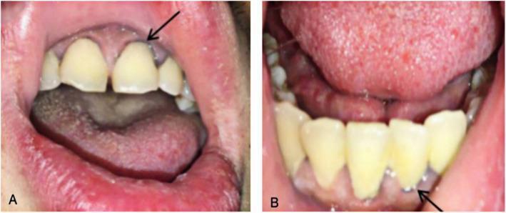
Two intraoral clinical photographs (A and B) demonstrate a characteristic physical finding of Chronic Lead Poisoning (CLP). Image A shows the maxillary gingiva where a black arrow points to a 'Burton line'—a distinct, thin, blue-grey or dark line occurring along the gingival margin at the base of the teeth. Image B displays the mandibular gingiva with a similar dark line indicated by an arrow. The surrounding oral tissues exhibit signs of poor dental hygiene, including yellowish dental discoloration, mild gingival inflammation, and a coated tongue. Burton lines are a classic clinical sign of lead toxicity, resulting from the reaction of circulating lead with sulfur-producing bacteria in the oral cavity, forming lead sulfide precipitates in the gum tissue. This visual evidence serves as a critical diagnostic clue for heavy metal exposure in patients presenting with systemic symptoms such as abdominal pain or anemia.
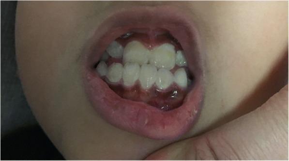
This clinical photograph shows an intraoral view of a pediatric patient exhibiting Burton’s line, a characteristic sign of chronic lead poisoning (plumbism). The image reveals a distinct, dark bluish-grey linear band of pigmentation along the gingival margins where the gums meet the teeth. This hyperpigmented line is visible on both the maxillary and mandibular gingiva. The dental examination shows yellowish-white primary or mixed dentition with evidence of mild crowding and plaque accumulation. The clinical significance of this finding lies in its association with lead exposure, where lead reacts with sulfur-producing bacteria in the oral cavity to form lead sulfide precipitates within the gingival tissue. This sign is typically more prominent in patients with poor oral hygiene. The photograph serves as a diagnostic visual for toxicology and pediatric dental education, illustrating the systemic manifestations of heavy metal toxicity in the oral cavity.
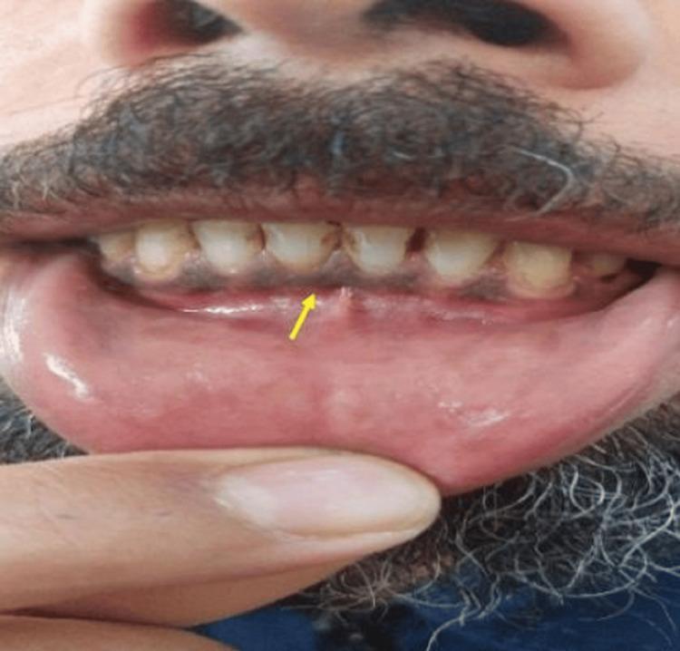
This clinical photograph displays intraoral imaging of a male patient’s lower oral cavity. The focal point, highlighted by a yellow arrow, is a distinct bluish-purple pigmented line along the marginal gingiva of the mandibular teeth. This finding is characteristic of a 'Burtonian line' (or Burton line), a clinical sign associated with chronic lead poisoning (plumbism). The pigmentation results from the reaction between circulating lead and sulfur-producing bacteria in the oral plaque, forming lead sulfide precipitates. The surrounding gingival tissues appear otherwise normal in texture, though some mild dental staining is visible at the cervical margins of the teeth. This image serves as a classic diagnostic aid in toxicology and occupational medicine, illustrating a characteristic physical manifestation of heavy metal toxicity in patients presenting with systemic symptoms like fatigue, abdominal pain, and metallic taste.
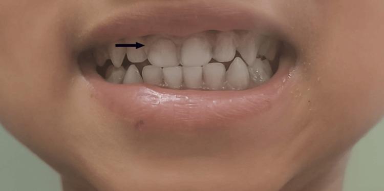
This clinical intraoral photograph displays the maxillary and mandibular anterior teeth and gingiva of a patient. The primary finding is a thin, bluish-purple hyperpigmented line appearing along the gingival margin of the upper teeth, classically known as Burton's line. A horizontal black arrow highlights this discoloration where the gingiva meets the dental enamel. The gums otherwise appear pale pink, and the lips show signs of mild pallor. This finding is a characteristic clinical sign of chronic lead poisoning (plumbism), resulting from the reaction of circulating lead with sulfur-producing bacteria in the oral cavity, which precipitates lead sulfide at the gum line. The image serves as a diagnostic reference for identifying heavy metal toxicity during a physical examination.
basophilic stippling erythrocytes lead poisoning blood smear
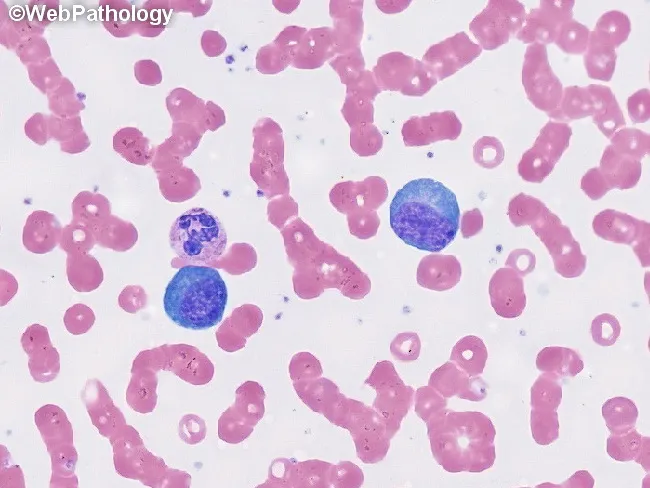
This is a peripheral blood smear prepared with Wright-Giemsa stain and examined under brightfield illumination at high magnification (approximately 1000x total, 100x objective with oil immersion). The smear displays predominantly erythrocytes with uniform pink cytoplasm and characteristic biconcave morphology, arranged singly with occasional rouleaux. Interspersed among the red cells are leukocytes with visible nuclei. Notably, two large lymphocyte-like cells or mononuclear leukocytes appear conspicuously larger than surrounding erythrocytes; these cells have round to oval, deeply basophilic nuclei with dense chromatin and scant, lightly basophilic cytoplasm. No visible granulocytic cytoplasm granules or multiple lobes are clearly resolved in this field, and there are no obvious nucleoli or cytoplasmic inclusions evident in these cells. The background lacks abnormal pigment or reticulocytosis. Overall cell morphology is consistent with a normal or mildly reactive hematologic smear; in a clinical context, additional data such as complete blood count, differential, and patient history would be needed to determine whether there is leukocytosis or blasts. This image serves as an educational reference for recognizing basic peripheral blood components, evaluating leukocyte size variation, and assessing RBC morphology for hematology training and diagnostic differential diagnoses. Additional cases and image contexts can reinforce recognition of hematologic normal variants and patterns.
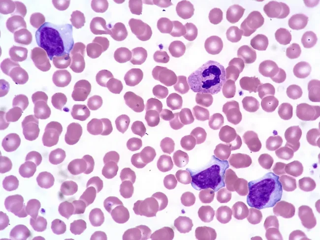
Comprehensive description: This is a brightfield, Wright-Giemsa stained peripheral blood smear examined by light microscopy at high magnification (oil immersion). The specimen shows several activated (atypical) lymphocytes in the peripheral blood, a hallmark of infectious mononucleosis. The lymphocytes have abundant basophilic cytoplasm and irregular, often indented nuclei with clumped chromatin and inconspicuous nucleoli. A normal segmented neutrophil is present for contrast. Erythrocytes exhibit occasional cytoplasmic membrane scalloping around red cells, a feature sometimes observed in EBV-related lymphocytosis. The background cytology is otherwise typical, with uniform erythrocytes and occasional platelets. The activated lymphocytes vary in size and cytoplasmic texture, some appearing plasmacytoid with vesicular basophilia, while others are more indented with coarse chromatin. These morphologic changes reflect an immune response to Epstein-Barr virus infection and are described as reactive lymphocytosis. Clinically, the combination of acute lymphadenitis, fever, a positive Monospot test, and EBV serology supports infectious mononucleosis. The image helps differentiate viral-induced lymphocytosis from bacterial lymphadenitis and from hematologic malignancies when integrated with clinical data. In summary, this smear illustrates the classical hematologic manifestation of EBV-associated mononucleosis. These atypical lymphocytes, sometimes named Downey cells, accompany fever and pharyngitis in EBV infection and aid differentiation from leukemia when combined with serology and clinical data.
wrist drop lead poisoning radial nerve palsy
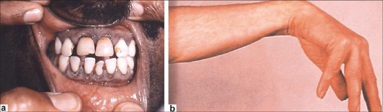
This composite clinical photograph displays two classic physical manifestations of chronic lead poisoning (plumbism). Figure 1a is an intraoral photograph showing the maxillary and mandibular gingiva. A prominent Burtonian line (lead line) is visible as a dark, bluish-gray discoloration along the gingival margin, resulting from the reaction of circulating lead with sulfur-producing bacteria. Secondary findings include dental caries and poor oral hygiene. Figure 1b is a clinical photograph of a patient's forearm and hand demonstrating 'wrist drop' (radial nerve palsy). The wrist is in a flexed position due to paralysis or significant weakness of the extensor muscles, an indication of lead-induced peripheral neuropathy. These images serve as educational tools for recognizing systemic heavy metal toxicity through dermatologic, oral, and neurological physical examination findings. The clinical context relates to occupational exposure and the resulting neuromuscular and mucosal pathology.
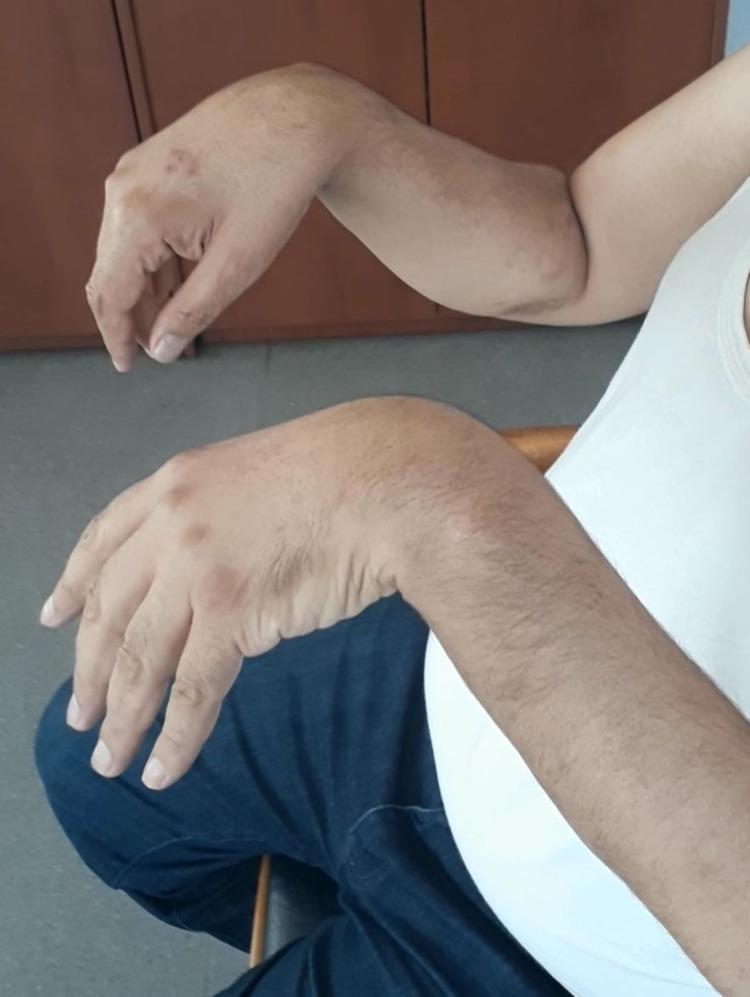
Clinical photograph showing a patient with bilateral wrist drop, a classic neurological sign of radial nerve palsy. The image captures both forearms in a pronated position with the hands hanging limply in passive flexion at the wrist joints. There is a visible inability to maintain extension of the wrists and digits against gravity, indicating significant weakness or paralysis of the extensor muscles (including the extensor carpi radialis longus/brevis, extensor digitorum communis, and extensor indicis proprius). The skin on the dorsal aspect of the hands and distal forearms shows scattered, hyperpigmented macules and patches. This presentation is characteristic of peripheral compressive neuropathy of the radial nerve, often occurring at the spiral groove of the humerus. The visual evidence supports a diagnosis of severe motor deficit in the radial nerve distribution, commonly associated with prolonged external pressure or 'Saturday night palsy' scenarios.
Lead Poisoning - Forensic Medicine and Toxicology
1. Introduction
2. Poisonous Compounds / Sources of Lead
| Salt | Common Name | Appearance |
|---|---|---|
| Lead acetate | Sugar of lead / Salt of Saturn | White crystals |
| Lead carbonate | Safeda / White lead | White crystalline powder |
| Lead chromate | Chrome yellow | Bright yellow powder |
| Lead monoxide | Litharge / Mudrasang | Pale brick-red or orange masses |
| Lead tetroxide | Red lead / Sindoor / Vermilion | Scarlet crystalline powder |
| Lead sulfide | - | Least toxic |
| Tetraethyl lead | - | Organic (volatile) |
- Storage batteries (car batteries) - most common industrial source
- Lead-based paints (most common source of exposure in children)
- Petrol / gasoline (tetraethyl lead as anti-knock agent)
- Solders, electric cable insulation
- Pottery and ceramic glazes
- Hair dyes
- Projectiles (bullets, fishing sinkers)
- Radiation shielding
- Lead glass (12-28% lead content)
- Plumbing (historically)
3. Mechanism of Action
- Inhibiting delta-aminolevulinic acid dehydrase (ALA-D) - a zinc-binding enzyme critical for heme biosynthesis, thus disrupting hemoglobin and cytochrome production
- Combining with sulfhydryl (-SH) groups - inhibits tissue enzyme systems broadly
- Causing hemolysis of mature red cells
- Depressing bone marrow - causing anemia, basophilic stippling of RBCs, anisopoikilocytosis, and thrombocytopenia
- Interfering with mitochondrial oxidative phosphorylation and ATPases
- Disrupting calcium-dependent messengers and enhancing oxidative stress and cell apoptosis
- Causing spasm of capillaries and arterioles - toxic effects result from fixation of lead in the brain and peripheral nervous system
4. Absorption, Distribution, and Excretion
- Inhalation (most dangerous - 10x more toxic than ingestion)
- Gastrointestinal tract (ingestion)
- Abraded skin / mucous surfaces
5. Fatal Dose and Fatal Period
| Parameter | Value |
|---|---|
| Fatal dose (absorbed lead) | ~0.5 g |
| Fatal dose (lead acetate) | ~20 g |
| Fatal dose (lead carbonate) | ~4 g (Parikh); 40 g (Essentials) |
| Tetraethyl lead | A drop or two of pure compound may cause serious symptoms |
| Fatal period (acute) | 1-2 days (Essentials); death may occur on 2nd-3rd day (Parikh) |
6. Acute Lead Poisoning
- Astringent/metallic taste, dry throat, thirst
- Nausea, vomiting
- Colic-like abdominal pain - relieved by pressure
- Constipation (more common than diarrhea); feces are black and offensive
- Urine is suppressed (oliguria/anuria)
- Cramps in legs, arthralgia (if protracted)
- Headache, drowsiness, insomnia, paresthesias
- Paralysis of limbs
- Peripheral circulatory collapse, depression, coma
- Cerebellar ataxia is common in children in acute poisoning
- Death (preceded by collapse)
Acute Lead Encephalopathy (especially with tetraethyl lead):
- Gastric symptoms absent or slight
- Headache, sleeplessness
- Tremors of eyes, mouth, and fingers
- Paralysis or loss of vision
- Hallucinations, excitement, delirium or apathy
- Convulsions
- Permanent mental changes or insanity
7. Chronic Lead Poisoning (Plumbism / Saturnism)
Sources / Causes:
- Inhalation of lead dust and fumes by white lead makers and users of lead paints
- Smelter workers, painters, plumbers
- Children eating lead paint peelings (pica - ages 18 months to 3 years, especially in tenement areas)
- Environmental and industrial exposure
Signs and Symptoms (System-wise):
- Lead colic (severe, intermittent abdominal pain)
- Constipation, anorexia, nausea
- Abdominal cramps, vomiting
- Burton's line (Lead line) - a dark blue-grey line along the gingival margin at the base of teeth, formed by reaction of circulating lead with sulfur-producing bacteria in oral plaque, precipitating lead sulfide in gum tissue. More prominent with poor oral hygiene.
- Wrist drop (most characteristic) - due to lead neuropathy affecting the radial nerve
- Foot drop
- Peripheral neuropathy (motor > sensory)
- Big joints generally affected; small joints escape
- Muscle weakness, paralysis
- Anemia (normocytic/hypochromic)
- Basophilic stippling of RBCs (pathognomonic)
- Anisopoikilocytosis
- Thrombocytopenia
- Hemolysis
- Nephropathy (proximal tubule damage)
- Lethargy, weight loss, weakness
- Pallor (from anemia)
8. Medico-legal Importance / Diagnosis
Clinical Diagnostic Clues:
- Burton's line on gums
- Wrist drop / foot drop
- Basophilic stippling of RBCs on blood smear
- Lead lines on X-ray - dense radiodense bands at ends of long bones along growth plates (especially in children)
- Eosinophilic intranuclear inclusions in hepatocytes and proximal renal tubule cells (histopathology)
Laboratory / Forensic Tests:
- Blood lead level (BLL): The gold standard
- CDC upper limit for children: 0.05 mg/L (5 µg/dL)
- 0.05-0.44 mg/L: close follow-up and investigation
-
0.45 mg/L: chelation therapy indicated
- Urinary ALA (delta-aminolevulinic acid): Elevated
- Blood smear: Basophilic stippling
- Atomic absorption spectrophotometry for precise blood/urine lead quantification
Medico-legal Significance:
- Primarily encountered as an industrial/occupational hazard rather than homicidal poisoning
- May arise as an issue in workmen's compensation and occupational health litigation
- Lead from bullets retained in body cavities can cause systemic toxicity
- Environmental exposure (contaminated water, soil) is a public health and legal matter
- Pica in children (ingestion of lead paint) is the most common cause of lead poisoning deaths in children in urban settings
9. Autopsy Findings
Acute Lead Poisoning:
- Signs of acute gastroenteritis
- Gastric mucosa: thickened, softened, eroded patches, covered with a greyish-white deposit
- Large intestine may contain black-coloured material (lead sulphide)
- Congested gastric mucosa with eroded patches
Chronic Lead Poisoning:
- Brain - most striking finding: massively swollen, flattening of gyri, extremely pale (almost white)
- PAS-positive, pink-staining homogeneous material in perivascular spaces of brain
- Blood smear: basophilic stippling of erythrocytes
- Eosinophilic intranuclear inclusions in hepatocytes and proximal renal tubule cells
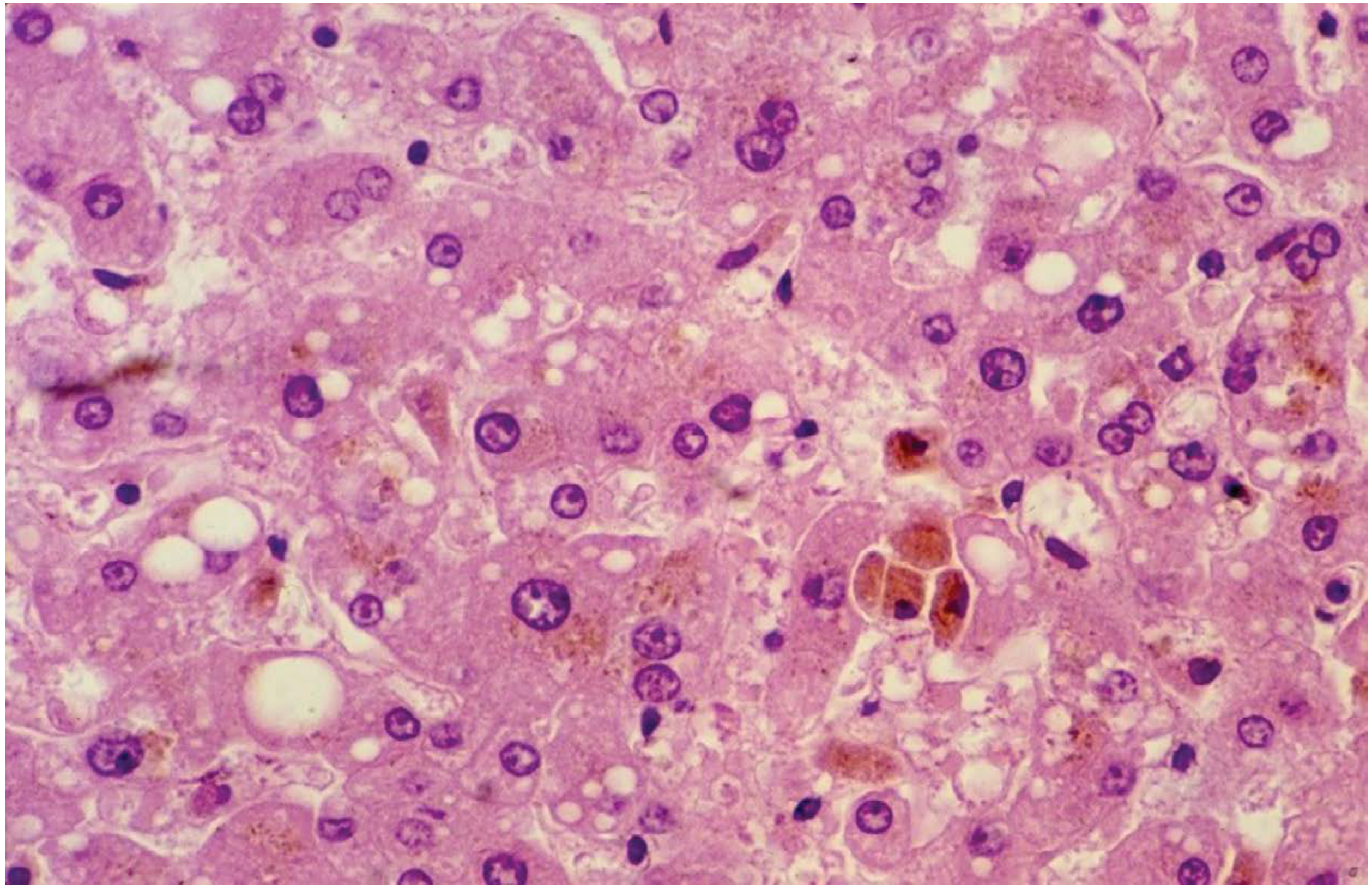
- Lead lines on X-ray at growth plates of long bones (in children)
10. Treatment
Acute Poisoning:
- Gastric lavage with 1% solution of sodium or magnesium sulphate (to convert lead to insoluble lead sulphate)
- Ample washing with plain water
- Demulcents
- BAL (British Anti-Lewisite / dimercaprol) + Calcium disodium EDTA (versenate) - combined chelation (most effective)
- DMSA (succimer) - oral chelating agent
- Penicillamine (D-penicillamine)
- Calcium chloride 5 mg of 10% solution IV - promotes deposition of lead from blood into skeleton
- Morphine and atropine for painful colic
- Peritoneal dialysis or hemodialysis in severe cases
- Symptomatic treatment
Tetraethyl Lead (Skin Exposure):
- Wash skin with kerosene within 15 minutes - removes the poison quantitatively
Antidotes Summary:
| Antidote | Route | Notes |
|---|---|---|
| BAL (dimercaprol) | IM | Used in combination with EDTA |
| Calcium disodium EDTA (versenate) | IV | Most effective when combined with BAL |
| DMSA (succimer) | Oral | Preferred for children with mild-moderate elevation |
| D-penicillamine | Oral | Second-line |
11. Quick Reference Summary Card
| Feature | Detail |
|---|---|
| Type | Heavy metal poison |
| Most toxic form | Volatile lead (inhaled); soluble salts > metallic lead |
| Most common source (adults) | Industrial/occupational exposure |
| Most common source (children) | Lead paint peelings (pica) |
| Pathognomonic sign | Burton's line (gums) + basophilic stippling |
| Motor sign | Wrist drop (radial nerve), foot drop |
| Fatal dose (acetate) | ~20 g |
| Fatal period (acute) | 1-2 days |
| Antidote | BAL + Ca-EDTA; DMSA; penicillamine |
| Autopsy hallmark | Pale swollen brain; eosinophilic intranuclear inclusions (liver/kidney) |
| Blood test | Blood lead level (BLL) by atomic absorption spectrophotometry |
| Child action level | BLL > 0.45 mg/L → chelation |