Listhesias x-rays
Reading File
Finding Sources
Finding Sources
Searching Images
spondylolisthesis x-ray radiograph
Reading File
Reading File
Loading Image
Loading Image
Loading Image
Here is a comprehensive overview of listhesis (spondylolisthesis) on X-ray.
Listhesis — Radiographic Overview
Listhesis refers to the displacement of one vertebra relative to an adjacent one:
- Anterolisthesis (spondylolisthesis) — forward displacement of the superior vertebra
- Retrolisthesis — backward displacement of the superior vertebra
X-Ray Views Used
| View | What It Shows |
|---|---|
| Lateral | Best view — directly shows the step-off/displacement |
| AP (anteroposterior) | Coronal alignment; less sensitive for slip |
| Oblique | Shows pars interarticularis defect ("Scotty dog" sign) — 15% of lysis lesions visible here that the lateral misses |
Plain lateral radiographs demonstrate 80% of spondylolysis lesions; oblique views add another ~15%. — Miller's Review of Orthopaedics 9e
What to Look For on Lateral X-Ray
- Step-off — anterior displacement of the vertebral body relative to the one below
- Disc space narrowing — especially at the affected level
- Facet joint degeneration — joint space loss, sclerosis (degenerative type)
- Spinous process malalignment — in degenerative spondylolisthesis, the spinous process moves anteriorly with the slipped vertebra (distinguishes it from isthmic type)
- Pars defect — lucency at the pars interarticularis (isthmic type)
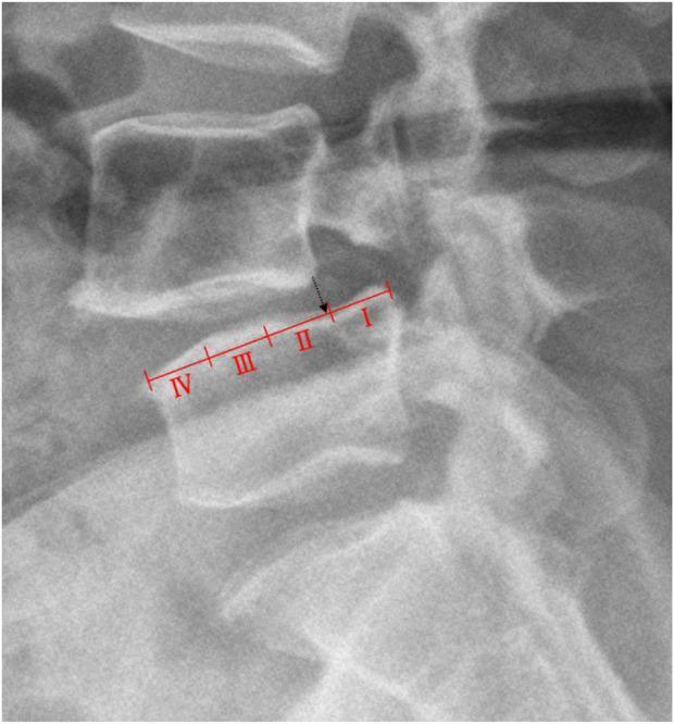
Meyerding Grading System
The superior endplate of the lower vertebra is divided into four equal quarters:
| Grade | Slip Amount |
|---|---|
| I | 0–25% |
| II | 25–50% |
| III | 50–75% |
| IV | >75% |
| V (spondyloptosis) | >100% |
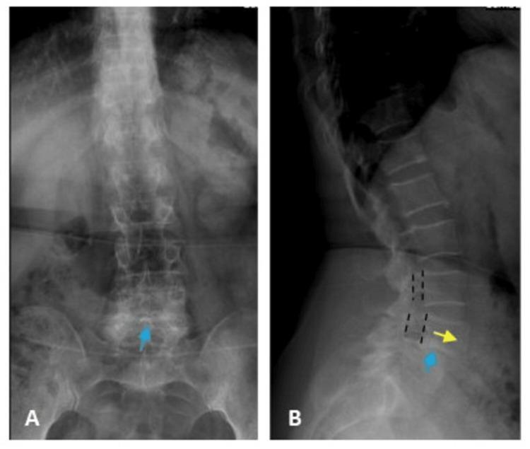
Two-View Example: Degenerative L4–L5 Spondylolisthesis
Types of Spondylolisthesis (Newman–Wiltse–McNab Classification)
| Type | Cause | Key X-ray Feature |
|---|---|---|
| I — Dysplastic/Congenital | Abnormal articular processes | Slip at L5–S1, no pars defect |
| II — Isthmic | Pars interarticularis defect | Oblique view: "Scotty dog" collar sign |
| III — Degenerative | Facet joint OA | Intact neural arch; anterior spinous process shift |
| IV — Traumatic | Neural arch fracture | Fracture line on plain film |
| V — Pathologic | Bone disorder (Paget's, tumor) | Generalized bone disease features |
| VI — Iatrogenic | Post-laminectomy | Surgical history |
Degenerative Type — Key Radiographic Points
- Most common cause of lumbar spondylolisthesis over age 50
- Most common level: L4–L5 (4× more common if L5 is sacralized)
- Women 4× more affected than men
- Lateral X-ray: anterolisthesis + facet joint narrowing/sclerosis + disc space narrowing
- The neural arch is intact — even a small slip can cause canal stenosis/cauda equina syndrome
- CT/MRI adds assessment of facet joint OA, foraminal narrowing, and ligamentum flavum hypertrophy
— Grainger & Allison's Diagnostic Radiology; Miller's Review of Orthopaedics 9e
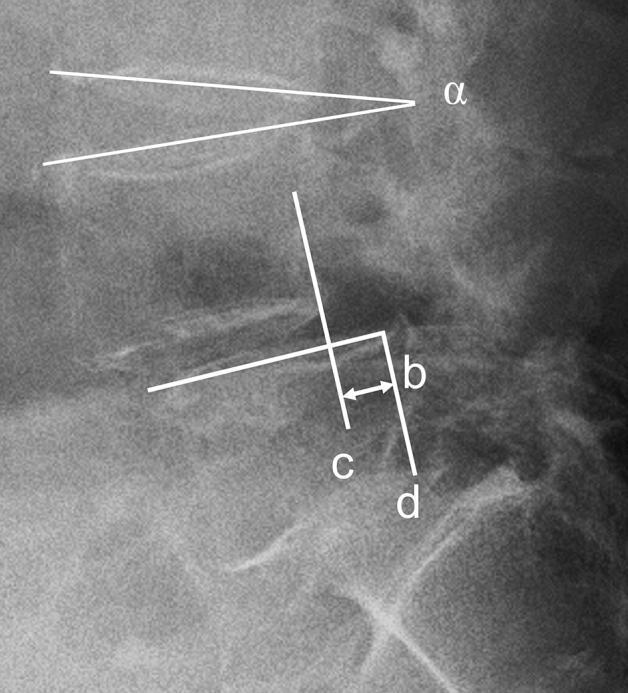
Additional Measurements on Lateral X-Ray
Beyond the Meyerding grade, the lateral film also allows assessment of:
- Slip angle (normally <0°, indicating lordosis at L5–S1)
- Sacral inclination (normally >30°)
- Pelvic incidence (normally ~50°) — used in surgical planning
This is a shared conversation. Sign in to Orris to start your own chat.