
Post op pain management
"postoperative pain" AND "multimodal analgesia"
multimodal analgesia postoperative pain management diagram
Post-Operative Pain Management
Core Principle: Multimodal Analgesia
"Multimodal analgesia combines different classes of medications that have different pharmacological mechanisms of action, resulting in additive or synergistic effects to reduce postoperative pain and its sequelae."
- Morgan and Mikhail's Clinical Anesthesiology, 7e
Assessment of Post-Op Pain
- Use a numeric pain scale (0-10) or visual analogue scale; document periodically throughout recovery (Joint Commission mandate)
- Pain severity varies among procedures and anesthetic techniques
- Both under- and over-estimation by staff are common; the patient's own perception remains the gold standard
- Vital signs (tachycardia, hypertension, sweating) are suggestive but unreliable alone; importantly, analgesic administration can precipitate hypotension in hypovolemic patients - Barash Clinical Anesthesia, 9e
Drug Classes in Multimodal Analgesia
1. NSAIDs
- Adding NSAIDs to systemic opioids diminishes pain intensity, reduces opioid requirements, and decreases opioid side effects (PONV, sedation, urinary retention)
- Risks: GI bleeding, wound healing impairment, renal dysfunction, possible anastomotic leak risk (controversial)
- COX-2 inhibitors (e.g., celecoxib): reduce platelet dysfunction and GI bleeding vs. non-selective NSAIDs; renal effects remain a concern; cardiovascular risk in vascular surgery patients is documented
- Morgan and Mikhail's Clinical Anesthesiology, 7e
2. Acetaminophen (Paracetamol)
- Available oral, rectal, or IV (parenteral)
- Analgesic effect ~20-30% less than NSAIDs, but safer pharmacological profile
- Significantly reduces pain intensity and spares opioid consumption after orthopedic and abdominal surgery
- Works synergistically with NSAIDs; may allow NSAIDs to be reserved for breakthrough pain
- Pediatric dosing: IV 10-15 mg/kg (neonates 10 mg/kg), oral/rectal 20-30 mg/kg rectal in infants
- Morgan and Mikhail's Clinical Anesthesiology, 7e; Barash Clinical Anesthesia, 9e
3. Gabapentinoids (Gabapentin, Pregabalin)
- Single preoperative dose decreases post-op pain and opioid consumption in the first 24 h
- Optimal dose and duration are still debated; may potentially reduce incidence of chronic post-surgical pain
- Key side effects: Sedation and dizziness, especially in older adults - can increase fall risk
4. NMDA Receptor Antagonists
- Perioperative low-dose ketamine (bolus or infusion) produces significant reduction in pain, opioid consumption, and PONV
- Particularly beneficial in patients on chronic opioids (opioid-tolerant patients)
- May reduce post-op pain and opioid consumption; optimal dosing remains uncertain
- Side effects include hypotension and potentiation of neuromuscular blockade
5. IV Lidocaine
- Growing evidence supports its use as a multimodal component
- In major abdominal surgery: associated with faster return of bowel function and shorter hospital stay
- Requires continuous cardiovascular monitoring (CNS and cardiac toxicity risk); currently limited to PACU, ICU, or monitored wards
- Dosing: 1.5 mg/kg IV bolus, then 1.5-2 mg/kg/h intraoperatively, followed by 1 mg/kg/h post-op
- Morgan and Mikhail's Clinical Anesthesiology, 7e
6. Opioids
- Remain a cornerstone of surgical pain management despite efforts to minimize use
- Patient-Controlled Analgesia (PCA) provides better pain control, greater patient satisfaction, and fewer side effects vs. nurse-administered PRN dosing
- Common parenteral opioids: morphine, fentanyl, hydromorphone
- Transition to oral opioids (oxycodone, hydrocodone) combined with NSAIDs or acetaminophen as patient recovers
- Opioid prescribing should be minimized: studies show most elective surgery patients use opioids for only 2 days or fewer; many achieve adequate control with over-the-counter medications
Regional Analgesia Techniques
| Technique | Notes |
|---|---|
| Epidural analgesia | Gold standard for thoracic/abdominal/lower limb surgery; combines local anesthetic (bupivacaine 0.1-0.125% or ropivacaine 0.1-0.2%) + opioid (fentanyl 2-2.5 mcg/mL) |
| Peripheral nerve blocks | Ultrasound-guided; single-shot or continuous catheter; excellent for orthopedic procedures |
| Local anesthetic wound infusion | Simple, effective for superficial wounds |
| Intrathecal (spinal) opioids | Long-lasting; requires monitoring for delayed respiratory depression |
- Ultrasound-guided nerve blocks combined with multimodal analgesia are a "reasonable alternative to epidural analgesia for postoperative pain management in low- to moderate-risk patients" - Miller's Anesthesia, 10e
Perioperative Analgesic Adjuvants - Quick Reference
| Drug | Dose (IV) | Timing | Setting |
|---|---|---|---|
| Lidocaine | 1.5 mg/kg bolus → 1-2 mg/kg/h infusion | Pre/Intra/Post | Abdominal, thoracic, orthopedic |
| Ketamine | 0.5-1 mg/kg bolus | Pre/Post (PCA add-on) | Opioid-tolerant, cardiac, orthopedic |
| Dexmedetomidine | Alpha-2 agonist adjunct | Intra/Post | Pediatric, ICU |
| Ketorolac | 15-30 mg IV (adults); 0.5-0.75 mg/kg (pediatric) | Post | Orthopedic, abdominal |
Patient-Controlled Analgesia (PCA)
- Adults: Morphine 1-2 mg q6-10 min lockout; hydromorphone 0.2-0.4 mg q6-10 min
- Pediatric (≥5 yrs): Morphine 20 mcg/kg/dose or hydromorphone 5 mcg/kg/dose; 10-min lockout
- Continuous background infusions increase respiratory depression risk - use with caution
- Morgan and Mikhail's Clinical Anesthesiology, 7e
Special Populations
Pediatric
- Regional techniques have greatly expanded in use
- Epidural: bupivacaine 0.1-0.125% or ropivacaine 0.1-0.2% + fentanyl 2-2.5 mcg/mL at 0.1-0.4 mL/kg/h
- Codeine is falling out of favor due to pharmacogenetic variability (ultra-rapid metabolizers)
- Ketorolac in neonates <21 days or <37 weeks corrected gestational age carries increased bleeding risk
Opioid-Tolerant Patients
- Standard doses are inadequate; require higher baseline doses
- Low-dose ketamine infusion is particularly beneficial in this group
Elderly
- Gabapentinoids carry higher sedation and fall risk
- Analgesic dosing should be reduced; renal function affects NSAID and opioid clearance
Opioid Stewardship
- Multimodal pain protocols to minimize opioid prescriptions
- Education on recognizing opioid abuse patterns
- Most elective surgical patients need opioids for ≤2 days post-op; many achieve control with OTC medications alone
- Enhanced recovery pathways (ERAS) incorporate opioid-sparing strategies as a standard of care
- Schwartz's Principles of Surgery, 11e
Recent Evidence (2024-2025)
- A 2025 meta-analysis (PMID 40319048) confirms multimodal analgesia significantly reduces post-hepatectomy wound pain and shortens hospital stay
- A 2025 systematic review (PMID 40937914) found epidural analgesia superior to systemic opioids for pain management after VATS (video-assisted thoracoscopic surgery)
- A 2024 network meta-analysis in British Journal of Anaesthesia (PMID 39366846) identified the most effective non-opioid adjuvants for reducing post-op opioid consumption in obese patients, supporting individualized multimodal regimens in this high-risk population
Extensor mechanism of knee
extensor mechanism knee quadriceps patella patellar tendon anatomy
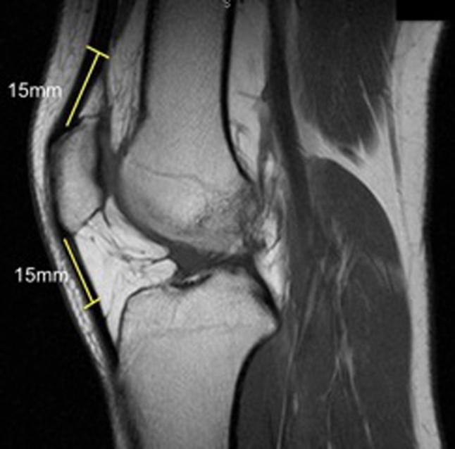
This diagnostic image is a sagittal proton-density magnetic resonance imaging (MRI) scan of the human knee joint, illustrating the anatomy and preoperative measurement protocol for ligament reconstruction. Key visible structures include the distal femur, the patella, the proximal tibia, and the extensor mechanism. The image specifically demonstrates a standardized method for assessing potential donor tissue thickness for autografts, such as Bone-Patellar Tendon-Bone (BPTB) or quadriceps tendon grafts. Two specific measurements are highlighted with yellow lines: a 15 mm segment extending proximally from the superior pole of the patella along the quadriceps tendon, and a 15 mm segment extending distally from the inferior pole of the patella along the patellar tendon. These markers indicate the anatomical locations where tendon thickness should be measured to evaluate graft suitability. The clinical focus of the image is orthopedic preoperative planning for Anterior Cruciate Ligament (ACL) reconstruction, providing a quantitative approach to individualizing surgical graft selection based on the patient's native anatomy.
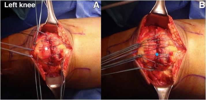
This clinical photograph displays a two-panel intraoperative view (A and B) of a surgical repair for a quadriceps tendon rupture in a left knee. The patient is in a supine position with the surgical site exposed via a midline longitudinal incision. In panel A, the peritendinous quadriceps tendon is being repaired using a figure-of-8 suture technique with No. 1-0 Vicryl sutures, indicated by a white star. The sutures approximate the anterior soft tissue and retinaculum over the superior pole of the patella. Panel B demonstrates the 'suture bridge' technique, where multiple suture limbs originating from the quadriceps tendon are gathered and secured at the inferior pole of the patella, marked by a blue star. This construct ensures the full thickness of the tendon is reattached to the patellar bone, restoring the extensor mechanism. Surgical retractors are visible, providing visualization of the patellar anatomy and the distribution of high-strength suture material across the repair site. The images illustrate key steps in transosseous tendon-to-bone fixation.
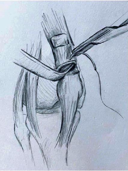
This medical illustration depicts a surgical technique for quadriceps tendon reconstruction using a synthetic mesh graft. The drawing shows the knee joint anatomy including the patella, patellar tendon, and the remnant of the quadriceps tendon. A tubularized mesh, such as a polypropylene mesh, is seen being secured to the proximal quadriceps tendon stump. A surgical needle holder or forceps is shown in the superior-right field, applying a running suture—specifically a Krackow suture technique—to achieve intrasubstance fixation. The mesh is integrated into a longitudinal tunnel within the tendon, providing a scaffold for repair in cases of chronic or recurrent rupture. The illustration focuses on the proximal fixation stage, emphasizing the mechanical anchoring of the graft to the native soft tissue to restore the extensor mechanism of the knee. This pedagogical diagram is intended for orthopaedic surgical education, demonstrating salvage procedures for complex tendon repairs.
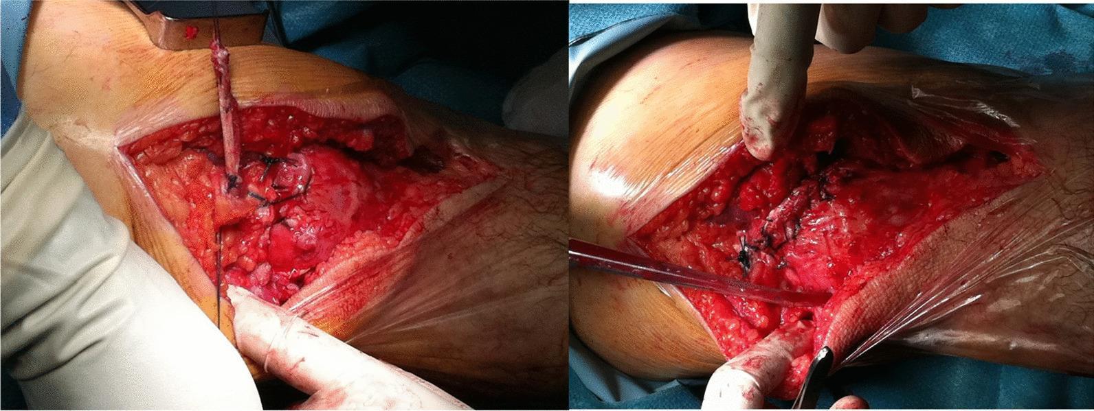
This clinical photograph set depicts an intraoperative view of an orthopedic surgical procedure, specifically a quadriceps tendon rupture repair. The image shows a large, open surgical site on the anterior aspect of the knee. The exposed anatomy includes the subcutaneous adipose tissue, the patella, and the proximal retracted stump of the quadriceps tendon. Key visual features involve the application of a tendon graft secured with multiple black, non-absorbable sutures. These sutures are visible anchoring the graft material to both the periosteum at the patellar tunnel exit holes and to the proximal tendon stump using a series of interrupted knots. Hemostasis is partially managed, with visible areas of clotted and fresh blood within the wound bed. The procedural focus illustrated is the terminal stage of a figure-of-eight graft augmentation used to bridge and reinforce the primary quadriceps tendon repair, demonstrating the method of attachment and suture patterns required for functional stability of the extensor mechanism.
patella anatomy sesamoid bone quadriceps vastus knee
Extensor Mechanism of the Knee
"The quadriceps muscles, quadriceps tendon, medial and lateral retinacula, patella, patellar tendon, and tibial tubercle comprise the extensor mechanism of the knee."
- Rosen's Emergency Medicine, 10e
Anatomy Diagram - Anterior Compartment of the Thigh
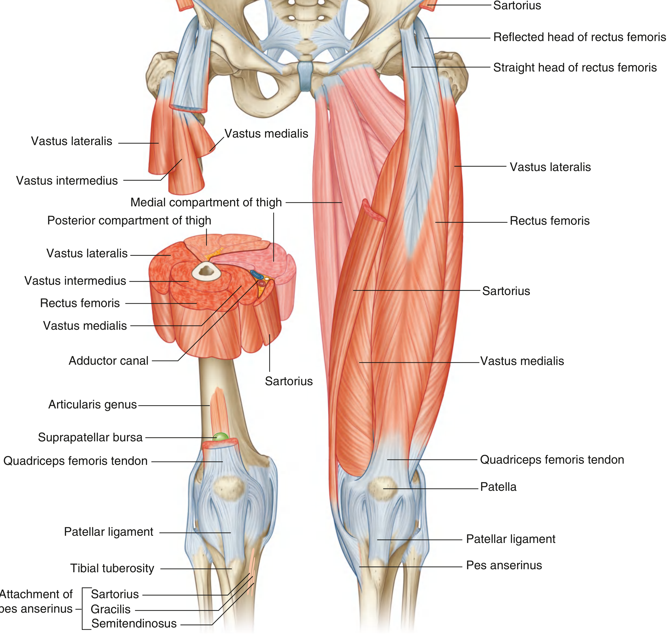
1. The Quadriceps Femoris Muscle
| Muscle | Origin | Key Notes |
|---|---|---|
| Rectus femoris | Anterior inferior iliac spine (straight head); ilium above acetabulum (reflected head) | Only head that crosses the hip joint - also flexes hip |
| Vastus medialis | Intertrochanteric line → medial lip of linea aspera → medial supracondylar line | Distal fibers form VMO (vastus medialis obliquus); critical for terminal extension and patellar tracking |
| Vastus lateralis | Intertrochanteric line → lateral lip of linea aspera (largest of the vastus muscles) | Pulls patella laterally; counterbalanced by VMO |
| Vastus intermedius | Upper 2/3 of anterior and lateral femur | Deepest head; merges into deep quadriceps tendon |
2. The Quadriceps Tendon
- Formed by convergence of all four quadriceps heads
- Inserts on the base (superior border) of the patella
- Has a trilaminar structure: superficial fibers from rectus femoris, middle from vastus medialis and lateralis, deep from vastus intermedius
- The more superficial fibers are continuous over the anterior surface of the patella and merge into the patellar ligament
3. The Patella
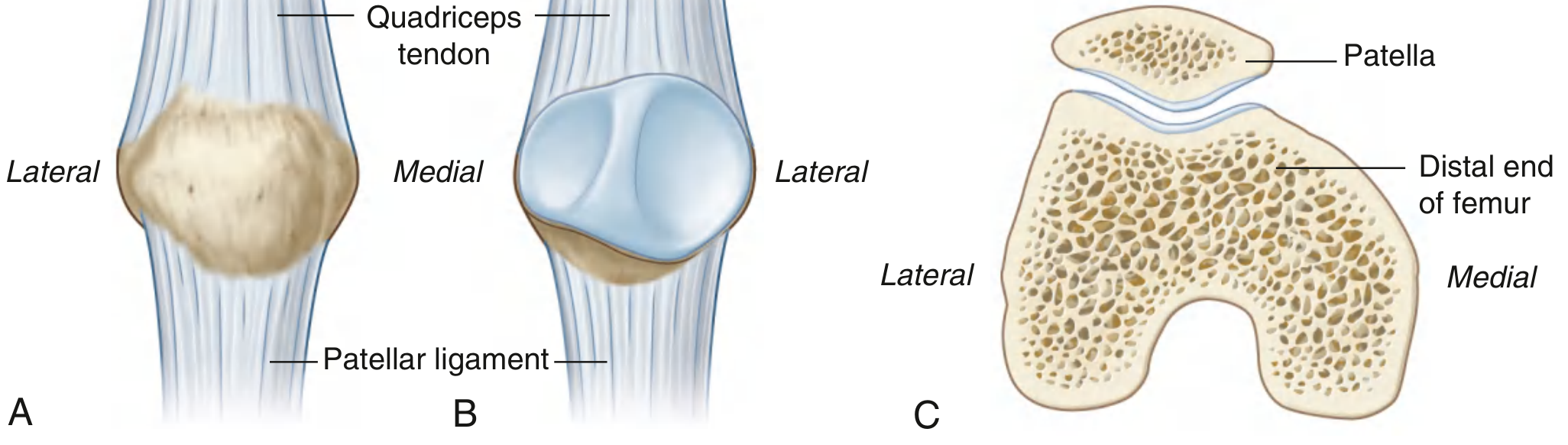
- The largest sesamoid bone in the body
- Shape: Triangular with apex pointing inferiorly
- Base (superior): Broad - attachment of quadriceps tendon
- Apex (inferior): Pointed - attachment of patellar ligament
- Posterior surface: Articulates with the femoral trochlea; has medial and lateral facets separated by a central ridge - the lateral facet is larger (corresponding to the larger lateral femoral condyle)
- Function of the patella: Provides anterior displacement of the quadriceps tendon, acting as a lever that increases the mechanical advantage (moment arm) of the quadriceps by ~30-50%, particularly in the 20-70° arc of flexion
4. The Patellar Ligament (Patellar Tendon)
- Functionally the continuation of the quadriceps tendon below the patella
- Attaches above to the apex and margins of the patella
- Attaches below to the tibial tuberosity
- The patellar ligament is parallel to the posterior cruciate ligament (PCL), and the quadriceps tendon is parallel to the ACL at 30° of knee flexion (important in ACL graft selection)
- Gray's Anatomy for Students; Campbell's Operative Orthopaedics, 15e
5. The Retinacula
Medial Retinacula (proximal to distal):
| Structure | Attachment | Role |
|---|---|---|
| Medial Patellofemoral Ligament (MPFL) | Patella (middle/superior 2/3) → medial femoral epicondyle | Primary static stabilizer against lateral patellar displacement |
| Medial Quadriceps Tendon Femoral Ligament (MQTFL) | Quadriceps tendon → medial femur | Proximal patellar restraint |
| Medial Patellotibial Ligament (MPTL) | Patella → medial tibia | Distal patellar restraint |
| Medial Patellomeniscal Ligament (MPML) | Patella → medial meniscus | Distal patellar restraint |
Lateral Retinacula:
- Lateral patellofemoral and lateral patellotibial ligaments
- Normally contribute to patella tracking; pathologic instability only with iatrogenic injury or surgical release
- Campbell's Operative Orthopaedics, 15e
6. Patellofemoral Joint and Tracking
- Balance between VMO pull (medially) vs. vastus lateralis (laterally)
- The Q-angle (quadriceps angle): angle between line from ASIS to patella center and patellar tendon - normally ~15° in males, ~18° in females; increased Q-angle predisposes to lateral patellar tracking problems
- Depth of the trochlear groove (shallow trochlea = dysplasia = instability risk)
- Integrity of MPFL
Sagittal MRI of the Extensor Mechanism
Clinical Significance - Disruptions of the Extensor Mechanism
- Patellar fracture - most common cause of extensor mechanism disruption
- Quadriceps tendon rupture - more common in patients >40 years; associated with systemic disease (lupus, DM, gout, hyperparathyroidism, uremia, obesity, steroids, fluoroquinolone use)
- Patellar tendon rupture - more common in patients <40 years, especially athletes
- Palpable gap in the quadriceps tendon
- Patella displaced inferiorly (quadriceps rupture) or superiorly (patellar tendon rupture)
- Inability to perform straight-leg raise
- Swelling and ecchymosis
- Audible pop at time of injury
Related Conditions of the Extensor Mechanism
| Condition | Location | Population | Notes |
|---|---|---|---|
| Patellar tendinopathy ("Jumper's knee") | Proximal patellar tendon | Athletes | Focal thickening + hypervascularity on US/MRI |
| Osgood-Schlatter disease | Distal patellar tendon / tibial tuberosity | Children 8-13 yrs | Traction apophysitis; TT enlargement or fragmentation |
| Chondromalacia patellae | Patellar cartilage | Adolescents/young adults | Post-traumatic or malalignment-related cartilage damage |
| Patellofemoral pain syndrome | Patellofemoral joint | Young active adults | VMO weakness, malalignment |