
Diabetic foot
Reading File
Now I'll search the medical library for diabetic foot content across multiple angles in parallel.
Finding Sources
Finding Sources
Searching PubMed
"diabetic foot"[MeSH Terms] AND management
Excellent results. Now let me read the most comprehensive sources in parallel - Bailey & Love (surgery), Goldman-Cecil (medicine), Rosen's (emergency), and Campbell's (orthopaedics).
Reading File
Reading File
Reading File
Reading File
Now let me read more from Bailey & Love (classification, management) and Campbell's (pathophysiology, Charcot, treatment) to get complete coverage.
Reading File
Reading File
Reading File
Reading File
Reading File
Reading File
I now have comprehensive information from multiple authoritative textbooks. Let me also fetch the clinical photo from Bailey & Love to include in the answer.
Loading Image
Loading Image
I now have all the content needed for a thorough, well-structured answer. Let me compile it.
Diabetic Foot
Epidemiology
Diabetes affects ~37.4 million Americans (11.3% of the population), rising from 25.8 million in 2010. Foot complications are the leading cause of hospital admissions in diabetic patients, with an annual incidence of 1-2% per year. A diabetic patient carries a lifetime risk of 15% for developing a foot ulcer. Up to one-fifth of ulcers result in amputation. Five-year mortality from diabetic foot complications is comparable to many cancers. - Campbell's Operative Orthopaedics 15e, p.5054
Pathophysiology
Three major mechanisms converge to cause diabetic foot disease:
1. Peripheral Neuropathy (most important)
Chronically elevated glucose leads to:
- Glycosylation of hemoglobin, proteins, and arterial walls
- Thickening of basement membranes and reduced endothelial nitric oxide activity
- Epineural vessel atherosclerosis and multifocal ischemic proximal nerve lesions
- Metabolic effects: sorbitol accumulation, enzyme deficiencies, increased oxygen-free radical activity
The resulting motor, sensory, and autonomic dysfunction manifests as:
- Sensory: Loss of protective sensation - the inability to feel a 10g Semmes-Weinstein monofilament is one of the most predictive risk factors for foot morbidity
- Motor: Muscle imbalance with weak intrinsic muscles overpowered by stronger extrinsic muscles → hammer toes, claw toes, distal migration of the fat pad; gastrosoleus contracture increases forefoot force loads
- Autonomic: Loss of skin oils → fissuring and skin breakdown; increased susceptibility to trauma
2. Peripheral Arterial Disease (ischemia)
- Diabetics are twice as likely to develop peripheral arterial disease
- Diabetics with PAD are nine times more likely to develop a foot ulcer
- Glycosylated end products deposit in arteriolar walls, impairing perfusion
- Results in ischemic ulcers and severely impairs healing
3. Immune Dysfunction
- Altered chemotaxis of polymorphonuclear cells
- Cell wall abnormalities increase susceptibility to infection
- Hyperglycemia impairs the normal immune response
Clinical Features
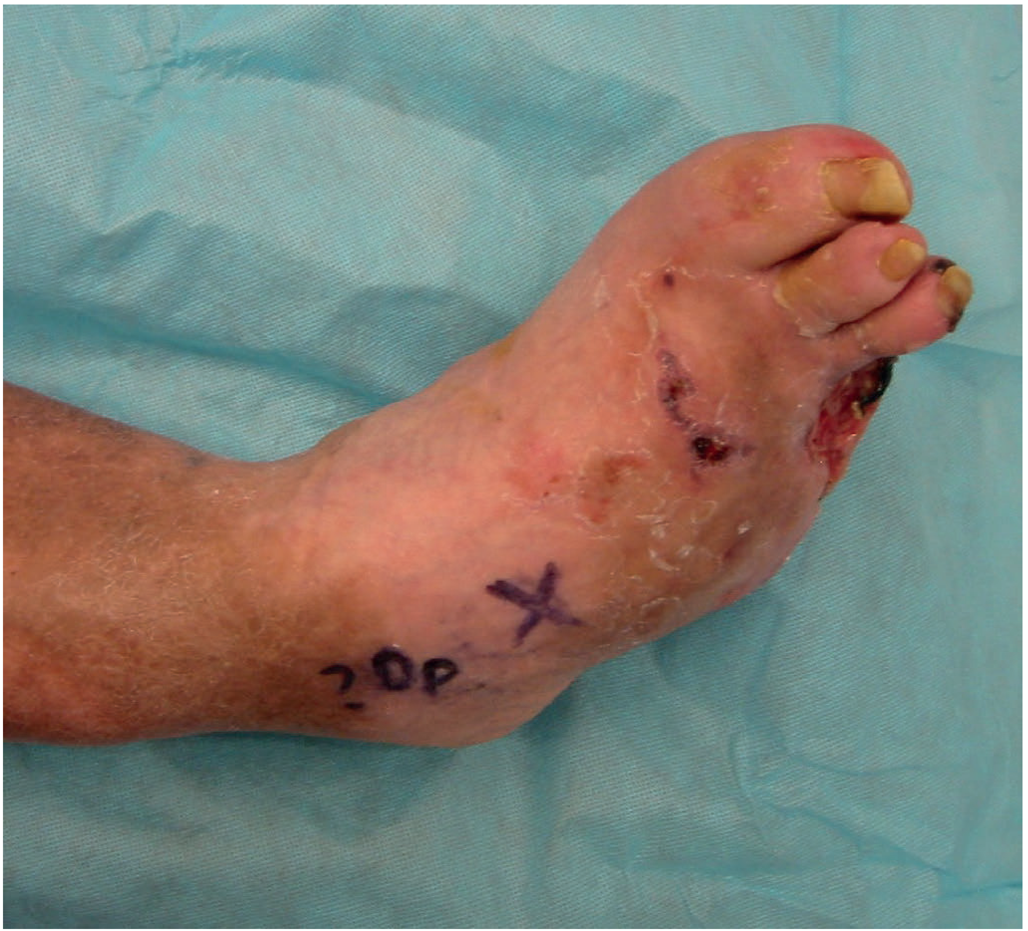
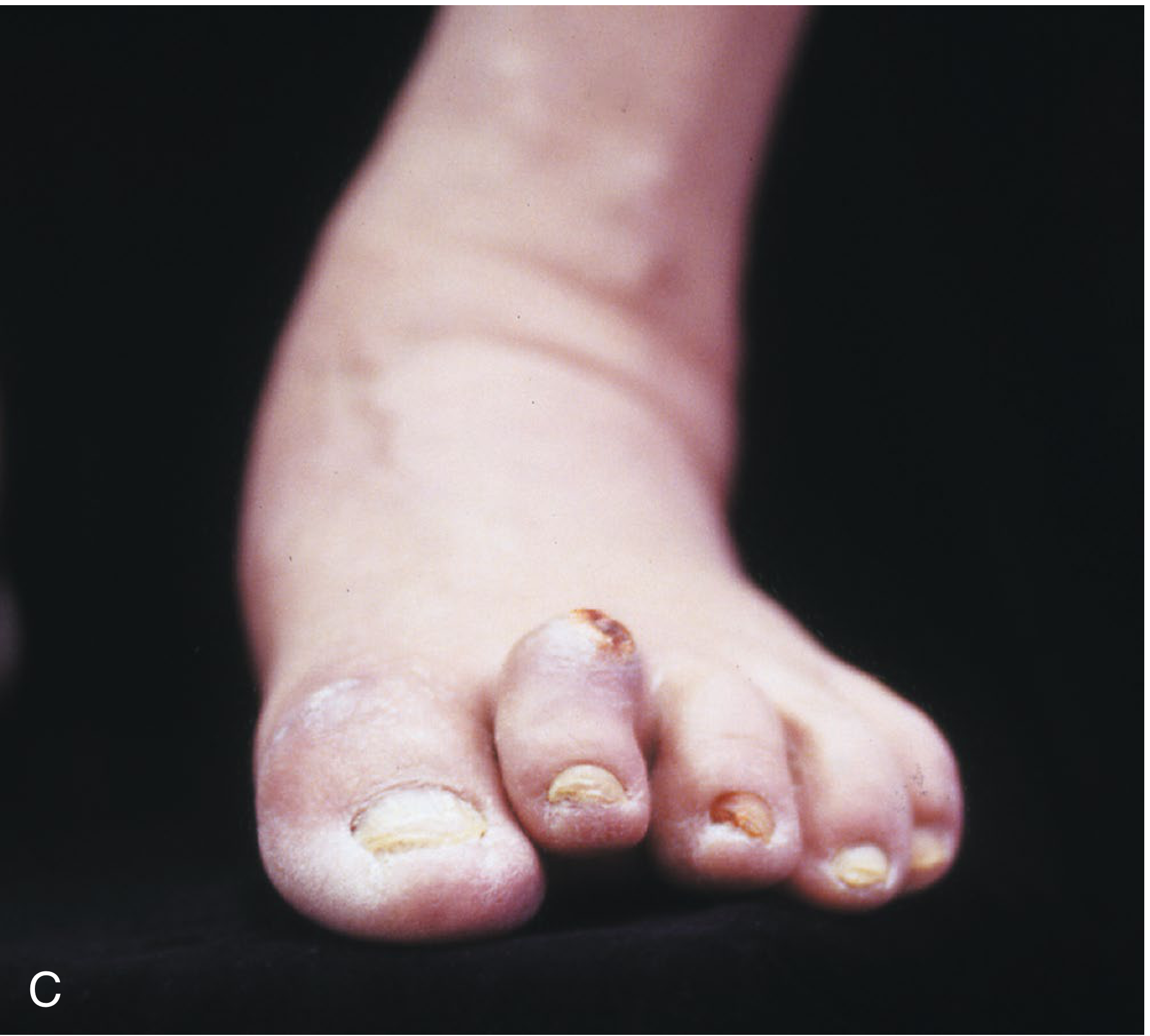
Common presentations:
- Plantar ulceration (typically under metatarsal heads) often surrounded by callus
- Ulceration of the calcaneum and forefoot bones
- Hammer/claw toes, skin fissuring
- Cellulitis, pus, crepitation (gas-producing organisms)
- Charcot foot (neuroarthropathy): repeated undetected fractures destroy normal foot architecture, causing the classic "rocker bottom" deformity
Key history to obtain: Episodes of ulceration, prior amputations, known neuropathic arthropathy, visual impairment, renal disease, paresthesias/numbness, claudication symptoms.
Ulcer Classification
Wagner Classification (most widely used)
| Grade | Description |
|---|---|
| 0 | Pre/post ulcerative lesion - intact skin |
| 1 | Superficial ulcer, not penetrating subcutaneous tissue |
| 2 | Deep ulcer to tendon, capsule, or bone |
| 3 | Deep ulcer with osteitis, osteomyelitis, or joint sepsis |
| 4 | Partial foot gangrene (forefoot/toe) |
| 5 | Whole foot gangrene |
Brodsky Depth-Ischemia Classification modifies Wagner by differentiating ischemic from neuropathic ulcers - ulcers that are both infected and ischemic are 90 times more likely to require amputation. An infected ulcer alone carries a 40-55% chance of some form of amputation. - Campbell's Operative Orthopaedics 15e, p.5056
Diagnosis and Investigations
Neurological Assessment
- Semmes-Weinstein 5.07 monofilament (10g): inability to feel this is a major risk factor
- Equivalent results can be obtained using a 4.5g monofilament under both first metatarsal heads
Vascular Assessment
- Ankle-Brachial Index (ABI): Normal 0.9-1.2; >1.3 suggests non-compressible calcified vessels; <0.5 indicates ulcer unlikely to heal without vascular intervention
- Ankle systolic pressure >60-90 mmHg is required for healing
- Toe pressures are more reliable when vessels are non-compressible (distal vessels often spared); absolute toe pressure >40 mmHg associated with wound healing; normal toe/brachial index >0.7
Lab Tests
- Blood tests are often unhelpful - inflammatory markers may be normal or only mildly raised
- CBC, ESR, CRP - useful for baseline in suspected infection
- HbA1c - higher levels are an independent risk factor for ulcer development; HbA1c >7% associated with higher surgical complications
- Blood glucose >200 mg/dL preoperatively increases risk of nonunion, infection, wound healing problems
Microbiology
- Superficial swabs from ulcers/sinus tracts are not reliable for identifying deep infection organisms
- Bone biopsy for culture should be considered in extensive or complex infection
- The "probe-to-bone" test combined with elevated inflammatory markers and abnormal plain radiographs confirms osteomyelitis
Imaging
| Modality | Use |
|---|---|
| Plain X-ray | First-line: look for osteomyelitis, gas in soft tissues, foreign bodies, bone destruction, Charcot changes - note: early osteomyelitis may be normal for weeks to months |
| MRI | Most sensitive for bone involvement and osteomyelitis - modality of choice for soft tissue assessment |
| CT | Useful for bone architecture detail, sequestra, cortical disruptions; weight-bearing CT guides bony surgical planning |
| Bone scan (Tc-99m) | 3-phase: cellulitis shows uptake in phases 1-2 only; osteomyelitis and Charcot show abnormal uptake in all 3 phases |
| Tagged WBC scan | May show false positive for infection in early Charcot (WBCs accumulate even without infection) |
Microbiology of Diabetic Foot Infections
- Mild infections: Gram-positive cocci predominate - S. aureus (including MRSA) and β-hemolytic streptococci
- Severe/deep infections: Polymicrobial - aerobic gram-positive cocci + gram-negative bacilli + anaerobes
- Pseudomonas is over-represented; empirical therapy for severe infections should cover it
- Anaerobes: consider adding metronidazole, particularly for abscesses and devitalized tissue
Management
General Principles
- Tight glycemic control (HbA1c <7%)
- Strict non-weight-bearing regimen
- Meticulous wound care
- Pressure off-loading
Wound Management
- Debridement of necrotic tissue is mandatory
- Pressure off-loading: special shoes, orthotics, or total-contact casting
- Wound size at 4 weeks is predictive: ≥50% reduction in 4 weeks = significantly higher healing at 12 weeks
- Ulcers <1 cm² heal without amputation in 96% of cases; ulcers >3 cm² only in 72%
Antibiotics
| Severity | Treatment |
|---|---|
| Mild | TMP-SMX 800/160 mg BD, OR Clindamycin 300 mg q8h, OR Cephalexin 500 mg QID |
| Moderate-Severe | Piperacillin-tazobactam 3.375g IV q8h + Vancomycin 15 mg/kg IV q12h |
Based on Rosen's Emergency Medicine 9e, Table 115.2
For osteomyelitis: antibiotics should be tailored by culture results (preferably bone biopsy, not surface swab). Duration typically 6 weeks for established osteomyelitis.
Vascular Intervention
- Full vascular assessment is mandatory in those with poor peripheral pulses
- Proximal angioplasty or bypass surgery to improve distal vascularity may be needed before wound surgery
- ABI <0.5 generally requires revascularization before ulcer management
Additional Treatments
- Negative-pressure wound therapy (NPWT): evidence supports use in diabetic foot ulcers (2024 meta-analysis, PMID: 39241769)
- Advanced wound dressings: a 2024 Italian guidelines meta-analysis evaluated various dressings vs. standard of care (PMID: 38864979)
- Bioengineered skin substitutes, autologous leukocyte/platelet preparations
- Hyperbaric oxygen: some efficacy in complicated infections, especially with anaerobic organisms
- Smart wearable technology for pressure monitoring is an emerging area (PMID: 40682082)
Surgical Management
- Debridement/drainage of collections, necrotic areas, or extensive osteomyelitis
- Distinguish superficial osteitis (loss of soft tissue cover) from deep osteomyelitis - in the former, antibiotics and biopsy may be less important than glycemic control + vascular supply + pressure relief
- Amputation: up to one-third of patients with deep infections eventually require amputation
- Excision must be adequate to remove all infected material
- Excess bone resection may be needed to allow tension-free closure
- If extensive peripheral neuropathy, below-knee amputation in a sensate zone may be more appropriate than foot surgery with poor wound healing
Charcot Neuroarthropathy
- Results from repeated undetected micro-fractures in the insensate foot
- Destroys normal foot architecture → classic "rocker bottom" or Charcot deformity
- X-ray is the imaging modality of choice to stage progression
- Midfoot joint pressures are 46% higher in diabetic cadaver feet during simulated walking
- Treatment: off-loading, total-contact casting, surgical stabilization in select cases
Prevention
- Routine foot examination at every medical visit for those with sensory loss
- Daily self-inspection for cracks, fissures, ulcers, or inflammation
- Avoid walking barefoot (even at home); wear protective footwear (avoid sandals)
- Avoid heating pads or hot-water bottles on the feet (thermal injury risk)
- Referral to foot care specialist for sensory loss, foot deformity, extensive callus, or nonhealing ulcers
- Maintain HbA1c <7% to reduce neuropathy progression
Sources:
- Bailey & Love's Short Practice of Surgery 28e, pp.628-629
- Campbell's Operative Orthopaedics 15e, pp.5054-5059
- Goldman-Cecil Medicine International Edition, p. (Diabetic Foot chapter)
- Rosen's Emergency Medicine 9e, pp.2540-2541
This is a shared conversation. Sign in to Orris to start your own chat.