
Hey orris Can u teach me enteric fever according to davidson book of medicine Also differentiate it from its differntials
Reading File
Now let me search the medical library for enteric fever content from Davidson's.
Finding Sources
Finding Sources
I don't see Davidson's in the results. Let me check what books are available and search specifically for Davidson's content on typhoid.
Finding Sources
Finding Sources
Davidson's is not in the library, but Harrison's and Goldman-Cecil are. Let me check if Davidson's is available under any name, and simultaneously pull the comprehensive content from Harrison's (the closest equivalent gold-standard internal medicine text).
Finding Sources
Reading File
Good content. Now let me read the next sections (pathogenesis, clinical features, complications, diagnosis, treatment) and also get the Goldman-Cecil content for differentials.
Reading File
Reading File
Now let me get the treatment, diagnosis and differentials sections from Harrison's:
Reading File
Finding Sources
Let me now get the differential diagnosis section and the treatment section from Harrison's:
Reading File
Reading File
I now have excellent, comprehensive content. Let me fetch the rose spots image and read the treatment section from Harrison's to complete the picture.
Reading File
I now have all the content I need. Let me compile the comprehensive answer.
Enteric Fever (Typhoid Fever) - A Complete Guide
Note: Davidson's Principles and Practice of Medicine is not in our textbook library. This answer is drawn from Harrison's Principles of Internal Medicine (22nd Ed., 2025) and Goldman-Cecil Medicine - both are equivalent gold-standard internal medicine references that cover the same content as Davidson's. The clinical content is virtually identical.
1. Definition
Enteric fever is a systemic febrile illness caused by dissemination of Salmonella enterica serovar Typhi (typhoid fever) or Paratyphi A, B, or C (paratyphoid fever). The term "enteric" was proposed in 1869 to distinguish it from typhus, based on its characteristic involvement of the intestinal lymphoid tissue (Peyer's patches). The two terms - typhoid fever and enteric fever - are used interchangeably.
- Harrison's Principles of Internal Medicine 22e, p. 1359
2. Etiology & Microbiology
| Organism | Disease |
|---|---|
| S. Typhi | Classic typhoid fever |
| S. Paratyphi A | Paratyphoid fever (usually milder) |
| S. Paratyphi B | Paratyphoid fever |
| S. Paratyphi C | Paratyphoid fever (rare) |
- Unlike NTS (non-typhoidal Salmonella), these organisms have no animal reservoir - humans are the only hosts.
- Transmission: fecal-oral route via contaminated water/food, or from chronic carriers.
3. Epidemiology
- 9.2-21 million cases of typhoid fever and 5 million cases of paratyphoid fever per year globally, with 110,000-280,000 deaths annually.
- Highest incidence: Indian subcontinent (India, Pakistan, Bangladesh, Nepal), Eastern Mediterranean, and Sub-Saharan Africa - exceeding 1000 cases/100,000 children in some urban areas.
- Risk factors: contaminated drinking water, street food, raw fruits/vegetables grown with sewage fertilizer, lack of hand hygiene, and H. pylori co-infection (reduces gastric acidity).
- Multidrug-resistant (MDR) strains emerged in the 1980s (resistant to chloramphenicol, ampicillin, trimethoprim). Since the 1990s, strains with decreased susceptibility to ciprofloxacin (DSC) have emerged, especially on the Indian subcontinent.
- Harrison's, p. 1359-1360
4. Pathogenesis (Week-by-Week)
| Week | Pathological Event |
|---|---|
| Incubation (5-21 days) | Bacteria ingested → invade M cells over Peyer's patches → pass to mesenteric lymph nodes → primary bacteremia |
| Week 1 | Bacteria seed reticuloendothelial system (liver, spleen, bone marrow) → multiply intracellularly → secondary bacteremia begins → fever rises in stepwise fashion |
| Week 2 | Sustained fever, rose spots appear; Peyer's patches hyperplastic and inflamed |
| Week 3-4 | Necrosis of Peyer's patches → risk of intestinal hemorrhage (6%) and perforation (1%) |
5. Clinical Features
Incubation Period
10-14 days (range: 5-21 days), depending on inoculum size, host immunity, and vaccination status.
Symptoms (from a series of 669 cases in Nepal)
| Symptom | Frequency |
|---|---|
| Prolonged fever (38.8-40.5°C) | >75% |
| Headache | 80% |
| Anorexia | 55% |
| Abdominal pain | 30-40% |
| Chills | 35-45% |
| Cough | 30% |
| Diarrhea OR constipation | 22-28% vs 13-16% |
| Nausea/vomiting | 18-24% |
| Sweating, myalgias | 20-25% |
Key Physical Signs
- Relative bradycardia (pulse-temperature dissociation) - at the peak of high fever, heart rate is disproportionately low; seen in up to 50% of patients
- Rose spots - faint, salmon-colored, blanching maculopapular rash, 2-4 mm, on the trunk and chest; appears in ~30% of patients at the end of week 1; lasts 2-5 days; Salmonella can be cultured from biopsies of these lesions. Important: hard to see in dark-skinned patients.
- Hepatosplenomegaly (~50% of patients)
- Coated tongue (51-56%)
- Stepwise rise in fever in the first week, then sustained high fever
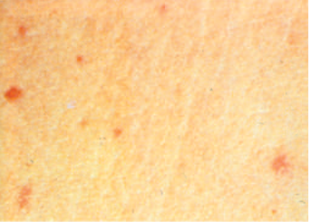
6. Complications (~27% of hospitalized patients)
Gastrointestinal (Weeks 3-4)
- Intestinal hemorrhage (6%) - from ulceration of Peyer's patches
- Intestinal perforation (1%) - life-threatening, mortality 10-32%; requires immediate surgical intervention
Neurological (2-40%)
- "Muttering delirium" or "coma vigil" (picking at bedclothes/imaginary objects)
- Meningitis, Guillain-Barré syndrome, neuritis
- Neuropsychiatric symptoms
Other (rare but important)
- DIC, hemophagocytic syndrome
- Myocarditis, endocarditis, pericarditis
- Hepatitis, pancreatitis, splenic abscess
- Orchitis, osteomyelitis, glomerulonephritis
Chronic Carriage
- 2-5% of untreated patients become chronic asymptomatic carriers (shedding S. Typhi in stool or urine for >1 year)
- More common in women, infants, those with biliary abnormalities or Schistosoma haematobium co-infection
- Chronic carriage is associated with increased risk of gallbladder cancer
- Harrison's, p. 1360-1361
7. Investigations
Laboratory (Non-specific)
| Test | Finding |
|---|---|
| CBC | Leukopenia + neutropenia (15-25% of cases); leukocytosis in children or with perforation |
| LFTs | Mildly elevated (transaminases, ALP) |
| CRP/ESR | Elevated |
| Widal test | Detects O and H agglutinins; high false-positive and false-negative rates |
Definitive Diagnosis: Culture (Gold Standard)
| Culture Site | Sensitivity | Notes |
|---|---|---|
| Blood culture | 40-60% | Lower in first week, with prior antibiotics; most sensitive in week 1 |
| Bone marrow culture | ~80% | Best single test; yield not reduced by 5 days of antibiotics |
| Stool culture | 30-40% in week 1; higher in week 3 | Useful if blood culture negative |
| Rose spot biopsy culture | Variable | Occasionally positive |
| Duodenal string test | High | Non-invasive; can be positive when bone marrow is negative |
| All three combined | >90% | Best approach |
Rapid Diagnostic Tests
- Tubex and Typhidot detect IgM/IgG to O and H antigens
- Sensitivity ~70-80%, specificity ~80-90% - useful at point-of-care but not reliable enough to replace blood culture
- PCR: sensitivity 40-100% depending on gene targets; increasingly available
8. Treatment
Antibiotic Therapy (Harrison's Table 171-1)
| Indication | Agent | Dose/Route | Duration |
|---|---|---|---|
| Empirical (uncomplicated) | Azithromycin | 1 g PO day 1, then 500 mg/day | 7 days |
| Fully susceptible strain | Ciprofloxacin | 500 mg PO BD | 10 days |
| DSC or MDR strain | Ceftriaxone | 2 g IV/IM once daily | 10-14 days |
| Severe/complicated | Ceftriaxone | 2 g IV once daily | 10-14 days |
| XDR strain | Azithromycin or carbapenem | As per susceptibility | 10-14 days |
Key point: Fluoroquinolones (ciprofloxacin) should NOT be used as first-line empiric therapy for travelers from South Asia due to high prevalence of DSC strains. Azithromycin or ceftriaxone is preferred empirically.
Supportive Care
- Dexamethasone (3 mg/kg initial dose, then 1 mg/kg every 6 hours x 8 doses) for severe toxemia with altered consciousness
Outcomes
- With prompt treatment: mortality <1%, fever resolves in 3-5 days
- Untreated: mortality 10-30%
- Relapse occurs in 5-10%, usually 2-3 weeks after fever resolution, caused by the same strain
- Harrison's, p. 1361-1362
9. Prevention
| Vaccine | Type | Protection | Notes |
|---|---|---|---|
| Ty21a (Vivotif) | Live attenuated oral | ~60-70% for S. Typhi | 4 oral doses; some protection against S. Paratyphi A & B |
| Vi polysaccharide (Typhim Vi) | Parenteral | ~60-70% for S. Typhi | Single IM injection; NO protection against paratyphoid (no Vi antigen on Paratyphi) |
| Typhoid conjugate vaccine (TCV) | Vi conjugated to tetanus toxoid | Better in children | WHO recommended; longer lasting immunity |
10. Differential Diagnosis - Detailed Comparison
This is the most clinically important section. The key differentials for enteric fever (a patient presenting with prolonged fever + abdominal symptoms from a tropical region) are:
A. Malaria
| Feature | Enteric Fever | Malaria |
|---|---|---|
| Fever pattern | Sustained/continuous, stepwise rise | Tertian (every 48h) or quartan (72h) in P. vivax/P. falciparum/P. malariae; P. falciparum often irregular |
| Rigors | Mild chills | Prominent rigors |
| Rash | Rose spots (30%) | Absent |
| Splenomegaly | Common (50%) | Very common |
| Relative bradycardia | Yes | No |
| Abdominal pain | Common | Less common (unless splenic rupture) |
| Neurological | Late/rare | Cerebral malaria with P. falciparum |
| Diagnosis | Blood culture | Peripheral blood film, RDT (HRP-2 antigen) |
| Key blood test | Leukopenia | Leukopenia + thrombocytopenia + hemolytic anemia |
B. Dengue Fever
| Feature | Enteric Fever | Dengue |
|---|---|---|
| Fever | Sustained, stepwise | Biphasic (saddleback) - brief improvement then return |
| Rash | Rose spots (faint, maculopapular on trunk) | Morbilliform or petechial rash, often spreads centrifugally, "islands of white in a sea of red" |
| Myalgia/bone pain | Mild | Severe ("breakbone fever") |
| Bleeding | Rare | Common (petechiae, epistaxis, gum bleeding) - dengue hemorrhagic fever |
| Relative bradycardia | Yes | No - often tachycardia |
| Leukopenia | Yes | Yes - but with thrombocytopenia (hallmark) |
| Liver involvement | Mild | Hepatomegaly, elevated transaminases |
| Diagnosis | Culture | NS1 antigen (early), dengue IgM/IgG |
C. Rickettsial Infections (Scrub Typhus, Typhus Group)
| Feature | Enteric Fever | Rickettsial (Scrub Typhus) |
|---|---|---|
| Eschar | Absent | Pathognomonic eschar at bite site (painless, black necrotic ulcer) - seen in scrub typhus |
| Rash | Rose spots (faint, trunk) | Maculopapular, spreads centrifugally from trunk to limbs |
| Lymphadenopathy | Absent | Prominent regional lymphadenopathy near eschar |
| Exposure | Contaminated food/water | Mite bite in scrub vegetation, lice/fleas |
| Relative bradycardia | Yes | Less typical |
| Response to doxycycline | Moderate | Dramatic and rapid (diagnostic test) |
| Weil-Felix reaction | Negative | Positive (OX-K in scrub typhus) |
| Diagnosis | Blood culture | Serology (IgM Weil-Felix, ELISA), PCR |
D. Brucellosis
| Feature | Enteric Fever | Brucellosis |
|---|---|---|
| Fever | Sustained/continuous | Undulant (waves of fever), or intermittent |
| Sweating | Mild | Drenching night sweats (characteristic) |
| Occupation/exposure | Food/water | Animal contact (cattle, goats, sheep, pigs, veterinarians, abattoir workers) or unpasteurized dairy |
| Spinal involvement | No | Vertebral osteomyelitis (spondylitis) - classic |
| Orchitis | Rare | Common (orchitis/epididymo-orchitis) |
| Hepatosplenomegaly | Common | Common |
| Duration | Usually <4 weeks | Months if untreated |
| Diagnosis | Blood culture | Blood culture (slow, needs biphasic media) + Brucella agglutination test (SAT) |
E. Visceral Leishmaniasis (Kala-azar)
| Feature | Enteric Fever | Kala-azar |
|---|---|---|
| Fever | 2-4 weeks | Months to years (prolonged, irregular) |
| Wasting | Mild | Progressive wasting, weight loss |
| Splenomegaly | Moderate | Massive splenomegaly - most prominent feature |
| Hepatomegaly | Moderate | Common |
| Skin changes | Rose spots (transient) | Darkening of skin (hence "kala-azar" = black fever) |
| Blood count | Leukopenia | Pancytopenia (anemia, leukopenia, thrombocytopenia) |
| Serology | Widal | rK39 RDT, bone marrow aspirate showing amastigotes |
| Exposure | Contaminated food/water | Sandfly bite (Phlebotomus sp.) |
F. Viral Hepatitis (A & E)
| Feature | Enteric Fever | Viral Hepatitis A/E |
|---|---|---|
| Jaundice | Absent or mild (hepatitis-like) | Prominent jaundice |
| Prodrome | Fever, headache, relative bradycardia | Fever, malaise, anorexia, dark urine, pale stools |
| Abdominal pain | Diffuse | Right upper quadrant tenderness (tender hepatomegaly) |
| Liver enzymes | Mildly elevated | Markedly elevated (AST/ALT often >500-1000 U/L) |
| Rash | Rose spots | Absent (urticaria sometimes in Hep B) |
| Diagnosis | Blood culture | HAV/HEV IgM serology |
G. Leptospirosis
| Feature | Enteric Fever | Leptospirosis |
|---|---|---|
| Biphasic illness | No | Classic biphasic: leptospiremic phase (4-7d) then immune phase |
| Conjunctival suffusion | Absent | Conjunctival injection/suffusion (classic sign) |
| Myalgia | Mild | Severe calf/thigh myalgia (hallmark) |
| Jaundice + renal failure | Rare | Weil's disease - severe jaundice + acute renal failure |
| Exposure | Contaminated food | Water exposure (floods, swimming), animal urine contact |
| Diagnosis | Blood culture | Microscopic Agglutination Test (MAT), ELISA, PCR |
H. Miliary Tuberculosis
| Feature | Enteric Fever | Miliary TB |
|---|---|---|
| Duration | Usually <4 weeks | Weeks to months |
| Chest X-ray | Normal | Miliary mottling (1-3 mm nodules throughout both lung fields) |
| Choroid tubercles | Absent | Choroid tubercles on fundoscopy (pathognomonic) |
| TB contacts/history | No | Usually yes |
| Fever character | Stepwise rise | Low-grade, persistent |
| Diagnosis | Blood culture | Sputum AFB, BAL, bone marrow biopsy, IGRA/Mantoux |
Quick Comparison Summary Table
| Feature | Enteric Fever | Malaria | Dengue | Scrub Typhus | Brucellosis | Kala-azar |
|---|---|---|---|---|---|---|
| Fever | Sustained/stepwise | Periodic/tertian | Biphasic/saddleback | Sustained | Undulant | Prolonged |
| Rose spots | Yes (30%) | No | No | No | No | No |
| Eschar | No | No | No | Yes | No | No |
| Relative bradycardia | Yes | No | No | Possible | Possible | No |
| Splenomegaly | Moderate | Marked | Mild | Present | Present | Massive |
| Thrombocytopenia | Mild/absent | Present | Marked | Present | Mild | Present |
| Rash type | Faint rose spots | Absent | Petechial/morbilliform | Maculopapular | Absent | Absent |
| Key diagnostic clue | Blood culture | Peripheral smear | NS1 antigen | Eschar + serology | Animal exposure | rK39/bone marrow |
| Drug of choice | Ceftriaxone/azithromycin | ACT/chloroquine | Supportive | Doxycycline | Doxycycline + rifampicin | Amphotericin B |
Sources: Harrison's Principles of Internal Medicine 22e (2025), pp. 1359-1362; Goldman-Cecil Medicine; Fitzpatrick's Dermatology 5e.
Note: Davidson's Medicine was not available in the library. The content above is clinically equivalent and drawn from Harrison's - which covers enteric fever in the same depth and approach as Davidson's. The clinical framework, week-by-week progression, complications, and differentials are all consistent with standard Davidson's teaching.
Teach me Meningoencephalitis wrt clinical features, investigation treatment and complications
Reading File
Finding Sources
Finding Sources
Good finds. Let me read key sections in parallel - from Rosen's Emergency Medicine (clinical), Plum & Posner (neurological detail), Harrison's (treatment), and Robbins (pathology/complications):
Finding Sources
Reading File
Reading File
Now let me read the comprehensive Rosen's clinical and Harrison's treatment sections in parallel:
Reading File
Reading File
Now let me get the clinical features + treatment + investigations + complications from Rosen's and the full bacterial meningitis Goldman-Cecil content:
Reading File
Reading File
Excellent. Let me now read the clinical features, CSF findings and treatment sections from Goldman-Cecil:
Reading File
Reading File
Excellent content. Let me get the treatment section and clinical features section from Goldman-Cecil:
Reading File
Reading File
Now let me read the treatment section and complications from Goldman-Cecil:
Reading File
I have all the content I need. Let me now compile the full comprehensive answer.# Meningoencephalitis - Complete Guide
Sources: Goldman-Cecil Medicine, Rosen's Emergency Medicine, Robbins Pathologic Basis of Disease, Plum & Posner's Diagnosis and Treatment of Stupor and Coma
1. Definition & Concept
Meningitis = inflammation of the arachnoid membrane, pia mater, and the intervening CSF (subarachnoid space + ventricles).
Encephalitis = inflammation of the brain parenchyma itself.
Meningoencephalitis = simultaneous involvement of both the meninges AND brain parenchyma. Viral encephalitis is almost invariably associated with meningeal inflammation, so in practice most encephalitides are meningoencephalitides.
2. Etiology by Category
Bacterial
| Organism | Age Group / Setting |
|---|---|
| Streptococcus pneumoniae | Most common in adults (>50% of cases); predisposed by otitis media, sinusitis, pneumonia, splenectomy, alcoholism |
| Neisseria meningitidis | Children, adolescents, young adults in close quarters (dorms, barracks) |
| Haemophilus influenzae type b | Mostly children; rare since HiB vaccine |
| Listeria monocytogenes | Neonates, elderly >60y, immunocompromised, pregnant women; food-borne |
| Group B Streptococcus / E. coli | Neonates (<6 weeks) |
| Gram-negative bacilli | Nosocomial / post-neurosurgery |
| S. aureus / CoNS | Post-neurosurgical, penetrating trauma |
| Mycobacterium tuberculosis | Subacute presentation; immunocompromised |
Viral (Most Common Overall)
| Organism | Notes |
|---|---|
| Enteroviruses | Most common viral cause overall |
| Herpes simplex virus 1 (HSV-1) | Most common cause of sporadic fatal encephalitis; temporal lobe predilection |
| Herpes simplex virus 2 (HSV-2) | Neonatal meningitis; recurrent aseptic meningitis in adults |
| VZV (varicella-zoster) | Reactivation in immunocompromised |
| Arboviruses | West Nile, Japanese encephalitis, Eastern/Western equine encephalitis - epidemic, vector-borne |
| CMV/EBV | Immunocompromised patients, HIV |
| Mumps, Measles | Rare in vaccinated populations; can be severe |
| Rabies | Retrograde axonal spread; invariably fatal |
| HIV | Acute seroconversion meningitis |
Fungal
| Organism | Notes |
|---|---|
| Cryptococcus neoformans/gattii | Most common fungal cause; HIV/immunocompromised |
| Aspergillus | Invasive, immunocompromised |
| Coccidioides immitis | Endemic (SW USA); subacute |
| Mucor | Diabetics; direct sinus invasion |
Others
| Parasitic | Naegleria fowleri (primary amebic meningoencephalitis - swimming in warm freshwater), Acanthamoeba (granulomatous AME), Toxoplasma |
| TB | Subacute/chronic; commonest in developing world |
| Spirochetal | Lyme disease (Borrelia), Syphilis, Leptospirosis |
| Rickettsial | Rocky Mountain Spotted Fever |
3. Pathogenesis
- Route of entry: Hematogenous spread (most common), direct extension from ENT infections (otitis media, sinusitis), post-neurosurgical/traumatic, retrograde axonal spread (HSV, rabies)
- Bacterial: Nasopharyngeal colonization → mucosal invasion → bacteremia → blood-brain barrier crossing → CSF entry → unimpeded bacterial multiplication (CSF lacks antibodies, complement, phagocytes) → cytokine storm → inflammation cascade → increased BBB permeability → cerebral edema → raised ICP → cerebral ischemia
- Viral: Neuroinvasion via viremia or axonal transport → neuronal/glial cell death → perivascular lymphocytic infiltration → microglial nodules → ± areas of necrosis (especially HSV in temporal lobes)
4. Clinical Features
Classic Triad of Meningitis
Fever + Headache + Neck stiffness - present together in only ~44% of patients, but at least one is present in virtually all
Full Clinical Syndrome
| Feature | Detail |
|---|---|
| Fever | High-grade; almost invariable |
| Headache | Severe, global, "worst headache of life" in acute onset |
| Neck stiffness (nuchal rigidity) | Resistance to passive flexion of the neck - due to meningeal irritation |
| Photophobia + Phonophobia | Classic triad with headache and neck stiffness |
| Altered consciousness | Indicates encephalitis component - drowsiness, confusion, delirium, coma |
| Seizures | More in encephalitis; ~30% of bacterial meningitis cases |
| Focal neurological deficits | Hemiparesis, aphasia, cranial nerve palsies (III, VI, VII, VIII) |
| Vomiting | Common |
| Rash | Petechial/purpuric non-blanching rash = pathognomonic of meningococcal meningitis (meningococcemia) |
Signs of Meningeal Irritation
| Sign | How to Elicit | Positive Result |
|---|---|---|
| Kernig's sign | Patient supine, hip flexed 90° - attempt to extend knee | Pain + resistance to extension beyond 135° |
| Brudzinski's sign | Passive neck flexion in supine patient | Involuntary flexion of hips and knees |
| Jolt accentuation | Patient rotates head horizontally at 2-3 Hz | Worsening of headache = positive |
| Neck stiffness | Passive chin-to-chest flexion | Resistance/pain |
Note: These signs may be absent in elderly, immunocompromised, and neonates - have a lower threshold for LP in these groups.
Features Specific to Encephalitis (vs. Pure Meningitis)
- Altered mental status / impaired consciousness - key distinguishing feature
- Behavioral and personality changes (especially HSV-1 - temporal lobe)
- Memory disturbance (HSV - limbic system)
- Seizures (focal or generalized)
- Movement disorders, tremors, ataxia
- Hallucinations (auditory, visual)
- Aphasia (HSV temporal lobe)
- Muttering delirium, picking at bedclothes
Age-Specific Presentations
| Age | Clinical Pattern |
|---|---|
| Neonates | Non-specific - poor feeding, irritability, bulging fontanelle, fever or hypothermia, high-pitched cry; neck stiffness often absent |
| Children | Classic triad more common; seizures prominent |
| Adults | Classic triad; rash if meningococcal |
| Elderly | Subtle - confusion, obtundation without fever/meningism; high mortality |
| Immunocompromised | Atypical, insidious onset; think fungal, TB, CMV |
Warning Features Requiring Urgent CT Before LP
- Papilledema
- Focal neurological signs
- New-onset seizures
- GCS <15 (altered consciousness)
- Immunocompromised state
- History of CNS disease (mass, stroke, focal infection)
5. Investigations
A. Blood Tests
| Test | Finding/Purpose |
|---|---|
| Blood cultures | Positive in ~50-80% of bacterial meningitis; obtain BEFORE antibiotics if possible but do NOT delay antibiotics |
| CBC | Leukocytosis (bacterial); leukopenia (viral, early bacterial) |
| CRP, Procalcitonin | Elevated in bacterial; help distinguish bacterial vs viral |
| Blood glucose | Required to interpret CSF glucose ratio |
| Coagulation screen | DIC in fulminant meningococcal disease |
| LFTs, RFTs | Baseline + metabolic causes of confusion |
| Serum lactate | Elevated in sepsis |
| HIV serology | All patients - risk stratification |
| Blood PCR (meningococcal, pneumococcal) | Useful if antibiotics given before LP |
B. Neuroimaging - CT/MRI Head
CT FIRST if ANY of the warning features above are present - then LP after.
| Finding | Significance |
|---|---|
| Normal CT | Majority of bacterial meningitis - safe to proceed to LP |
| Hypodense temporal lobe lesions | HSV encephalitis (CT relatively insensitive - MRI preferred) |
| Ring-enhancing lesion | Brain abscess, toxoplasmosis |
| Hydrocephalus | TB meningitis, cryptococcal meningitis |
| Meningeal enhancement (contrast CT/MRI) | Bacterial/fungal meningitis |
| MRI (FLAIR/DWI) | Gold standard for encephalitis - shows temporal lobe changes in HSV earlier than CT |
Critical rule: Do NOT delay antibiotics to wait for CT or LP. Start antibiotics within 1 hour of presentation ("door-to-antibiotic <60 minutes").
C. Lumbar Puncture (LP) - Gold Standard
When NOT to do LP first:
- Signs of raised ICP (papilledema, Cushing's triad, altered GCS)
- Focal neurological signs
- Do CT first - if normal, proceed to LP
CSF Analysis - The Core Table:
| Parameter | Normal | Bacterial | Viral | TB | Fungal (Crypto) |
|---|---|---|---|---|---|
| Appearance | Crystal clear | Turbid/purulent | Clear/slightly turbid | Clear/ground-glass | Clear/slightly turbid |
| Opening pressure (cm H₂O) | 10-20 | ↑↑ (200-300+) | Normal/slightly ↑ | ↑ | ↑↑ (very high) |
| WBC (cells/μL) | <5 | 1000-10,000 (PMN dominant) | 10-1000 (lymphocyte dominant) | 100-500 (lymphocyte) | 20-500 (lymphocyte) |
| Cell type | - | Neutrophils (PMN) | Lymphocytes | Lymphocytes (± early PMN) | Lymphocytes |
| Protein (mg/dL) | 20-40 | ↑↑ (100-500) | Mildly ↑ (50-100) | ↑↑ (100-500) | ↑ (50-200) |
| Glucose (mg/dL) | 50-80 (2/3 serum) | ↓↓ (<40) | Normal | ↓ (<45) | ↓ |
| CSF:serum glucose ratio | >0.6 | <0.4 | >0.6 | <0.5 | <0.5 |
| Gram stain | - | Positive 60-80% | Negative | Negative (AFB occasionally) | Negative |
| India ink | - | - | - | - | Positive (Cryptococcus) |
| Culture | Sterile | Positive (gold standard) | Negative | Positive on Lowenstein Jensen (slow) | Positive |
Additional CSF Tests Based on Suspicion:
- HSV PCR - gold standard for HSV encephalitis (sensitivity ~98%)
- Enterovirus PCR - viral meningitis
- AFB smear + culture - TB meningitis
- Cryptococcal antigen - immunocompromised/HIV
- VDRL - neurosyphilis
- Cytology - malignant meningitis
- Oligoclonal bands - ADEM/MS
- Opening pressure - always measure, especially in cryptococcal meningitis (can be extremely high)
D. EEG
- Indicated in encephalitis, especially if seizures suspected
- HSV encephalitis: periodic lateralizing epileptiform discharges (PLEDs) over temporal lobes - characteristic
- Helps distinguish encephalitis from metabolic encephalopathy
E. Brain Biopsy
- Rarely needed; reserved for diagnostically uncertain cases where treatment hinges on tissue diagnosis (e.g., ruling out CNS lymphoma vs. encephalitis)
6. Treatment
Step 1: Emergency Stabilization
- Airway, breathing, circulation
- IV access, blood cultures
- Antibiotics within 60 minutes (before LP if CT needed, before CT if CT not needed)
- Corticosteroids with or before first antibiotic dose
Step 2: Empiric Antibiotic Therapy
| Age/Setting | Likely Organisms | Empiric Therapy |
|---|---|---|
| <3 months | GBS, E. coli, Listeria | Ampicillin + Cefotaxime (± Gentamicin) |
| 3 months - 50 years | N. meningitidis, S. pneumoniae | Ceftriaxone 2g IV BD + Vancomycin |
| >50 years / immunocompromised | S. pneumoniae, Listeria, gram-negatives | Ceftriaxone + Vancomycin + Ampicillin |
| Post-neurosurgery / trauma | S. aureus, CoNS, Pseudomonas | Vancomycin + Cefepime (or meropenem) |
| Penicillin allergy | - | Vancomycin + Meropenem (or TMP-SMX for Listeria) |
Key principle: Drug levels in CSF must be 10-20x the MBC. Continue full IV dosing throughout treatment - do NOT reduce dose as patient improves (normalization of BBB reduces CSF penetration).
Step 3: Adjunctive Dexamethasone
Dexamethasone 0.15 mg/kg IV q6h x 4 days - give with or BEFORE first antibiotic dose.
- Reduces mortality and neurological sequelae in bacterial meningitis, especially pneumococcal
- Mechanism: Reduces cytokine-mediated inflammation, cerebral edema, and BBB disruption
- Do NOT give in viral meningitis (unless ADEM/autoimmune suspected)
- Caution: May reduce CSF penetration of vancomycin - ensure adequate dosing
Step 4: Antiviral (if encephalitis/viral suspected)
Acyclovir 10 mg/kg IV q8h x 14-21 days - START EMPIRICALLY if viral encephalitis is suspected.
- Do NOT wait for PCR results - delays worsen outcomes
- Acyclovir has reduced HSV encephalitis mortality from 70% → 9%
- Covers HSV-1, HSV-2, VZV
- Adjust dose for renal impairment
Step 5: Definitive/Specific Treatment
| Organism | Antibiotic | Duration |
|---|---|---|
| S. pneumoniae (sensitive) | Ceftriaxone 2g BD | 10-14 days |
| S. pneumoniae (resistant) | Ceftriaxone + Vancomycin ± Rifampicin | 10-14 days |
| N. meningitidis | Ceftriaxone 2g BD (or Penicillin G if sensitive) | 5-7 days |
| H. influenzae | Ceftriaxone | 7 days |
| Listeria | Ampicillin + Gentamicin | 21 days |
| HSV encephalitis | Acyclovir 10 mg/kg q8h IV | 14-21 days |
| TB meningitis | RIPE (Rifampicin, INH, Pyrazinamide, Ethambutol) + Dexamethasone | 12 months |
| Cryptococcal meningitis | Amphotericin B + Flucytosine (induction 2 weeks) → Fluconazole (consolidation 8 weeks) | Long-term |
Step 6: Supportive Care
- Isolation (droplet precautions) for meningococcal disease - until 24h of antibiotics
- Head-of-bed elevation 30° to reduce ICP
- Avoid hypotonic fluids (worsen cerebral edema)
- Seizure management (benzodiazepines acutely, anti-epileptic drugs)
- Treat raised ICP: mannitol 0.5-1g/kg IV, hyperventilation (temporary), neurosurgery if needed
- Monitor for SIADH (restrict fluids) vs. cerebral salt wasting (give fluids + salt)
- Nasogastric feeding if GCS low
Chemoprophylaxis (Meningococcal Contacts)
- Rifampicin 600 mg BD x 2 days OR Ciprofloxacin 500 mg single dose OR Ceftriaxone 250 mg IM single dose
- For close contacts within 7 days of onset
7. Complications
Acute/Early Complications
| Complication | Mechanism | Notes |
|---|---|---|
| Raised ICP / Brain herniation | Cerebral edema | Most dangerous; can cause uncal or tonsillar herniation → death |
| Seizures | Cortical irritation, metabolic, ischemia | 30% in bacterial; 50-70% in HSV encephalitis |
| Septic shock | Gram-negative endotoxin, cytokine storm | Especially meningococcal septicemia |
| Waterhouse-Friderichsen syndrome | Bilateral adrenal hemorrhage due to meningococcemia | Fulminant; rapidly fatal without steroids/support |
| DIC | Endotoxin activating coagulation | Purpura fulminans in meningococcemia |
| SIADH | Cytokine effect on hypothalamus | Hyponatremia → worsens cerebral edema |
| Cerebral venous sinus thrombosis | Thrombophlebitis spreading to dural sinuses | Focal deficits, papilledema, worsening despite treatment |
| Subdural empyema | Extension of infection | Urgent neurosurgical drainage |
| Brain abscess | Focal parenchymal infection | Ring-enhancing on CT; surgical + antibiotics |
| Hydrocephalus (acute) | Obstruction of CSF flow by exudate | Communicating (more common) or obstructive; needs ventricular drainage |
| Ventriculitis | Spread to ependymal lining | Poor prognosis |
Late / Neurological Sequelae
| Sequela | Frequency | Notes |
|---|---|---|
| Sensorineural hearing loss | 10-30% - most common sequel | Due to labyrinthitis/cochlear inflammation; commonest in pneumococcal meningitis; dexamethasone reduces risk |
| Cognitive impairment / intellectual disability | 10-20% | Memory, attention, executive function |
| Epilepsy | 5-10% | Especially if cortical involvement |
| Focal neurological deficits | 10-20% | Hemiplegia, aphasia, visual field defects |
| Hydrocephalus (chronic) | 5-10% | Communicating type from arachnoid fibrosis; requires VP shunt |
| Behavioral / psychiatric sequelae | Common after encephalitis | Especially HSV (limbic injury) - personality change, aggression, hypersexuality (Kluver-Bucy syndrome) |
| Visual impairment | Rare | Optic neuritis, cortical blindness |
| Motor disability | Variable | Cerebral palsy-like picture in neonates |
| Death | Bacterial: ~15%; Untreated HSV: ~70%; TB meningitis: ~25-50%; Listeria: up to 27% | Higher in extremes of age, delayed treatment, pneumococcal and listerial disease |
Complication of TB Meningitis (specifically)
- Hydrocephalus (communicating) - very common
- Cerebral infarction (vasculitis of lenticulostriate arteries)
- Cranial nerve palsies (especially VI, VII)
- Spinal block (adhesive arachnoiditis)
- Up to 25% of patients need neurosurgical intervention (VP shunt)
8. Quick Summary Framework
MENINGOENCEPHALITIS
        |
   ┌────┴────┐
Bacterial    Viral
   │          │
Urgent LP   Urgent LP
Blood Cx    HSV PCR
CT if ICP   EEG
   │          │
Ceftriaxone + Acyclovir
Vancomycin
+/- Ampicillin
+ Dexamethasone
        │
Monitor for:
- Raised ICP
- Seizures
- Hearing loss
- SIADH
- Herniation
9. Red Flags Demanding Immediate Action
- Non-blanching purpuric rash = meningococcemia - give benzylpenicillin IM/IV IMMEDIATELY even before hospital
- GCS falling = impending herniation - antibiotics + mannitol + ICU
- Papilledema = do NOT LP; CT first
- Bilateral fixed dilated pupils = herniation - neurosurgical emergency
- Fever + altered consciousness + temporal lobe signs = empiric acyclovir NOW
Sources: Goldman-Cecil Medicine (Chapter 381); Rosen's Emergency Medicine (Chapter 95); Robbins Pathologic Basis of Disease (Chapter 28); Plum & Posner's Diagnosis and Treatment of Stupor and Coma (Chapter)
Give its differentials
The differentials for meningoencephalitis are conditions that can mimic it - either with fever + headache + altered consciousness, or with CNS signs + CSF changes. Let me pull the textbook content on this.
Searching sources...
Finding Sources
Finding Sources
Reading File
Reading File
Reading File
Finding Sources
Finding Sources
Reading File
Finding Sources
Reading File
Finding Sources
I now have enough content for a thorough differentials section. Let me compile the answer.
Differentials of Meningoencephalitis
The differential is best approached by asking: "What else can give fever + headache + altered consciousness + meningeal signs?" Broadly, the differentials fall into infectious and non-infectious categories.
CATEGORY 1: Infectious - Within the CNS
These mimic meningoencephalitis most closely because they are CNS infections themselves.
1. Brain Abscess
| Feature | Meningoencephalitis | Brain Abscess |
|---|---|---|
| Onset | Acute (hours-days) | Subacute (days-weeks) |
| Fever | Prominent | Present but often low-grade |
| Headache | Severe, diffuse | Severe, focal, progressive |
| Focal neuro signs | Late/variable | Prominent, early |
| Neck stiffness | Yes | Absent or mild (unless rupture) |
| Seizures | Present | Common (focal seizures) |
| Papilledema | Raised ICP | More common (space-occupying) |
| CSF | Pleocytosis, ↑protein | Avoid LP if suspected (herniation risk) |
| CT/MRI | Diffuse enhancement | Ring-enhancing lesion with surrounding edema |
| Predisposition | None specific | Otitis media, sinusitis, dental infection, IV drug use |
- Key clue: Ring-enhancing lesion on CT/MRI + focal signs + history of dental/ENT infection = brain abscess until proven otherwise.
2. Subdural Empyema
| Feature | Meningoencephalitis | Subdural Empyema |
|---|---|---|
| Site | Diffuse meninges/parenchyma | Pus between dura and arachnoid |
| Onset | Acute | Subacute/acute |
| Fever | Yes | Yes |
| Focal signs | Variable | Rapid progression of hemiplegia, aphasia |
| Meningism | Yes | Yes (irritation of underlying meninges) |
| Seizures | Variable | Common, often focal |
| Source | Hematogenous / contiguous | Extension from sinusitis, otitis media, mastoiditis |
| MRI | Diffuse enhancement | Crescentic/lenticular extraaxial collection hyperintense on DWI (restricted diffusion) |
| Treatment | Antibiotics | Emergency neurosurgical drainage + antibiotics |
- Key clue: Sinusitis/mastoiditis + rapidly worsening focal neurological deficit + fever = subdural empyema until proven otherwise.
3. Cerebral Malaria (P. falciparum)
| Feature | Meningoencephalitis | Cerebral Malaria |
|---|---|---|
| Fever | Sustained/stepwise | High fever, rigors, cyclical |
| Altered consciousness | Yes | Yes - rapidly deepening coma |
| Seizures | Present | Very common, especially in children |
| Neck stiffness | Present | Usually absent (key differentiator) |
| Jaundice | Absent | Common (haemolysis) |
| Anaemia | Absent | Severe haemolytic anaemia |
| Blood film | Normal | Parasitized RBCs visible (ring forms, P. falciparum) |
| CSF | Pleocytosis | Normal or minimal pleocytosis |
| Travel history | Any | Travel to malaria-endemic area |
| Treatment | Antibiotics/antivirals | IV Artesunate |
- Key clue: Absence of neck stiffness + malaria travel history + positive blood film = cerebral malaria.
4. Tuberculous Meningitis
| Feature | Pyogenic Meningoencephalitis | TB Meningitis |
|---|---|---|
| Onset | Acute (hours-days) | Subacute/chronic (weeks-months) |
| Fever | High | Low-grade, prolonged |
| Headache | Severe | Progressive, worsening |
| Cranial nerve palsies | Occasional | Very common (VI, III, VII, VIII) |
| CSF cells | PMN neutrophils | Lymphocytes |
| CSF glucose | Very low (<40) | Low (<45) |
| CSF protein | Very high (100-500) | Markedly elevated |
| CSF appearance | Turbid/purulent | Cobweb clot / ground-glass |
| AFB smear | Negative | Positive (low sensitivity) |
| Hydrocephalus | Uncommon early | Common (communicating type) |
| HIV/immunosuppression | Risk factor | Strong association |
| Treatment | 3rd-gen cephalosporins | RIPE x 12 months + steroids |
- Key clue: Subacute onset + cranial nerve palsies + lymphocytic CSF + hydrocephalus = TB meningitis until proven otherwise. Always test for TB in developing world patients.
5. Cryptococcal Meningitis
| Feature | Bacterial Meningoencephalitis | Cryptococcal Meningitis |
|---|---|---|
| Onset | Acute | Indolent, weeks to months |
| Host | Any | Immunocompromised (HIV CD4 <100, transplant) |
| Fever | High | Low-grade or absent |
| Headache | Yes | Severe, prominent, often the main symptom |
| Meningism | Prominent | Mild or absent |
| CSF pressure | Elevated | Very elevated (can be >400 mm H₂O) |
| CSF cells | PMN pleocytosis | Minimal cells (very few lymphocytes) |
| CSF protein | High | Mildly elevated |
| CSF glucose | Very low | Low |
| India ink | Negative | Positive (encapsulated yeast, 60-80%) |
| Cryptococcal Ag | Negative | Positive (>95% sensitivity) |
| Treatment | Antibiotics | Amphotericin B + Flucytosine → Fluconazole |
- Key clue: HIV patient + subacute headache + minimal CSF cells + very high opening pressure + India ink positive = cryptococcal meningitis.
6. Viral Encephalitis subtypes (within the viral differential)
These can be confused with each other - key distinguishing features:
| Virus | Key Clinical Clue | CSF | MRI Finding |
|---|---|---|---|
| HSV-1 | Temporal lobe signs (memory loss, bizarre behavior, olfactory hallucinations, aphasia) | Lymphocytic, RBCs (hemorrhagic) | Temporal + insular FLAIR hyperintensity |
| West Nile Virus | Flaccid paralysis (polio-like), elderly, summer, mosquito | Lymphocytes | Substantia nigra / thalamic lesions |
| Japanese Encephalitis | Endemic area (South/SE Asia); Parkinson-like features, tremor | Lymphocytes | Bilateral thalamic lesions on MRI |
| Rabies | Animal bite → hydrophobia, aerophobia, ascending paralysis | Normal early | Brainstem involvement |
| CMV | HIV/transplant + periventricular enhancement | Low glucose | Periventricular enhancement |
| EBV (mononucleosis) | Sore throat, cervical LN, maculopapular rash, splenomegaly | Lymphocytes | - |
- Key rule: Any encephalitis with temporal lobe predilection = treat as HSV until PCR proves otherwise.
CATEGORY 2: Non-Infectious CNS Conditions
7. Autoimmune Encephalitis (Anti-NMDAR and others)
This is an increasingly recognized and clinically important differential - especially in young women.
| Feature | Viral Meningoencephalitis | Autoimmune Encephalitis |
|---|---|---|
| Age/Sex | Any | Young females predominant (NMDAR) |
| Onset | Acute | Subacute (days-weeks) |
| Prodrome | Viral URTI | Viral-like prodrome (headache, fever, malaise) - can be identical |
| Psychiatric symptoms | Variable | Prominent - psychosis, behavioral change often the presenting feature |
| Seizures | Present | Refractory, often multifocal |
| Movement disorders | Rare | Orofacial dyskinesias, choreoathetosis (NMDAR) |
| Autonomic instability | Rare | Prominent (NMDAR) - tachycardia, hyperthermia, hypersalivation |
| Memory loss | Variable | Prominent (limbic encephalitis) |
| CSF | Pleocytosis, PCR-negative | Lymphocytic pleocytosis, viral PCR negative |
| MRI | Temporal lobe (HSV) | FLAIR hyperintensity in limbic structures (hippocampus, amygdala) |
| EEG | PLEDs (HSV) | "Extreme delta brush" pattern (NMDAR) |
| Tumour association | None | Ovarian teratoma (NMDAR), thymoma, lung Ca |
| Treatment | Acyclovir | IV methylprednisolone + IVIG + Rituximab + tumour removal |
Key antibodies to know:
| Antibody | Syndrome | Association |
|---|---|---|
| Anti-NMDA receptor | Psychosis + seizures + orofacial dyskinesias + autonomic instability | Ovarian teratoma (young females) |
| Anti-LGI1 | Faciobrachial dystonic seizures (pathognomonic), hyponatraemia | Elderly males, thymoma |
| Anti-CASPR2 | Cognitive decline + sleep disturbance + ataxia (Morvan syndrome) | Elderly males |
| Anti-GABA-B | Refractory seizures + ataxia | Small cell lung Ca |
| Anti-AMPA | Refractory seizures, psychiatric | Lung/breast/thymoma |
- Key clue: Young woman with new-onset psychosis + seizures + abnormal movements + CSF negative for organisms → screen for anti-NMDAR antibodies in blood AND CSF.
8. ADEM (Acute Disseminated Encephalomyelitis)
| Feature | Viral Meningoencephalitis | ADEM |
|---|---|---|
| Trigger | Primary infection | Post-infectious or post-vaccination (1-4 weeks after) |
| Onset | Acute | Monophasic, acute |
| White matter involvement | Limited | Multifocal white matter demyelination |
| MRI | Focal/temporal (HSV) | Bilateral, asymmetric white matter FLAIR lesions |
| CSF | Pleocytosis | Oligoclonal bands, myelin basic protein elevated |
| Recurrence | Uncommon | Monophasic (vs MS which is relapsing) |
| Treatment | Antivirals | High-dose IV corticosteroids |
- Key clue: Neurological deficit appearing 1-4 weeks after viral illness or vaccination + multifocal MRI lesions = ADEM.
9. Subarachnoid Hemorrhage (SAH)
| Feature | Bacterial Meningitis | SAH |
|---|---|---|
| Onset | Acute | Hyperacute - "thunderclap headache" (worst headache of life, reaches peak in seconds) |
| Fever | Yes (early or delayed) | Absent initially (fever can develop 24-48h later from blood irritation) |
| Neck stiffness | Yes | Yes (blood in subarachnoid space irritates meninges) |
| Altered consciousness | Progressive | Sudden LOC at onset in many |
| Photophobia | Yes | Yes |
| CT head | Normal/diffuse | Hyperdense blood in basal cisterns, Sylvian fissure |
| CSF | Purulent, ↑WBC | Uniformly blood-stained + xanthochromia (yellow pigment) on spectrophotometry |
| CSF WBC | Elevated | Elevated only due to blood (low ratio) |
| Treatment | Antibiotics | Neurosurgical - coiling/clipping of aneurysm |
- Key clue: Thunderclap onset + blood-stained CSF + xanthochromia = SAH, not meningitis.
10. Non-Convulsive Status Epilepticus (NCSE)
| Feature | Encephalitis | NCSE |
|---|---|---|
| Fever | Present | Usually absent |
| Altered consciousness | Progressive | Fluctuating, persistent alteration |
| Overt seizures | May be present | Absent by definition |
| EEG | Slowing / PLEDs | Continuous seizure activity without clinical correlate |
| CSF | Pleocytosis | Normal |
| Response to anticonvulsants | Partial | Dramatic improvement |
- Key clue: Unexplained altered consciousness with no clear infectious cause → get EEG urgently before diagnosing encephalitis.
11. Metabolic / Toxic Encephalopathy
Conditions like hepatic encephalopathy, uraemic encephalopathy, hyponatraemia, hypoglycaemia, hypertensive encephalopathy, drug toxicity (serotonin syndrome, neuroleptic malignant syndrome):
| Feature | Meningoencephalitis | Metabolic Encephalopathy |
|---|---|---|
| Fever | Yes | Variable |
| Neck stiffness | Yes | Absent (key differentiator) |
| CSF | Pleocytosis | Normal |
| Blood tests | Normal metabolic panel | Abnormal (↓Na, ↓glucose, ↑ammonia, ↑urea) |
| Onset | Subacute/acute | Acute, often correlates with metabolic derangement |
- Key clue: Confused patient with NO neck stiffness + deranged bloods = metabolic cause first. BUT always check glucose at bedside immediately.
12. Cerebral Venous Sinus Thrombosis (CVST)
| Feature | Meningoencephalitis | CVST |
|---|---|---|
| Fever | Yes (infectious cause) | Variable (septic CVST has fever) |
| Headache | Diffuse | Progressive, severe, often worsened by straining |
| Focal signs | Late | Focal deficits related to venous territory |
| Seizures | Present | Very common |
| Papilledema | Late | Common (venous outflow obstruction) |
| Risk factors | Infection | OCP, pregnancy, thrombophilia, dehydration, mastoiditis (septic) |
| MRI + MRV | - | "Empty delta sign" on MRV (filling defect in dural sinus) |
| CSF | Pleocytosis | Elevated pressure, otherwise relatively normal |
| Treatment | Antibiotics | Anticoagulation (even with haemorrhage) |
Summary Table: The Big Differentials at a Glance
| Condition | Fever | Neck Stiffness | Focal Signs | CSF Cells | CT/MRI Clue | Key Distinguishing Feature |
|---|---|---|---|---|---|---|
| Bacterial Meningitis | High | +++ | Late | PMN ↑↑↑ | Normal or diffuse enhancement | PMN pleocytosis + positive Gram stain/culture |
| Viral Encephalitis | Yes | + | Variable | Lymphocytes | Temporal lobe (HSV) | HSV PCR, PLEDs on EEG |
| TB Meningitis | Low-grade | + | CN palsies | Lymphocytes | Basal enhancement + hydrocephalus | Subacute onset, AFB, cobweb clot |
| Cryptococcal Meningitis | Low/absent | + | Rare | Minimal | Normal | HIV + India ink + CrAg positive |
| Brain Abscess | Mild | - | +++ | Avoid LP | Ring-enhancing lesion | Focal signs + ring lesion |
| Subdural Empyema | Yes | + | +++ | - | Crescentic extraaxial DWI bright | Sinusitis + rapid focal deficit |
| SAH | Absent/late | +++ | Variable | Blood | Subarachnoid blood | Thunderclap onset, xanthochromia |
| Cerebral Malaria | High rigors | Absent | Yes | Normal | Normal | Blood film positive |
| Autoimmune Encephalitis | Variable | - | + | Lymphocytes | Limbic FLAIR | Antibodies (NMDAR), young female |
| ADEM | Post-viral | - | Yes | Oligoclonal bands | Bilateral WM lesions | Post-infectious, multifocal WM |
| NCSE | Absent | - | - | Normal | Normal | EEG shows continuous seizures |
| Metabolic Encephalopathy | Variable | Absent | - | Normal | Normal | Abnormal metabolic bloods |
| CVST | Variable | - | Yes | ↑ pressure | Empty delta sign | OCP/pregnancy + venous territory signs |
Sources: Adams and Victor's Principles of Neurology 12e; Bradley and Daroff's Neurology in Clinical Practice; Goldman-Cecil Medicine; Rosen's Emergency Medicine; Robbins Pathologic Basis of Disease
This is a shared conversation. Sign in to Orris to start your own chat.