

7a21266b-f30c-4aaa-811f-062e1b744ba4.jpeg
I have this black line seens last 3 year and more,what is its diagnosis
analyse_media
Reading File
Finding Sources
Searching PubMed
longitudinal melanonychia subungual melanoma diagnosis
Reading File
Reading File
Reading File
The image analysis and textbook sources give a clear, well-supported answer. Here it is:
Diagnosis: Longitudinal Melanonychia
The image shows a dark brown-black vertical band running the full length of the great toenail - this is the classic appearance of longitudinal melanonychia (also called melanonychia striata).
What is it?
Longitudinal melanonychia is caused by melanin production by melanocytes in the nail matrix (the growth zone at the base of the nail). The pigment travels up through the nail plate as it grows, creating a longitudinal stripe. - Dermatology 2-Volume Set 5e, Goldman-Cecil Medicine
Three main underlying causes (in order of concern):
| Cause | Examples | Risk |
|---|---|---|
| Melanocyte activation (most common) | Trauma/friction, racial pigmentation, drugs, fungal infection, pregnancy | Low - benign |
| Benign melanocyte proliferation | Nail matrix nevus (mole), lentigo | Low - benign but monitor |
| Malignant proliferation | Subungual melanoma | Serious - must be ruled out |
Why your case needs evaluation
Several features in your case warrant a dermatology review:
- Duration of 3+ years - a stable narrow band is reassuring, but melanoma can be slow-growing
- Single digit involvement - involvement of just one nail in an adult is more concerning than multiple nails
- Great toe location - the hallux is the most common site for subungual melanoma
- Dark/black color - darker pigmentation raises suspicion compared to light brown
What the doctor will look for - "ABCDEF" rule for nail melanoma:
- A - Age (peak 5th-7th decade) / African-American, Asian, or Native American ancestry
- B - Band that is Black/brown, Blurred border, Breadth >3 mm
- C - Change in the band or lack of Change after treatment
- D - Digit involved (thumb > hallux > index finger)
- E - Extension of pigment onto the nail fold (Hutchinson's sign - a red flag for melanoma)
- F - Family or personal history of melanoma
From the image, there is no obvious Hutchinson's sign (pigment spreading onto the skin around the nail), which is a good sign. The band appears narrow and with relatively sharp, parallel borders - features more consistent with a nail matrix nevus than melanoma.
What you should do
See a dermatologist. This cannot be fully assessed from a photo alone.
The dermatologist will:
- Perform dermoscopy (a magnified, polarized light examination of the nail)
- Look for irregular lines, Hutchinson's sign, or other high-risk features
- If there is any doubt, perform a nail matrix biopsy - histopathology is the gold standard for diagnosis
Do NOT ignore this simply because it has been present for 3 years. Subungual melanoma is often delayed in diagnosis precisely because people assume it is benign. Early detection dramatically improves outcomes. - Conway et al., 2023 (PMID 36980308)
Bottom line: The most likely diagnosis is a nail matrix nevus causing longitudinal melanonychia - a benign pigmented mole at the base of your nail. However, subungual melanoma must be actively excluded by a dermatologist with dermoscopy and possibly biopsy. Please make an appointment.
Subungual melanoma
Subungual melanoma is a rare but serious melanoma arising from the nail matrix, most commonly affecting the great toe or thumb. It typically presents as longitudinal melanonychia and is often diagnosed late.
Searching sources...
Finding Sources
Searching PubMed
subungual melanoma diagnosis management
Finding Sources
Reading Article
PMID: 36980308
Reading File
Reading File
Reading File
Loading Image
Here is a detailed overview of subungual melanoma from authoritative dermatology textbooks and recent literature.
Subungual Melanoma
Definition & Classification
Subungual melanoma is a type of acral lentiginous melanoma (ALM) - the most common melanoma subtype in dark-skinned and Asian populations - arising from melanocytes of the nail matrix, nail bed, or periungual skin. It accounts for approximately 0.7-3.5% of all melanomas overall, but a disproportionately higher percentage in people with darker skin. - Andrews' Diseases of the Skin
Epidemiology
- Peak incidence: 40-70 years of age
- Higher proportion in African Americans, Asians, and Native Americans (as a % of their total melanomas)
- In Black patients, 60% of melanomas occur on the foot - subungual or plantar
- No association with UV exposure (unlike other melanoma types)
- Personal or family history of melanoma increases risk
- Most commonly affects: Thumb > Great toe > Index finger
Clinical Presentation
Pigmented (majority ~75%)
- Begins as longitudinal melanonychia - a brown-black band running the full length of the nail
- Proximal widening of the band indicates rapid matrix involvement
- Nail dystrophy (crumbling, splitting, distortion of the nail plate) develops with advanced disease
- Hutchinson sign - pigmentation spreading onto the proximal or lateral nail fold skin - is highly suspicious for melanoma
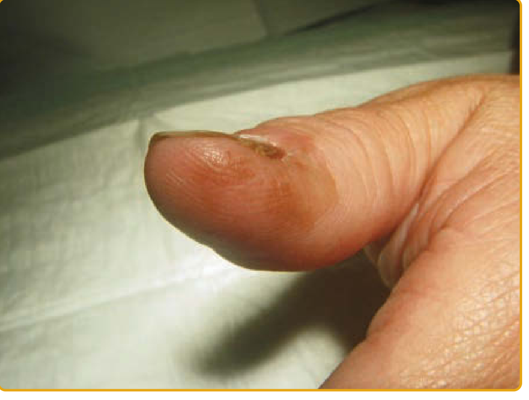
Amelanotic (25-33%)
- No pigment - a major diagnostic pitfall
- Arises from the nail bed, causes onycholysis with oozing
- Frequently misdiagnosed as pyogenic granuloma, ingrown nail, wart, or onychomycosis
- Carries worse prognosis due to even greater diagnostic delay
Differential Diagnosis
Subungual melanoma is commonly misdiagnosed as: - Andrews' Diseases of the Skin
| Mimic | Key distinguishing feature |
|---|---|
| Subungual hematoma | Moves distally as nail grows; history of trauma |
| Onychomycosis | Fungal culture positive; no linear band |
| Pyogenic granuloma | Fleshy, bleeds easily; no pigment band |
| Verruca vulgaris | Rough surface; no matrix origin |
| Glomus tumor | Intense point tenderness; subungual location |
| Kaposi sarcoma | Multiple lesions; known HIV/immunosuppression |
| Chronic paronychia | Inflammation around fold; no longitudinal band |
Diagnostic Criteria - The ABCDEF Rule
(Levit et al., J Am Acad Dermatol 2000) - Fitzpatrick's Dermatology
| Letter | Criterion |
|---|---|
| A | Age 40-70 years; African-American, Asian, Native American ethnicity |
| B | Band: Brown-black, breadth >3 mm, blurred or irregular border |
| C | Change: Rapid increase in width, color change, nail dystrophy not improving with treatment |
| D | Digit: Thumb > big toe > index; single digit involvement |
| E | Extension: Hutchinson sign (pigment on proximal/lateral nail fold) |
| F | Family or personal history of melanoma or dysplastic nevi |
Diagnosis Workup
- Clinical examination - look for Hutchinson sign, band characteristics
- Dermoscopy - reveals micro-Hutchinson sign, irregular lines, loss of parallel pattern
- Nail matrix biopsy - histopathology remains the gold standard
- Shows large atypical dendritic melanocytes with irregular junctional nests
- Frequent association with subungual onycholemmal cysts (do not confuse with SCC)
- Staging - same as cutaneous melanoma (Breslow thickness, sentinel node biopsy for invasive lesions)
Treatment
| Stage | Treatment |
|---|---|
| In situ / early invasive | Wide local excision with digit preservation |
| Advanced invasive | Distal digit amputation (remains standard) |
| Hutchinson sign (in situ spread) | Topical imiquimod immunotherapy |
| Metastatic / advanced | Targeted therapy (BRAF/MEK inhibitors if BRAF-mutated), immune checkpoint inhibitors (anti-PD-1, anti-CTLA-4) |
Note: BRAF mutations are less common in acral/subungual melanoma than in cutaneous melanoma, so BRAF inhibitors benefit fewer patients. KIT mutations are more common and may respond to imatinib. - Conway et al., 2023
Prognosis
- In situ and early invasive: Very good prognosis
- Advanced / metastatic: 5-year survival rate of only 15-24% - Fitzpatrick's Dermatology
- The poor prognosis is not because the tumor is inherently more aggressive, but because diagnostic delay of years to decades is common - leading to late-stage presentation
- Postoperative follow-up depends on Breslow thickness and staging
Key Takeaway
Subungual melanoma is "not particularly aggressive - it is the delay in diagnosis and treatment that makes the prognosis so poor." - Fitzpatrick's Dermatology
Any single-digit longitudinal melanonychia in an adult, especially on the hallux or thumb, must be evaluated with dermoscopy and low threshold for biopsy. The 3-year history of the black line in your image makes a dermatology review urgent.
Image
It looks like your message just says "Image" but no image was attached. Could you please re-upload the image you'd like me to look at?

1000120229.jpg
What is you probably diagnosis
analyse_media
This is a closer, clearer view of the same great toenail. Here is my detailed assessment:
Clinical Analysis of This Image
What is visible:
| Feature | Finding | Significance |
|---|---|---|
| Band type | Longitudinal, base to tip | Classic melanonychia |
| Color | Very dark brown-black | Higher suspicion |
| Width | ~15-20% of nail width | >3 mm = ABCDEF "B" criterion met |
| Borders | Sharp, parallel, well-defined | Somewhat reassuring |
| Color uniformity | Solid and uniform | Could be benign nevus |
| Nail plate | Intact, no dystrophy | Reassuring - not advanced |
| Proximal nail fold | Possible small pigmented smudge at cuticle | Possible Hutchinson sign - concerning |
Probable Diagnosis
Most likely: Nail matrix nevus (benign melanocytic nevus)
- The band has sharp, parallel, regular borders
- Uniform dark pigmentation throughout
- No nail dystrophy or crumbling
- Present and stable for 3 years without reported change
- These features together favor a benign nail matrix nevus
However - cannot exclude: Subungual melanoma (in situ)
Several features warrant serious concern:
- Width >3 mm - meets the ABCDEF "B" criterion
- Dark near-black color - higher risk than light brown
- Great toe location - highest risk digit
- Single nail involvement
- Possible pigment at the cuticle - if confirmed on examination, this is Hutchinson's sign, the single most important clinical red flag for subungual melanoma
What This Means for You
| Scenario | Action needed |
|---|---|
| Nail matrix nevus | Monitoring every 6 months, possible biopsy for certainty |
| Subungual melanoma in situ | Wide excision with digit preservation - excellent cure rate |
| Invasive melanoma | Surgical amputation of distal digit + staging |
You cannot safely distinguish a nevus from early subungual melanoma by clinical appearance alone - not even dermatologists can without dermoscopy and biopsy.
The possible pigmentation at the proximal nail fold (Hutchinson sign) in this image makes this a high-priority case. A 3-year history without change is somewhat reassuring, but subungual melanoma can be slow-growing and still be life-threatening if missed.
Urgent Recommendation
See a dermatologist as soon as possible - ideally within the next 2-4 weeks, not months. Request:
- Nail dermoscopy to evaluate the band pattern and confirm/exclude Hutchinson sign
- Nail matrix biopsy - if any doubt remains after dermoscopy, biopsy is the only way to get a definitive answer
Early-stage subungual melanoma treated promptly is curable. The danger lies only in delay.
This is a shared conversation. Sign in to Orris to start your own chat.