This is my seminar presentation on sutures , needles and suturing techniques This is my seminar on suturing technique, sutures, needles. I want to add some topics in detail in this. So give me detail on which sutures are used with their indications, secondary suturing of burst abdomen.

Reading File
Finding Sources
Finding Sources
Finding Sources
Finding Sources
Finding Sources
Finding Sources
Finding Sources
Finding Sources
Reading File
Reading File
Searching the Web

secondary suturing burst abdomen wound dehiscence surgical technique management

Reading File
Reading Web Page

secondary suturing burst abdomen technique retention sutures steps

Reading File
Reading File
Now I have everything needed to give a thorough, well-structured answer for your seminar. Here it is:

Sutures - Types, Indications & Secondary Suturing of Burst Abdomen


PART 1: SUTURE MATERIALS AND THEIR INDICATIONS

Classification Overview

Suture materials are divided into two broad categories based on durability:
  • Absorbable - lose tensile strength within 60 days, degraded by hydrolysis or proteolytic enzymes
  • Non-absorbable - resistant to hydrolysis and enzymatic degradation; maintained indefinitely or removed electively
Both categories exist as monofilament (single strand) or multifilament (braided/twisted), each with distinct infection risk, handling, and knot security profiles. Braided sutures handle better and tie more securely, but carry a higher infection risk due to bacteria becoming entrapped between filament strands. Monofilaments have a low coefficient of friction and glide easily through tissue, but require extra throws to secure knots.

ABSORBABLE SUTURES

1. Surgical Gut (Catgut)

  • Source: Collagen from sheep or bovine submucosa
  • Type: Monofilament, natural
  • Absorption: Plain gut - 70 days; Chromic gut (treated with chromic salts to delay absorption) - 90 days; Fast-absorbing gut - 42 days
  • Tensile strength retention: Plain gut loses ~50% at 7 days; chromic gut retains strength slightly longer
  • Tissue reactivity: HIGH - degraded by proteolytic enzymes, causes the greatest inflammatory response of all sutures
  • Indications:
    • Fast-absorbing gut (5-0 or 6-0): skin grafts, head and neck reconstructions that are well approximated with buried sutures, pediatric scalp wounds (avoids need for removal under sedation)
    • Chromic gut: oral mucosal closures, episiotomies, superficial lacerations in children where suture removal would be difficult
    • Plain gut: rarely used today due to unpredictable absorption

2. Polyglactin 910 (Vicryl®, Polysorb®)

  • Type: Braided multifilament, synthetic (copolymer of glycolide and lactide)
  • Absorption: ~56-70 days by hydrolysis
  • Tensile strength retention: ~75% at 2 weeks, ~50% at 3 weeks
  • Tissue reactivity: Low (synthetic), but slightly more reactive than monofilament synthetics due to braiding
  • Indications:
    • Deep/buried dermal sutures - the most widely used absorbable suture for this purpose
    • Face repairs under low tension: 4-0 or 5-0
    • Larger truncal and extremity defects: 3-0 or 4-0
    • Subcuticular running closure (4-0, buried in place) - shown superior scar appearance vs. polypropylene in RCT
    • Mucosal closure (oral, vaginal, bowel anastomosis)
    • Vermilion lip repair
    • NOT ideal for wounds under significant tension (loses strength faster than PDS)
  • Vicryl Rapide: Partially hydrolyzed form, acts like fast-absorbing gut; used for surface sutures on the face (falls off spontaneously, no removal needed)

3. Poliglecaprone 25 (Monocryl®)

  • Type: Monofilament, synthetic (copolymer of glycolide and epsilon-caprolactone)
  • Absorption: 90-120 days
  • Tensile strength retention: ~50% at 1 week, ~20-30% at 2 weeks (relatively rapid loss)
  • Tissue reactivity: Very low
  • Indications:
    • Buried dermal sutures - very popular due to rapid absorption and reduced rate of suture extrusion (spitting)
    • Face (4-0 or 5-0) and trunk (3-0 or 4-0) deep closures
    • Running subcuticular closure (buried in place, no removal needed)
    • NOT suitable for wounds under significant tension - loses tensile strength too quickly

4. Polydioxanone (PDS® II)

  • Type: Monofilament, synthetic (polyester)
  • Absorption: 180-210 days
  • Tensile strength retention: ~70% at 2 weeks, ~50% at 4 weeks - the longest-lasting of all absorbable sutures
  • Tissue reactivity: Very low
  • Indications:
    • Wounds under significant or prolonged tension - best absorbable choice here
    • Fascial closure (abdominal fascia, where prolonged support is needed)
    • Pediatric cardiovascular surgery (where permanent sutures are avoided)
    • Tendon repair
    • Comfortable enough for intraoral mucosa (can be left to absorb or removed at 5-7 days)
    • Deep closures where extended tensile strength is required

5. Polyglyconate (Maxon®)

  • Type: Monofilament, synthetic (copolymer of glycolic acid and trimethylene carbonate)
  • Absorption: Similar to PDS
  • Tensile strength retention: Retains ~80% at 2 weeks - similar to PDS
  • Indications: Same as PDS - wounds under significant tension, fascial closures

NON-ABSORBABLE SUTURES

6. Nylon (Ethilon®, Monosoft®)

  • Type: Monofilament (most common) or braided (Nurolon®)
  • Tensile strength: High; slowly degrades ~15-20% per year
  • Key properties: High memory (springy - tends to return to original shape), low tissue reactivity, low coefficient of friction
  • Handling: Stiff; requires extra throws (at least 3) to prevent knot unravelling
  • Indications:
    • Standard skin/cutaneous closure (5-0 or 6-0 on face, 4-0 on trunk/extremities)
    • Scalp lacerations
    • Areas where cosmesis matters and sutures will be removed at 5-14 days
    • NOT comfortable near the lips (stiffness)

7. Polypropylene (Prolene®, Surgipro®)

  • Type: Monofilament
  • Key properties: Lowest coefficient of friction of all sutures (extremely slippery), high plasticity (stretches to accommodate edema then resumes tension), high elasticity, does not degrade in tissue over time, very low tissue reactivity
  • Handling: Requires multiple throws (4-5) due to poor knot security
  • Indications:
    • Running subcuticular closure - ideal choice; easily removable even after 1 year due to minimal tissue adherence
    • Cardiovascular surgery (vascular anastomoses - non-degradable, maintains strength permanently)
    • Hernia repairs (when permanent synthetic support is needed)
    • Skin closure where sutures must stay long-term (e.g., infected fields)
    • Wound closure in areas of post-operative edema (plasticity allows accommodation)

8. Silk

  • Type: Braided multifilament, natural (silkworm protein)
  • Key properties: Lowest memory, most pliable, best handling and knot security of all sutures; however, highest tissue reactivity among non-absorbables; technically non-absorbable but slowly degrades over 2+ years
  • Indications:
    • Oral mucosal sutures near the lips - most comfortable material for patients
    • Drain anchoring sutures
    • Vessel ligation ties (ease of handling)
    • Avoid in contaminated wounds (braided structure harbors bacteria)

9. Polyester (Mersilene®, Dacron®, Ethibond®)

  • Type: Braided multifilament; Ethibond is coated with polybutilate to reduce friction
  • Key properties: High tensile strength, good handling, low tissue reactivity; better knot stability than nylon or polypropylene
  • Indications:
    • Cardiac and vascular surgery (heart valve repair, vascular prosthesis attachment)
    • Vermilion lip repair (combines soft feel of silk with low reactivity)
    • Oral mucosal approximation
    • Areas where Ethibond's improved handling is preferred over nylon despite slightly more tissue reaction

10. Polybutester (Novafil®)

  • Type: Monofilament
  • Key properties: High elasticity (accommodates tissue swelling, then maintains tension as edema resolves), good handling, low friction, low reactivity
  • Indications:
    • Wounds with anticipated postoperative edema (e.g., joint surfaces, trauma)
    • Situations where dynamic tension adjustment is beneficial

11. Steel (Stainless Steel Wire)

  • Type: Monofilament or twisted multifilament
  • Key properties: Maximum tensile strength; completely non-reactive; not absorbed; uncomfortable
  • Indications:
    • Sternal closure after cardiac surgery (sternotomy)
    • Orthopedic cerclage wires
    • Tendon repair (particularly hand surgery)
    • Areas needing maximum retention (contaminated wounds with retention sutures)

Site-Based Suture Selection Guide

SiteDeep/Buried SutureSurface Suture
Face4-0/5-0 polyglactin 910 or poliglecaprone 255-0/6-0 nylon or polypropylene; or 6-0 fast-absorbing gut
Neck / distal extremities4-0 polyglactin 910 or poliglecaprone 254-0/5-0 nylon or polypropylene
Trunk / proximal extremities3-0/4-0 polyglactin 910 or poliglecaprone 253-0/4-0 nylon or running subcuticular polypropylene/poliglecaprone 25
ScalpRarely needed4-0/3-0 nylon or skin staples
Oral mucosa / lipsPolyglactin 910 or fast-absorbing gutSilk (comfort) or chromic gut
Fascia (abdominal)PDS, polyglyconate, or polyglactin 910N/A
Skin graftsN/A5-0/6-0 fast-absorbing gut or staples
CardiovascularN/APolypropylene (permanent, non-degradable)
(Source: Dermatology 2-Volume Set 5e, p. 2949; Rosen's Emergency Medicine, p. 772)

PART 2: SECONDARY SUTURING OF BURST ABDOMEN

Definition

Burst abdomen (abdominal wound dehiscence / disruption) refers to the postoperative separation of the layers of a laparotomy wound. It ranges from superficial skin-only dehiscence to complete dehiscence (burst abdomen) with evisceration of bowel or omentum through the disrupted wound.

Incidence and Predisposing Factors

Occurs in approximately 1% of all laparotomies (higher in patients with malignancy, malnutrition, or serious illness).
Predisposing factors:
Systemic:
  • Malnutrition and hypoproteinaemia (impairs wound healing)
  • Anaemia
  • Obesity
  • Diabetes mellitus
  • Corticosteroid use (impairs collagen synthesis)
  • Malignancy
  • Immunosuppression
Local/Technical:
  • Poor surgical technique - inadequate fascial bites, excessive tension
  • Wound infection / surgical site infection
  • Suture failure (wrong material, inadequate knot)
  • Dead space or haematoma formation
Postoperative:
  • Ileus/abdominal distension (increased intra-abdominal pressure)
  • Persistent coughing, vomiting
  • Inadequate analgesia leading to splinting and coughing

Clinical Presentation

  • Early sign: Sudden increase in serous, pink or serosanguinous wound drainage ("salmon-pink fluid") - this should be considered deep dehiscence until proven otherwise
  • Superficial dehiscence: Skin edges separate; fascial layer remains intact
  • Full/complete dehiscence (burst abdomen): Fascial layer disrupts; protrusion of omentum or bowel through wound
  • Typically occurs 5-10 days post-surgery (during the phase when wound tensile strength is only 7-60% of normal)

Immediate Management (Emergency Measures)

  1. A-E assessment - patients with burst abdomen can deteriorate rapidly
  2. IV access, IV fluids, resuscitation
  3. Broad-spectrum IV antibiotics immediately
  4. Cover the wound and any protruding viscera with saline-soaked gauze as a temporizing measure - NEVER let bowel lie dry; do NOT use plain dry gauze (can erode into bowel lumen and create fistula); petroleum jelly-soaked gauze preferred for exposed bowel
  5. Adequate analgesia
  6. Urgent return to operating theatre

Surgical Management - Secondary Closure

This is performed under general anaesthesia in the operating theatre.

Step-by-Step Technique:

1. Preparation
  • Open all of the skin incision fully and inspect the fascia carefully
  • Locate the previous fascial closure suture and trace it back to intact, non-dehisced fascia
  • Debride all necrotic/devitalized tissue at fascial edges to expose clean anatomy
2. Exploration of the Abdomen
  • Gently examine intra-abdominal contents for signs of injury, perforation, or collections
  • Assess the wound discharge - purulent fluid suggests infected collections, feculent/bilious fluid indicates enterocutaneous fistula (requires more complex management - washout, ostomy)
  • If no purulent or feculent discharge, exploration should be gentle and limited to avoid iatrogenic bowel injury
  • Lavage the peritoneal cavity with warm saline
3. Placement of Retention Sutures (Before Fascial Closure)
  • Place full-thickness retention sutures using thick non-absorbable monofilament or braided suture (e.g., No. 1 or No. 2 PDS, or non-absorbable nylon/polypropylene)
  • Pass sutures 3-4 cm from the fascial edge, passing full-thickness through: skin → subcutaneous fat → anterior rectus sheath → rectus muscle → posterior sheath → peritoneum
  • Visualize the needle at all times to avoid iatrogenic bowel injury
  • Do NOT tie retention sutures yet - they will be tied after fascial closure
4. Closure of the Fascia
  • Begin fascial closure with a suture at each end of the wound, then meet in the middle (reduces tension distribution)
  • Use large interrupted sutures with adequate fascial bites (mass closure technique preferred)
  • Each suture is placed under direct visualization with a malleable retractor protecting bowel
  • The final sutures are placed without tension on previously placed sutures so the needle is always visible
  • Suture material of choice: PDS (polydioxanone) or long-acting synthetic absorbable; alternatively a non-absorbable suture such as nylon for permanent mass closure
5. Tying Retention Sutures
  • Once fascial closure is complete, tie the retention sutures
  • Pass each retention suture through sterile IV tubing strips (bolsters) before tying - this distributes tension and prevents the sutures from cutting through the skin
  • Tie without excessive tension - the purpose is to reduce strain on the fascial repair, not strangulate tissue
6. Skin Closure
  • Close skin very loosely (or leave partially open) to allow drainage and prevent tension
  • In cases of infection or gross contamination: leave skin open to heal by secondary intention, or delayed primary closure after 4-5 days of wound hygiene

Special Situations

SituationModification
Gross intra-abdominal sepsis / necrotizing fasciitisPrimary re-closure unsafe - manage as open abdomen using negative pressure wound therapy (wound VAC)
Enterocutaneous fistulaResuscitation → CT/ultrasound assessment → operative washout + repair/diversion
Colostomy present near woundPlace retention sutures closer to midline; preserve at least 3 cm of flat skin around stoma for appliance adherence
Superficial dehiscence only (fascia intact)Conservative: abdominal binder + wound packing/dressings → healing by secondary intention; OR secondary closure after 4-5 days of granulation (shown superior to secondary intention alone in RCTs)

Postoperative Care After Re-closure

  • Aggressive nutritional support: high-protein diet, multivitamins - nutritional status directly impacts fascial healing
  • Retention sutures are left in place for up to 6-8 weeks (until skin wound has been well healed for 3-4 weeks)
  • Patients will complain of discomfort from retention sutures - counsel them in advance
  • Monitor for wound infection, seroma, and re-dehiscence
  • Abdominal binder support during recovery

Why Secondary Suturing is Superior to Conservative Management

Clinical evidence supports secondary closure over healing by secondary intention for patients with significant dehiscence:
  • One prospective study (Dodson et al.) showed mean healing time of 17.6 ± 7.2 days with secondary closure vs. 61.2 ± 35.8 days with secondary intention healing (p <0.001)
  • Debridement and immediate re-closure further reduces healing time to ~20 days median
  • Fewer post-operative visits, less disability, lower cost
(Sources: Rosen's Emergency Medicine 9e, p. 772; Dermatology 2-Volume Set 5e, pp. 2946-2949; VUMC Global Surgical Atlas - Closure of Laparotomy Wound Dehiscence; StatPearls - Wound Dehiscence; TeachMeSurgery)

These two sections - suture types with indications and secondary suturing of burst abdomen - are ready to be inserted directly into your seminar. Let me know if you want these formatted as slides, want any section expanded further (e.g., specific suture sizes per specialty, interrupted vs. continuous techniques), or want a diagram/table added.

Give secondary suturing of burst abdomen in detail with relavent images and line diagram

Reading File
Finding Sources
Finding Sources
Searching the Web

burst abdomen secondary suturing technique retention sutures diagram clinical images

Reading File
Reading File
Loading Image
Loading Image
Reading File
Loading Image
Loading Image
Loading Image
Searching the Web

burst abdomen clinical photograph evisceration bowel postoperative wound dehiscence

Loading Image
Loading Image
Generating Image
Loading Image
Generating Image
Loading Image
All images are now in hand. Here is the complete, detailed, fully illustrated section on secondary suturing of burst abdomen:

Secondary Suturing of Burst Abdomen - Detailed Account


1. DEFINITION

Burst abdomen (acute abdominal wound dehiscence / abdominal wound disruption) is the postoperative separation of the musculo-aponeurotic layers of a laparotomy wound. It ranges from partial (superficial, skin-only) to complete dehiscence with evisceration - protrusion of abdominal viscera (bowel or omentum) through the disrupted incision.
"Significant wound dehiscence occurs in approximately 1% of all laparotomies. In patients with malignant disease or other serious illness, the incidence may be much greater." -- Sabiston Textbook of Surgery 21e

2. ANATOMY OF THE ABDOMINAL WALL (Why It Bursts)

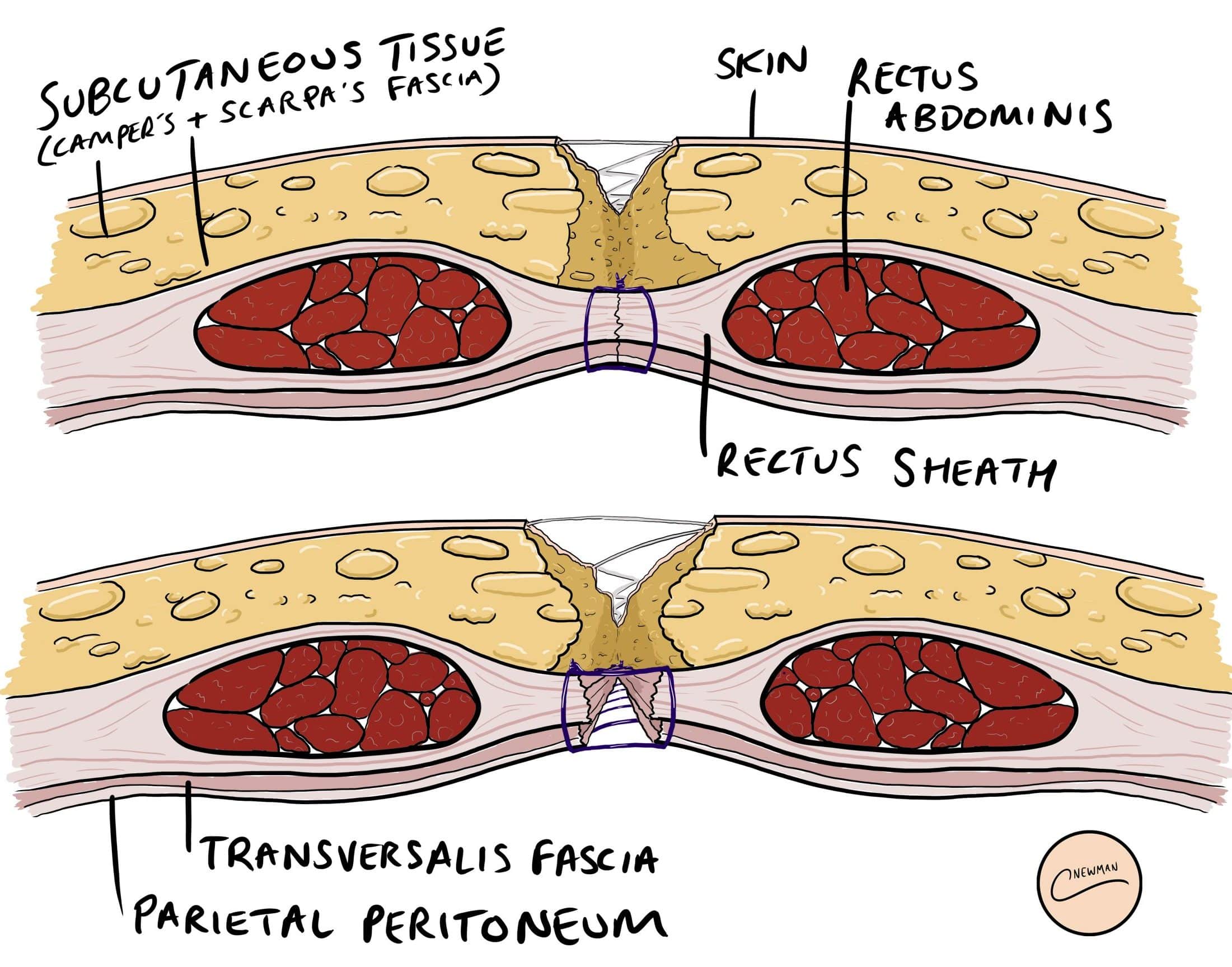
Understanding the layers is critical to the repair. A midline laparotomy incision disrupts these layers from outside in:
Cross-section of abdominal wall layers showing wound dehiscence - top panel shows fascial disruption, bottom panel shows deeper layer separation
Abdominal wall cross-section: Skin → Subcutaneous tissue (Camper's + Scarpa's fascia) → Anterior rectus sheath → Rectus abdominis → Posterior rectus sheath → Transversalis fascia → Parietal peritoneum
In burst abdomen, it is the fascial layer (rectus sheath / linea alba) that is the critical failure point. When fascia gives way, nothing prevents evisceration.

3. INCIDENCE AND TIMING

  • Overall incidence: ~1% of all laparotomies (up to 10-15% in high-risk patients)
  • Typical timing: Day 5-10 post-surgery (peak Day 7-8)
  • At this time wound tensile strength is only 7-60% of native bursting pressure - the wound is at its most vulnerable while still healing

4. PREDISPOSING FACTORS

Patient (Systemic) Factors

FactorMechanism
Malnutrition / HypoproteinaemiaImpaired collagen synthesis and wound healing
AnaemiaReduced oxygen delivery to healing wound
ObesityExcess intra-abdominal pressure, poor tissue vascularity
Diabetes mellitusImpaired neutrophil function, delayed healing
Corticosteroid useSuppresses fibroplasia and collagen cross-linking
MalignancyCachexia + immune compromise
JaundiceImpaired collagen synthesis, endotoxaemia
UraemiaImpaired platelet function and healing
ImmunosuppressionReduced wound defence

Local / Technical Factors

FactorMechanism
Wound infection / SSICollagenase-mediated collagen breakdown
Haematoma / SeromaActs as culture medium + prevents apposition
Poor techniqueInadequate fascial bites, excessive tension, wrong suture
Wrong suture materialFast-absorbing suture loses strength before fascia heals
Dead spaceIncreases fluid collection and infection risk

Postoperative Factors

FactorMechanism
Ileus / Abdominal distensionIncreases intra-abdominal pressure on suture line
Persistent coughing (chronic lung disease)Repeated stress on wound
VomitingSudden spikes in intra-abdominal pressure
Inadequate analgesiaReflex muscle spasm and coughing

5. PATHOPHYSIOLOGY

During the first 1-2 weeks post-surgery:
  • Wound tensile strength is only 7-10% of normal
  • At 5 weeks: ~60% of normal
  • After 1 month: dehiscence from normal activity becomes unlikely
The critical window is Days 5-10: enough time for infection to set in and degrade collagen, but wound not yet strong enough to resist the forces of postoperative ileus and patient activity.

6. CLINICAL PRESENTATION

Warning Sign (Premonitory Sign)

  • Sudden increase in serous, pink or serosanguinous wound drainage - the so-called "salmon-pink fluid"
  • This must be treated as deep fascial dehiscence until proven otherwise
  • Occurs 24-48 hours before visible dehiscence in ~50% of cases

Grades of Dehiscence

GradeFeatures
SuperficialSkin and subcutaneous tissue open; rectus sheath intact; no evisceration
Deep (fascial)Rectus sheath disrupted; new wound bulging; heavy serosanguinous discharge
Complete / Burst abdomenFull-thickness disruption; omentum or bowel visibly protrudes through wound

7. IMMEDIATE MANAGEMENT (Pre-Theatre)

This is a surgical emergency. Take these steps immediately on the ward:
1. CALL the surgical team and anaesthesia immediately
2. A-E assessment and resuscitation
3. IV access + IV fluid resuscitation
4. IV broad-spectrum antibiotics (e.g. piperacillin-tazobactam)
5. Adequate IV analgesia (reduces straining)
6. Cover the wound:
      - Moist (saline-soaked) gauze over the wound AND any protruding viscera
      - NEVER dry gauze directly on exposed bowel — it erodes into bowel lumen, causing fistula
      - Petroleum jelly gauze is preferred for exposed bowel
7. Do NOT attempt to reduce eviscerated bowel on the ward
8. Urgent transfer to operating theatre — patient supine, knees slightly bent to relax abdominal wall
9. NBM (nil by mouth)
10. Mark for stoma if likely required

8. OPERATIVE MANAGEMENT - SECONDARY SUTURING (STEP BY STEP)

Setting: Operating theatre, general anaesthesia. Cannot be done adequately under local or spinal.

PRE-OPERATIVE PLANNING

Before operating, assess:
  • Is there gross intra-abdominal sepsis or feculent discharge? (Will need full exploration + washout, possibly ostomy)
  • Is fascial closure technically feasible? (Is there loss of domain? Is there ischemic/necrotic fascia?)
  • What are the patient's risk factors for re-dehiscence? (Guides use of retention sutures)

STEP 1: OPEN THE WOUND AND ASSESS FASCIA

  • Open all of the skin incision fully (not just the visibly dehisced segment)
  • Carefully inspect the entire fascial closure from one end to the other
  • Locate where the previous fascial suture is intact and trace back from the dehisced area
  • Debride all necrotic/devitalised tissue at the fascial edges until clean, viable fascia is reached
  • This step is essential - closing over dead tissue guarantees re-dehiscence

STEP 2: ABDOMINAL EXPLORATION AND LAVAGE

  • Gently examine intra-abdominal contents:
    • Clear serous fluid → limited, gentle exploration; avoid iatrogenic bowel injury
    • Purulent discharge → search for undrained collections and address
    • Feculent/bilious fluid → enterocutaneous fistula suspected → extensive washout + repair/diversion
  • Apply gentle downward pressure on bowel for limited visualization
  • Lavage the peritoneal cavity with warm saline (1-3 litres)
  • If in doubt, do not over-explore - the priority is closure, not comprehensive exploration

STEP 3: PLACE RETENTION SUTURES (BEFORE FASCIAL CLOSURE)

This is the key distinguishing step in secondary suturing of burst abdomen.
Purpose of retention sutures:
  • They do NOT prevent fascial dehiscence
  • They DO prevent evisceration if the fascial closure fails again
  • They distribute tension over a wide area of the abdominal wall, sparing the suture line
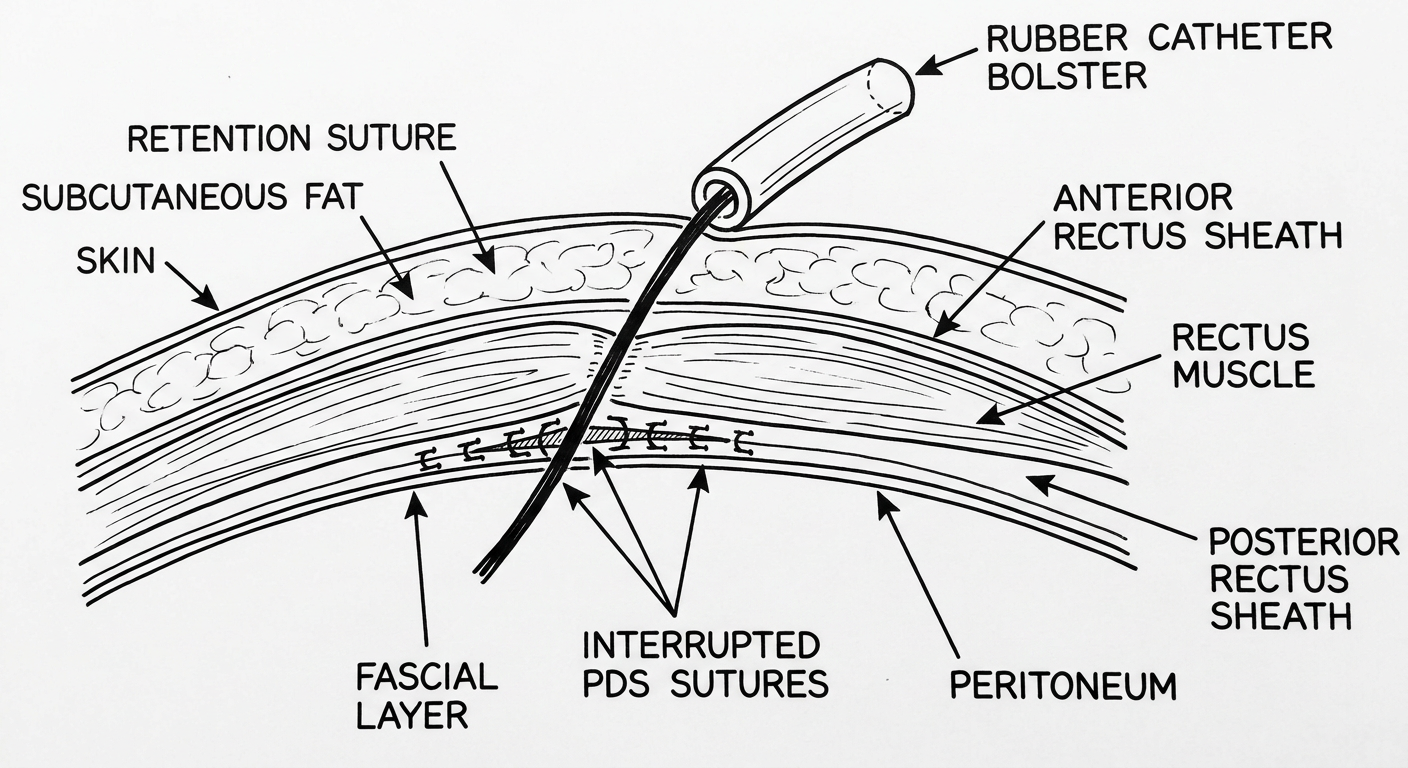
Technique:
Line diagram showing retention suture passing through all layers of abdominal wall with rubber catheter bolster on skin surface, alongside interrupted PDS fascial closure sutures
Line diagram: Retention suture passing full-thickness through skin → subcutaneous fat → anterior rectus sheath → rectus muscle → posterior sheath (extraperitoneal) with rubber catheter bolster on skin surface. Note the interrupted PDS sutures closing the fascia separately.
Placement details:
  • Suture material: No. 1 or No. 2 heavy non-absorbable - nylon, polypropylene, or braided (e.g. nylon, PDS No. 1/2)
  • Position: 3-4 cm from the fascial edge, on both sides of the wound
  • Spacing: 4-6 cm apart from each other along the wound length
  • Depth: Full-thickness passing through: skin → subcutaneous fat → anterior rectus sheath → rectus muscle → ideally extraperitoneal (posterior sheath, preperitoneal space) - avoiding intraperitoneal placement to prevent visceral entrapment
  • Visualise the needle at all times - place a malleable retractor under the fascia to protect bowel
  • Do NOT tie the retention sutures yet - leave them long and marked; they are tied after fascial closure is complete
Types of retention sutures:
Diagram showing four types of abdominal retention sutures - A: simple interrupted, B: horizontal mattress, C: far-near-near-far (Smead-Jones), D: figure-of-eight
Types of retention/through-and-through sutures: (A) Simple interrupted, (B) Horizontal mattress, (C) Figure-of-eight, (D) With external bolster - showing different configurations used in clinical practice

STEP 4: CLOSE THE FASCIA (MASS CLOSURE / INTERRUPTED)

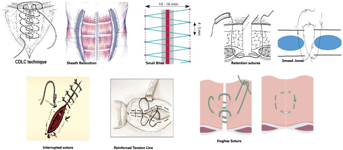
This is the definitive repair.
Comparison of closure techniques: CDLC technique, small bites technique, retention sutures diagram, Smead-Jones far-near-near-far suture, Hughes suture, interrupted suture pattern
Overview of abdominal closure techniques - from standard interrupted to small-bites technique to retention/Smead-Jones configurations
Preferred technique for secondary closure:
  • Interrupted mass closure is preferred over continuous suture for secondary closure
    • A meta-analysis (Gupta et al., 23 studies, N=10,900) showed interrupted closure was associated with significantly less dehiscence than continuous closure
    • A 2019 RCT (Bansiwal et al., 300 patients) showed burst abdomen rate of 20.1% with continuous vs. 5.4% with interrupted (p<0.05) in emergency laparotomy
    • With continuous closure, a single stitch failure or area of fascial necrosis opens the entire wound; with interrupted, only that segment fails
Suture material of choice:
  • Polydioxanone (PDS) No. 1 - preferred (monofilament, resists infection, retains tensile strength for 40-90 days - matches fascial healing timeline)
  • Alternatively: Polyglyconate (Maxon), polypropylene, or nylon
  • Avoid fast-absorbing sutures (polyglactin alone) - insufficient tensile strength duration for fascial healing (half-life only 3 weeks); associated with higher dehiscence rates (52% vs. 16% with PDS in one study)
  • Barbed sutures (if available): provide even tension distribution without knots
Technical principles:
  • Start with sutures at each end of the wound then meet in the middle (distributes tension)
  • Take adequate fascial bites - not too shallow (won't hold) and not excessively deep (excess tension + ischaemia)
  • Elective recommendation: small bites (5mm tissue, 5mm apart) - associated with lower incisional hernia rates
  • Emergency/re-closure: larger bites acceptable to secure repair
  • Maintain direct visualization throughout - malleable retractor protecting bowel under fascia
  • Do NOT close the peritoneum separately - no benefit and adds operating time

STEP 5: TIE RETENTION SUTURES OVER BOLSTERS

Now tie the retention sutures that were placed in Step 3, but only after fascial closure is complete.
  • Thread each retention suture through a bolster before tying:
    • Options: Red rubber catheter segments (most commonly used, as shown in textbook image), sterile IV tubing strips, plastic bridges, or commercial retention bridges
    • Bolsters distribute the tension over a wider skin area, preventing the heavy suture from cutting through the skin (cheese-wiring effect)
  • Tie without excessive tension - the aim is to reduce wound strain, not strangulate tissue
  • Avoid pulling too tightly - sutures will dimple the skin and cause pressure necrosis

STEP 6: SKIN CLOSURE

  • Close skin loosely - do not aim for tight skin apposition
  • Options:
    • Loose interrupted skin sutures or staples - allow drainage
    • Delayed primary closure (Day 4-5) if contaminated - leave skin open initially, pack with wet dressings, close when wound is clean and granulating
    • Leave skin open in gross contamination or infection - heal by secondary intention or vacuum-assisted closure (VAC therapy)

9. THE FOUR-STEP PROCESS - ILLUSTRATED SUMMARY

Four-panel line diagram showing secondary suturing steps: Panel 1 wound debridement with exposed bowel, Panel 2 retention sutures placed through all layers not yet tied, Panel 3 fascial closure with interrupted sutures, Panel 4 final closure with retention sutures tied over bolsters
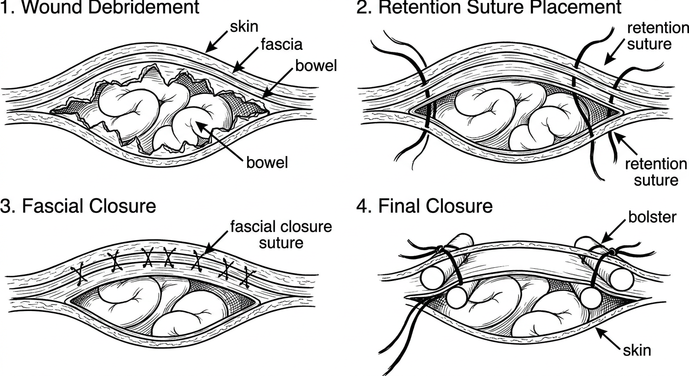
Step-by-step line diagram of secondary suturing of burst abdomen: 1. Wound debridement - fascial edges cleared, bowel visible 2. Retention sutures placed full-thickness through all layers, NOT yet tied 3. Fascial closure with interrupted PDS/mass closure sutures 4. Retention sutures tied over bolsters, skin loosely approximated

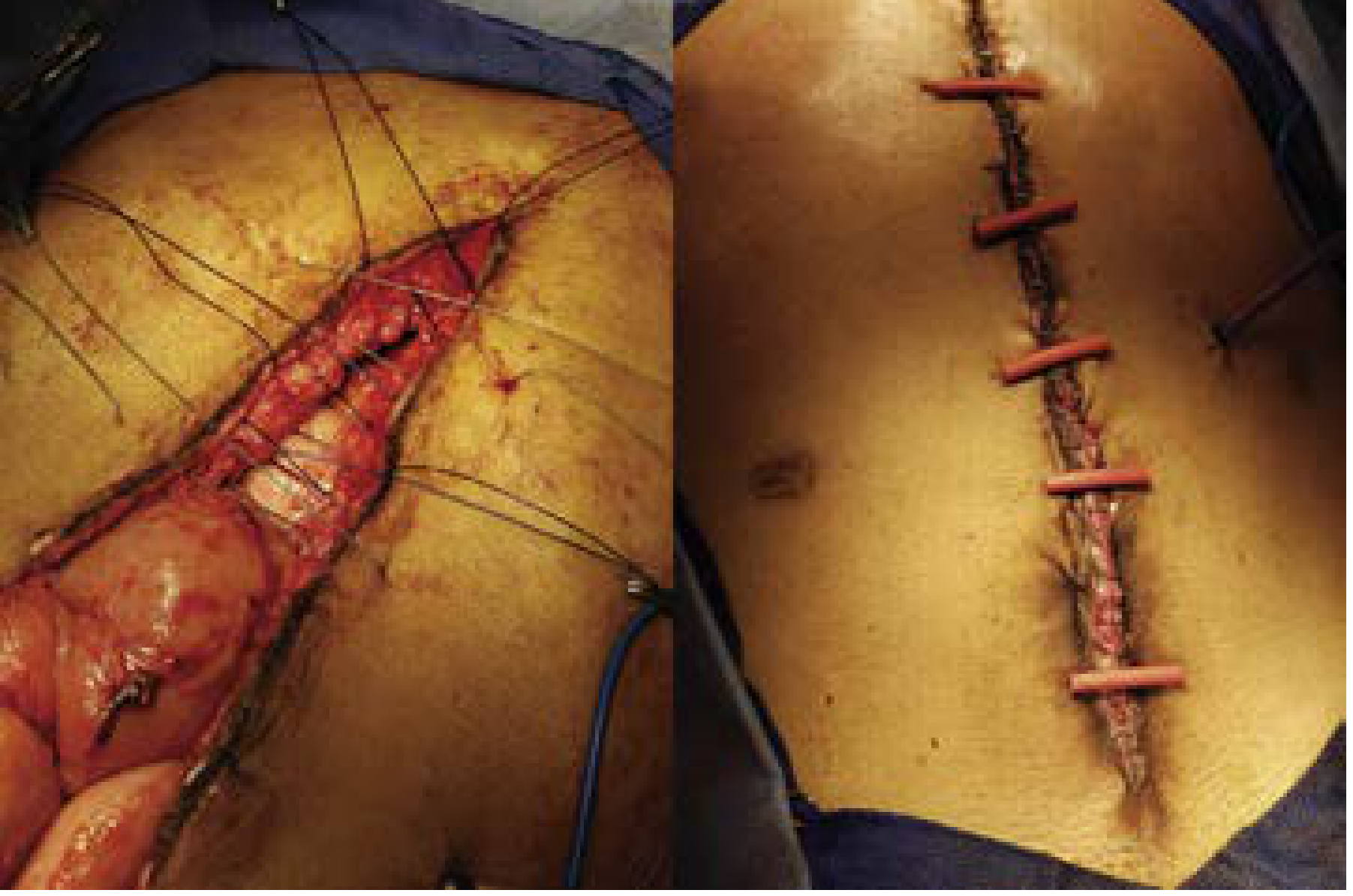
10. CLINICAL PHOTOGRAPH - INTRAOPERATIVE

Left panel: intraoperative photograph showing fascial closure with long retention sutures placed through all layers, yet to be tied, with eviscerated wound visible. Right panel: postoperative photograph showing retention sutures tied over red rubber catheter bolsters running parallel along the abdominal wound
Left: Intraoperative view - fascial closure in progress with long heavy retention sutures passed through all layers lateral to the wound, awaiting tying. Right: Postoperative view - retention sutures tied down over red rubber catheter bolsters, distributing pressure over the skin surface. (Source: Current Surgical Therapy 14e, Fig. 8)

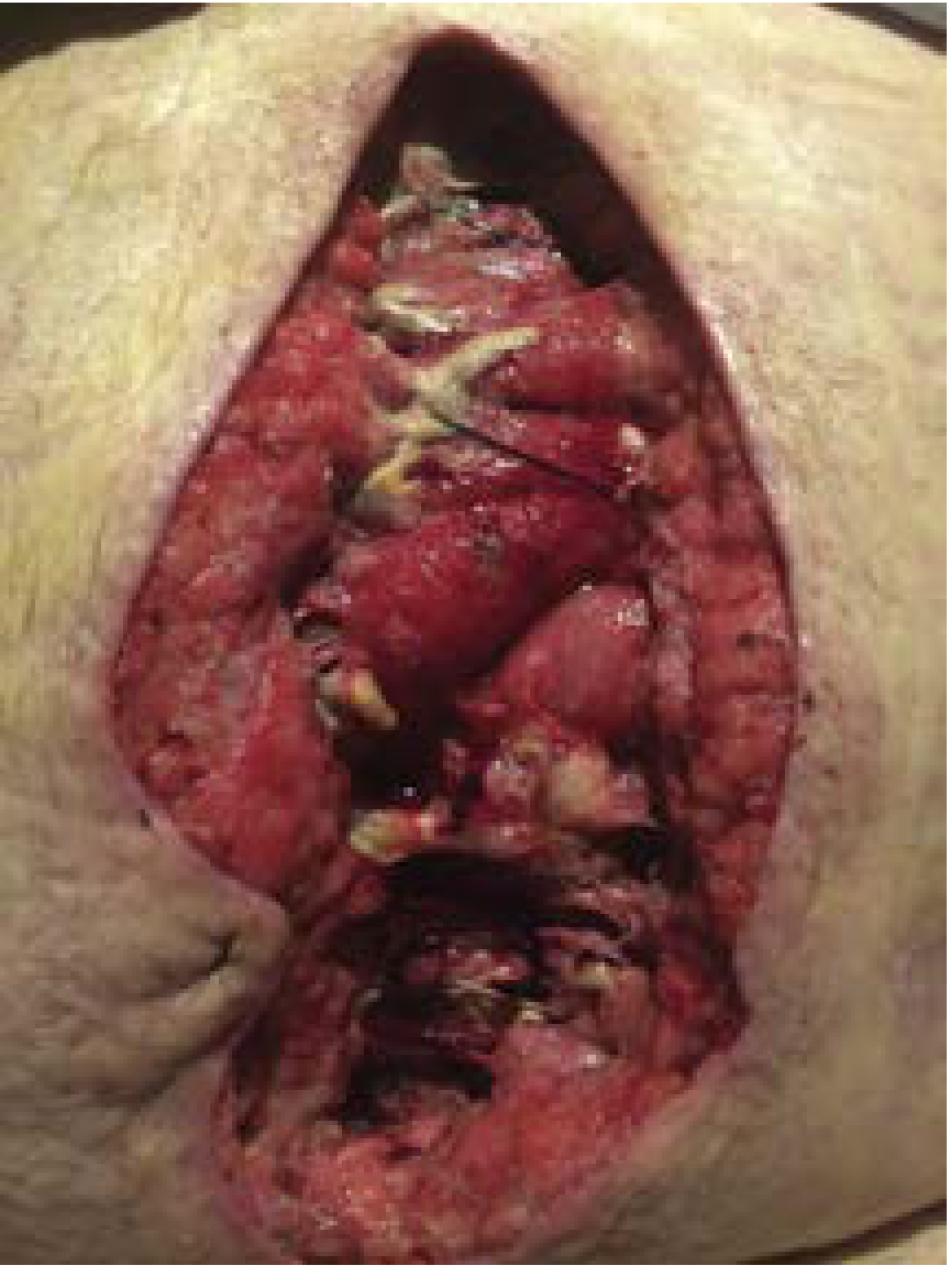
11. COMPLETE FASCIAL DEHISCENCE - APPEARANCE

Clinical photograph of complete fascial dehiscence after failure of continuous running abdominal closure - deep wound with exposed bowel and failure of fascia visible
Complete fascial dehiscence after partial failure of a continuous running primary abdominal closure - exactly the scenario that requires secondary suturing. Note the exposed bowel and completely disrupted fascia. (Source: Current Surgical Therapy 14e, Fig. 7)

12. SPECIAL SCENARIOS AND MODIFICATIONS

When Primary Re-closure is NOT Possible

SituationManagement
Gross abdominal sepsis / necrotizing fasciitis of abdominal wallOpen abdomen management - negative pressure wound therapy (wound VAC/Barker pack)
Loss of domain (fascial edges cannot be approximated without tension)Bridging mesh repair (biological or synthetic) + possible component separation
Bowel edema preventing closureBogota bag (sterile IV fluid bag used as temporary silo) or wound VAC
Enterocutaneous fistulaResuscitate → CT scan → operative washout → repair/diversion (ileostomy or colostomy)
Colostomy adjacent to woundPlace retention sutures closer to midline; preserve 3 cm of flat skin around stoma; avoid pulling sutures tight near stoma
Superficial dehiscence only (fascia intact)Abdominal binder + wound packing; OR secondary closure after 4-5 days granulation

13. POSTOPERATIVE CARE

MeasureDetail
NutritionHigh-protein diet + multivitamins immediately postoperatively; albumin infusion if severely hypoalbuminaemic
Retention suture durationLeave in for 6-8 weeks minimum; remove only after skin wound is well healed for 3-4 weeks
Patient counsellingWarn about discomfort from retention sutures - common complaint
Abdominal binderContinued support during recovery
AntibioticsComplete course for wound infection if present
Monitor for complicationsRe-dehiscence, seroma, wound infection, pressure necrosis under bolsters

14. COMPLICATIONS OF RETENTION SUTURES

ComplicationMechanismPrevention
Skin pressure necrosisHeavy suture cutting through skinBolsters (rubber catheter/IV tubing)
Pain / discomfortConstant tension on skinAdequate analgesia; avoid over-tightening
Wound complicationsSuture-skin interface inflammationWound hygiene; timely removal
Bowel trapping (if intraperitoneal)Suture loops around visceraKeep sutures extraperitoneal (preperitoneal space)
Difficulty with stoma applianceSutures near stoma sitePlace sutures 3 cm from stoma edge

15. PREVENTION OF BURST ABDOMEN (PRIMARY)

Preventing the condition is better than treating it.
  • Use slow-absorbing monofilament suture for fascial closure (PDS No. 1 preferred over polyglactin in high-risk patients)
  • Mass closure technique for fascial repair; interrupted preferred in emergency settings
  • Small-bites technique (5 mm bites, 5 mm apart) in elective cases - reduces hernia rate
  • Barbed sutures - even tension distribution, no knot as nidus for infection
  • Optimise nutrition preoperatively
  • Control blood glucose perioperatively
  • Correct anaemia
  • Use prophylactic retention sutures in selected very high-risk patients (though not routinely advocated due to complications)

16. OUTCOME DATA

ManagementMean Healing Time
Secondary intention (conservative dressings only)61.2 ± 35.8 days
Secondary re-closure (after 4 days granulation)17.6 ± 7.2 days
Debridement + immediate re-closure~20 days (median)
(Dodson et al., prospective study, p<0.001)
Secondary re-closure is consistently superior to conservative management in terms of healing time, outpatient visits, duration of disability, and cost.

(Sources: Sabiston Textbook of Surgery 21e, p. 718; Current Surgical Therapy 14e, p. 1422-1423; VUMC Global Surgical Atlas - Closure of Laparotomy Wound Dehiscence; StatPearls - Wound Dehiscence; TeachMeSurgery; Gupta meta-analysis 2008; Bansiwal et al. 2019)
This is a shared conversation. Sign in to Orris to start your own chat.