
Why in habitual patella dislocation, patella dislocates in flexion and reduced in extension
- The quadriceps (especially vastus lateralis) pulls the patella laterally
- In flexion, the patella is not yet seated in the trochlear groove, so lateral forces dominate
- In extension, the patella rides back into the groove and reduces passively
habitual patella dislocation mechanism flexion dislocation extension reduction biomechanics
https://musculoskeletalkey.com/types-of-patellar-instability…
habitual patellar dislocation flexion knee lateral
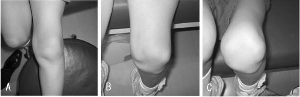
Type: Clinical photographs illustrating habitual patellar dislocation during knee flexion. Anatomical Region: Left knee joint. Description: A series of three monochrome clinical images (A, B, and C) demonstrating the progression of patellar tracking as the knee moves from extension into flexion. Image A shows the knee in extension, where the patella maintains a seemingly normal anatomical alignment within the trochlear groove. Image B captures the knee at approximately 90 degrees of flexion, revealing a clear lateral subluxation of the patella. Image C shows the knee at 120 degrees of flexion, where the patella is significantly dislocated laterally, creating a prominent soft tissue protrusion on the lateral aspect of the knee and an abnormal joint contour. Educational Focus: This series demonstrates the clinical manifestation of flexion-dependent patellar instability or habitual dislocation, highlighting how dynamic physical examination is essential when static extension views appear normal. Clinical Relevance: Useful for diagnosing patellofemoral tracking disorders, medial parapatellar instability, or high sulcus angles.
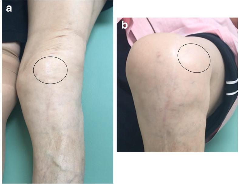
This composite of two clinical photographs illustrates dynamic patellar instability in a patient with a history of total knee arthroplasty (TKA). Image 'a' shows the left knee in a state of full extension. In this position, the patella (highlighted by a black circle) appears reduced and centered within the trochlear groove. Image 'b' demonstrates the same knee during flexion, where the patella (highlighted by a black circle) exhibits a clear lateral dislocation, moving out of its normal anatomical alignment toward the lateral aspect of the joint. Long surgical scars are visible on the anterior aspect of the knee and proximal tibia in both views, consistent with previous orthopedic procedures. Surface anatomy also reveals prominent venous vascularity in the lower leg and skin tension changes between extension and flexion. These findings are clinically characteristic of habitual patellar dislocation, often associated with component malalignment or soft tissue imbalance following knee replacement surgery.
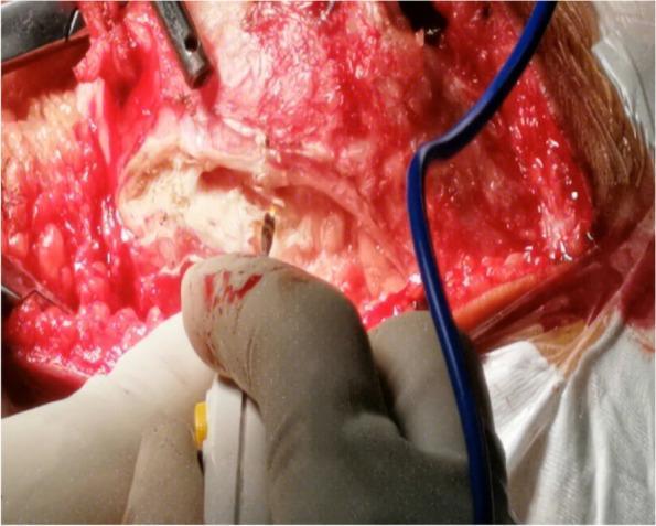
This clinical photograph captures an intraoperative view of an open orthopedic surgical procedure on the knee joint, specifically demonstrating a lateral soft tissue release for the treatment of habitual patellar dislocation. The surgical field shows a deep midline incision with significant exposure of the underlying musculoskeletal structures. The patella and patellar tendon are visible, surrounded by dissected soft tissues including vastus muscle fibers and connective fascia. Evidence of active surgical manipulation is present through the visible bone surfaces and blood-tinged tissues. A gloved hand is shown in the foreground holding an electrosurgical unit (bovie) to maintain hemostasis or perform further dissection. Additional metallic surgical instruments and a blue suction or monitoring tube are positioned within the operative site. This image serves as a high-level educational example of the complex soft tissue releases required to achieve anatomical patellar reduction in adolescent patients.
Habitual (Obligatory) Patellar Dislocation - Why Flexion Dislocates and Extension Reduces
The Core Problem: Quadriceps Mechanism Contracture
"The underlying pathologic condition of congenital or habitual dislocation of the patella is contracture of the quadriceps mechanism" - specifically the vastus lateralis, iliotibial band, and lateral capsule.
Why the Patella DISLOCATES in Flexion
-
Patella is "free" before the groove captures it
- In early flexion (0-30°), the patella is still above the trochlear groove - it has not yet engaged the bony walls of the groove. At this point there is no osseous constraint holding it in place.
-
Contracted lateral structures become taut in flexion
- As the knee flexes, the quadriceps-patellar complex is pulled proximally and laterally by the contracted vastus lateralis and iliotibial band. These structures physically cannot lengthen, so they drag the patella outward.
- Think of it like a tight bowstring on the lateral side: it shortens further as the knee bends and yanks the patella laterally.
-
Q-angle vector worsens in flexion
- In flexion, the tibial tuberosity moves relatively lateral (tibia externally rotates), increasing the effective Q-angle. This increases the lateral pull vector on the patella.
-
Trochlea cannot hold it
- Because the lateral retinaculum and iliotibial band are contracted and the medial structures (MPFL, VMO) are stretched and weak, the net force pulls the patella off the lateral femoral condyle. The patella jumps out laterally - often at 30-90° of flexion.
Why the Patella REDUCES in Extension
-
Quadriceps pull reverses direction in extension
- As the knee extends, the quadriceps contract to straighten the knee. This creates a straight superior pull on the patella along the long axis of the femur - not a lateral vector. This force is directed right into the trochlear groove.
-
Trochlear groove "catches" the patella
- Near full extension, the patella descends back into the trochlear groove. Even a shallow or dysplastic groove can reduce the patella when the lateral deforming forces are momentarily relieved.
-
Contracted structures are relatively "relaxed" in extension
- In the extended position, the lateral retinaculum and iliotibial band are at their most relaxed length. The tight lateral band that was dragging the patella sideways in flexion no longer has enough tension to override the groove.
-
Gravity and the straight-leg position assist
- With the leg extended, there is no flexion moment pulling the lateral structures taut. The patella passively falls back into position.
Visual Summary
Analogy to Remember
Key Pathological Anatomy (Campbell's Operative Orthopaedics, Block 11)
| Structure | Abnormality | Effect |
|---|---|---|
| Vastus lateralis | Contracted, fibrotic | Pulls patella laterally in flexion |
| Iliotibial band | Tight, abnormally attached to patella | Acts as lateral tether |
| Lateral retinaculum | Contracted | Resists medial translation |
| VMO (vastus medialis oblique) | Weak, stretched | Inadequate medial counter-force |
| MPFL | Attenuated | No passive medial restraint |
| Trochlear groove | May be shallow (dysplastic) | Poor bony containment |