
Overview of glaucoma
"glaucoma"[MeSH Terms]
glaucomatous optic disc cupping
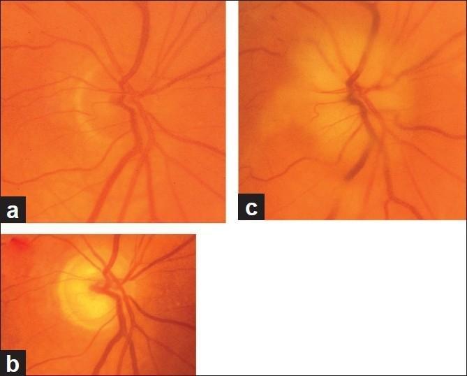
This series of fundus photographs illustrates the longitudinal progression of Arteritic Anterior Ischemic Optic Neuropathy (A-AION) in a right eye. Panel (a) shows the baseline state before disease onset, characterized by a pinkish-orange optic disc with a 'disc at risk' morphology, featuring a shallow or absent physiological central cup. Panel (b) depicts the acute phase one week after the onset of A-AION, demonstrating classic 'chalky white' optic disc edema. The disc is significantly swollen and elevated, with blurred margins and complete obscuration of the central cup. Panel (c) shows the chronic state four months later, following the resolution of edema. The optic disc now exhibits severe optic atrophy and prominent glaucomatous-like cupping. The cup-to-disc ratio has increased significantly to approximately 0.8, representing a deep central excavation and pallor of the neuroretinal rim. This sequence highlights the characteristic visual evolution from acute infarction to pseudo-glaucomatous cupping typical of A-AION.
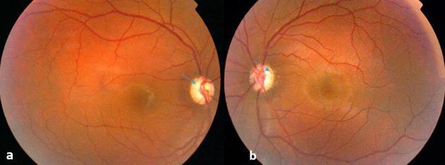
This image consists of two clinical fundus photographs showing the right (a) and left (b) eyes of a patient with methanol toxicity. The primary diagnostic finding is bilateral, near-total optic disc cupping, indicated by large, deep central excavations within the optic nerves (blue arrows). Despite the significant cupping, the neuroretinal rims retain a pinkish hue, indicating a lack of overt pallor or classic optic atrophy at this stage. The retinal background shows a normal orange-red coloration with well-defined vasculature. The retinal vessels appear to emerge from the periphery of the optic disc due to the deep cupping, a phenomenon known as 'bayoneting' or displacement. There are no visible retinal hemorrhages, exudates, or cotton-wool spots. This visual is an educational example of toxic optic neuropathy where structural damage (cupping) precedes or presents differently than typical glaucomatous or atrophic changes, highlighting the clinical significance of methanol's impact on the optic nerve head.
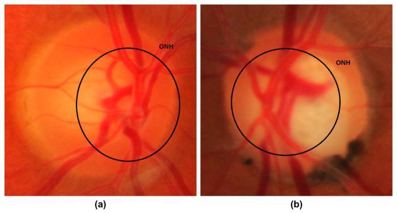
This medical visual consists of two side-by-side color fundus photographs focused on the optic nerve head (ONH), illustrating a comparison between a normal eye and a glaucomatous eye. Image (a) depicts a healthy optic disc with clear margins, a healthy neuroretinal rim, and a standard cup-to-disc ratio (CDR). The retinal vasculature exhibits a normal branching pattern radiating from the center. Image (b) shows signs of glaucoma, characterized by significant optic disc 'cupping.' The central optic cup is noticeably enlarged and pale, resulting in an increased CDR. The margins appear less defined, and the vessel arrangement shows lateral displacement, typical of glaucomatous remodeling. There are visible peripapillary changes, including darker irregular areas at the lower portion of the disc, which may represent peripapillary atrophy (PPA) or pigmentary changes. This comparison serves as an educational tool for ophthalmology to identify key diagnostic clinical signs of glaucomatous optic neuropathy, specifically the enlargement of the optic cup and the thinning of the neuroretinal rim.
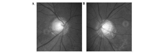
This diagnostic image consists of two grayscale fundus photographs (labeled A and B) showing the bilateral optic discs of a patient with optic atrophy following a basal ganglia hemorrhage. Panel A (right eye) demonstrates significant optic disc pallor and marked excavation of the optic cup. There is a focal loss of the neural rim, particularly evident in the nasal and superior-nasal sectors. Panel B (left eye) reveals a similar pattern of advanced cupping and diffuse pallor, but with a characteristic loss of the neuroretinal rim localized to the temporal region. In both images, retinal vasculature emerges from the deeply excavated cups, though the vessels themselves appear patent. This presentation of non-glaucomatous optic disc cupping and selective rim loss (nasal in one eye, temporal in the other) is clinically significant for localizing neurological lesions along the visual pathway, such as those causing homonymous hemianopia. The visual illustrates the morphological differences between typical glaucomatous changes and retrograde axonal degeneration secondary to intracranial pathology.
Definition and pathophysiology
- The main site of damage is the lamina cribrosa, where the optic nerve axons leave the eye. - Goldman-Cecil Medicine
- The ciliary body's nonpigmented epithelium makes aqueous humor. It drains through the trabecular meshwork and the uveoscleral pathways. When drainage doesn't match production, IOP rises. If the pressure is high enough or lasts long enough, ganglion cells die. - Goldman-Cecil Medicine
- Axon loss shows up at the optic disc as a larger cup, which is recorded as a higher cup-to-disc ratio. - Goldman-Cecil Medicine
- IOP is not the whole story. About 10% of glaucoma occurs with normal IOP. Other proposed mechanisms are mechanical stress on the nerve, reduced optic nerve perfusion, and blocked axoplasmic flow at the lamina cribrosa. - Textbook of Family Medicine 9e, p. 375
Risk factors
- Higher IOP (above about 21 mm Hg is "ocular hypertension", but no pressure is fully protective)
- African ancestry
- Older age
- Low blood pressure
- Family history or genetic predisposition
- Myopia
- Disc hemorrhage
- Anterior segment abnormalities
- Diabetes
- Corticosteroid use
Epidemiology
- Glaucoma causes at least 10% of blindness in the US. It is four times more common in African Americans, who are also eight times more likely to go blind from it. - Textbook of Family Medicine 9e
- Primary open-angle glaucoma (POAG) makes up about two thirds of US cases. Angle-closure is the most common type in Asia. - Textbook of Family Medicine 9e
Main types
| Type | Key features |
|---|---|
| Primary open-angle (POAG) | The angle is open. It is painless and gradual. Peripheral field is lost first, and central vision is lost late. Early symptoms can be trouble reading, reduced contrast sensitivity, or glare. |
| Angle-closure | The anatomic barrier to outflow is relative or absolute. It tends to occur in hyperopic eyes with shallow anterior chambers. Acute attacks bring severe pain, nausea and vomiting, a red eye, and a mid-dilated, nonreactive pupil. IOP can exceed 60 mm Hg. Pupil dilation can trigger it. |
| Secondary | Causes include pseudoexfoliation (the most common), steroids, trauma, inflammation, tumors, hemorrhage, and elevated venous pressure (e.g., Sturge-Weber). Topiramate can cause acute glaucoma through uveal effusions. |
| Congenital/developmental | Examples are congenital glaucoma, aniridia, and Sturge-Weber. These often need surgery, such as drainage devices. |
Diagnosis
- Tonometry: Goldmann applanation is the standard. Corneal thickness can distort the reading.
- Optic disc examination: Look for cupping, a high cup-to-disc ratio, and rim thinning.
- Automated perimetry: The characteristic visual field loss confirms the diagnosis. - Goldman-Cecil Medicine
- Gonioscopy assesses the angle. This comes from my own knowledge, not the textbook passages I pulled.
- OCT of the retinal nerve fiber layer is commonly used to follow progression. This also comes from my own knowledge.
- Family medicine texts recommend tonometry at least every 3 years starting at age 35. The US Preventive Services Task Force considers the evidence insufficient to assess the benefit of screening for open-angle glaucoma. - Textbook of Family Medicine 9e; Goldman-Cecil Medicine
Management
- Prostaglandin analogues: latanoprost, travoprost, once daily
- Beta-blockers: timolol
- Carbonic anhydrase inhibitors: dorzolamide
- Alpha-agonists: brimonidine
- Rho-kinase inhibitors: netarsudil
- Laser trabeculoplasty can be considered first-line. It often controls IOP for years without drops.
- In resistant cases, surgery is trabeculectomy or a tube shunt.
- Give topical timolol and systemic acetazolamide (250 to 500 mg IV, or oral if IV is unavailable) to lower IOP.
- Perform laser peripheral iridotomy to bypass the pupillary block.
- Treat the fellow eye prophylactically with iridotomy.
- Lens extraction is another option, because age-related lens growth contributes to angle closure.
Recent evidence check
- A 2026 systematic review of cancer and glaucoma (PMID 42228376)
- A 2026 systematic review on variability in how glaucoma is diagnosed in randomized trials (PMID 41474870). It argues for standardized diagnostic criteria.
- A 2024 meta-analysis linking glaucoma to Alzheimer's disease risk (PMID 39465755)
Explain the types of glaucoma
open-angle versus angle-closure glaucoma anterior chamber angle diagram
acute angle-closure glaucoma
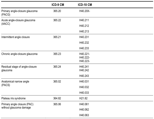
<table><thead><tr><th></th><th>ICD-9 CM</th><th>ICD-10 CM</th></tr></thead><tbody><tr><td>Primary angle-closure glaucoma (PACG)</td><td>365.20</td><td>H40.20X-</td></tr><tr><td>Acute angle-closure glaucoma (AACC)</td><td>365.22</td><td>H40.211<br>H40.212<br>H40.213</td></tr><tr><td>Intermittent angle closure</td><td>365.21</td><td>H40.231<br>H40.232<br>H40.233</td></tr><tr><td>Chronic angle-closure glaucoma</td><td>365.23</td><td>H40.221-<br>H40.222-<br>H40.223-</td></tr><tr><td>Residual stage of angle-closure glaucoma</td><td>365.24</td><td>H40.241<br>H40.242<br>H40.243</td></tr><tr><td>Anatomical narrow angle (PACS)</td><td>365.02</td><td>H40.031<br>H40.032<br>H40.033</td></tr><tr><td>Plateau iris syndrome</td><td>364.82</td><td>H21.82</td></tr><tr><td>Primary angle closure (PAC) without glaucoma damage</td><td>365.06</td><td>H40.061<br>H40.062<br>H40.063</td></tr></tbody></table>
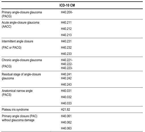
ICD-10 CM
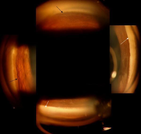
A composite clinical photograph showing post-operative gonioscopy of the iridocorneal angle in a patient with a history of acute angle-closure glaucoma and microspherophakia. The image depicts four quadrants of the anterior chamber angle of the right eye. In the inferior and nasal quadrants (indicated by black arrows), the drainage angle is partially opened, showing increased patency compared to the pre-operative state, though normal anatomical structures remain somewhat indistinct. Conversely, the superior and temporal quadrants (indicated by white arrows) demonstrate persistent synechial closure, characterized by peripheral anterior synechiae (PAS) where the iris tissue is adhered to the trabecular meshwork or cornea, obscuring the angle recess. This visual documentation illustrates the uneven anatomical recovery of the angle following surgical intervention (pars-plana lensectomy and vitrectomy) and is significant for monitoring intraocular pressure and assessing the risk of chronic angle-closure glaucoma.
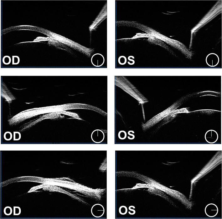
Six-panel ultrasound biomicroscopy (UBM) cross-sectional images of the right eye (OD) and left eye (OS) demonstrating secondary angle-closure glaucoma pathophysiology. The images show bilateral ciliochoroidal effusions, characterized by hypoechoic fluid-filled spaces between the ciliary body and the sclera. Key anatomical alterations include the complete disappearance of the ciliary sulcus and significant anterior displacement of the lens-iris diaphragm. This forward shift of internal ocular structures results in a shallow anterior chamber and complete closure of the anterior chamber angle (iridocorneal angle). The UBM modality highlights the underlying mechanisms of pupillary and non-pupillary block in the anterior segment. Such findings are clinically relevant for diagnosing conditions like uveal effusion syndrome, drug-induced angle closure (e.g., topiramate), or malignant glaucoma, where the posterior segment pressure or fluid accumulation forces the iris and lens forward, risking acute intraocular pressure elevation.
Classification at a glance
| Open-angle | Angle-closure | |
|---|---|---|
| Primary | POAG, including normal-tension glaucoma | Primary angle-closure (acute, subacute, chronic) |
| Secondary | Pseudoexfoliation, pigmentary, phacolytic, ghost cell, steroid, raised episcleral venous pressure | Neovascular, tumor-related, lens-induced, malignant glaucoma, topiramate-related |
| Congenital/developmental | Primary congenital glaucoma, aniridia, Sturge-Weber | Not covered here |
The mechanism that defines each group
- Open-angle: Aqueous humor has full access to the trabecular meshwork. IOP rises because resistance to outflow is increased. The cause is unknown in primary disease, and genetics probably contributes. GWAS has identified about 40 associated genes. - Robbins, Cotran & Kumar Pathologic Basis of Disease
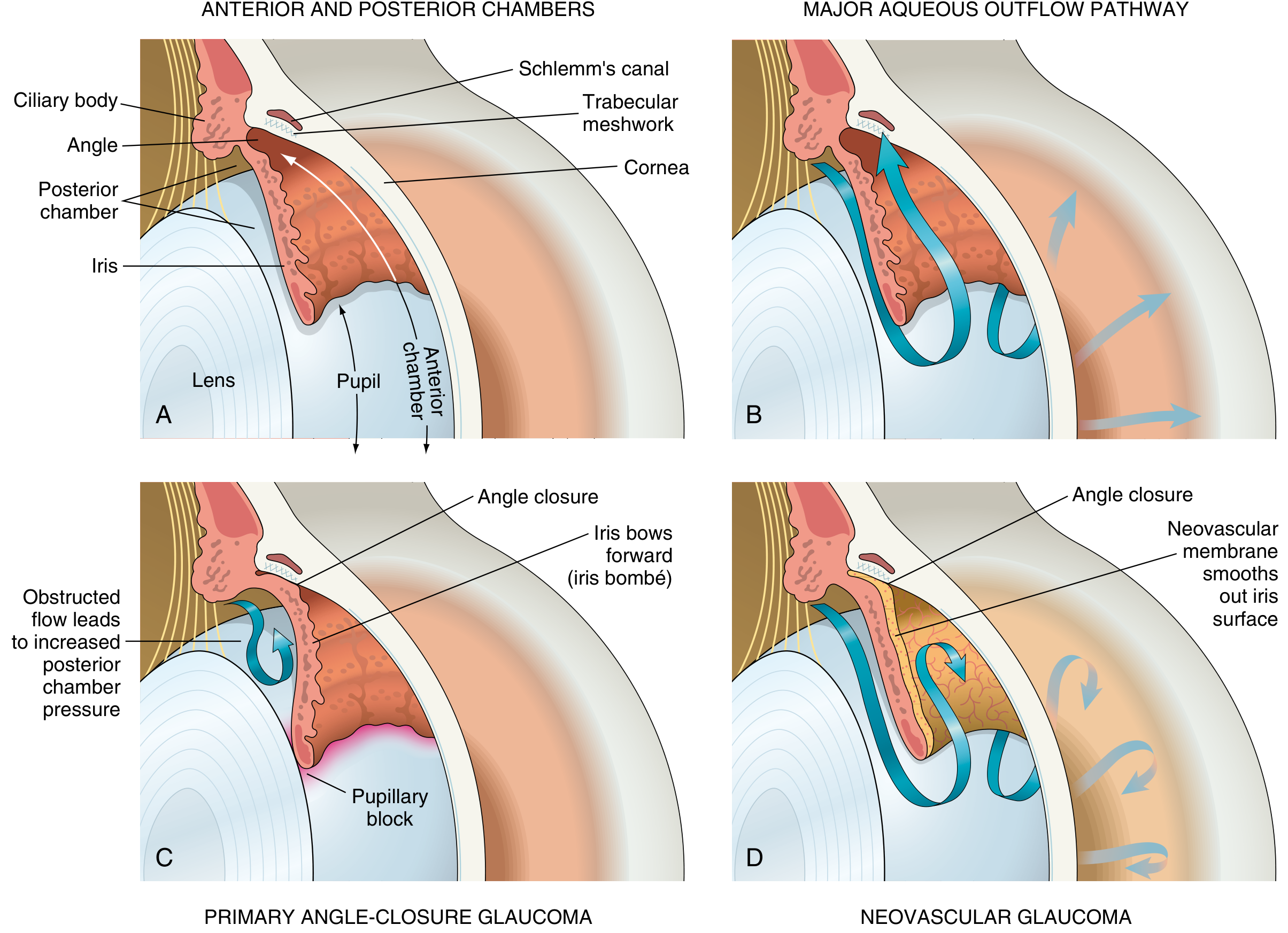
- Angle-closure: The peripheral iris sits against the trabecular meshwork and physically blocks outflow. - Robbins, Cotran & Kumar Pathologic Basis of Disease
1. Primary open-angle glaucoma (POAG)
- Definition: A chronic, progressive optic neuropathy of adult onset. It has an open angle, retinal nerve fiber layer thinning, glaucomatous disc damage, and typical field loss. There are no signs of a secondary cause. - Kanski's Clinical Ophthalmology
- Epidemiology: It is the most common form in White, Hispanic/Latino and Black individuals, and prevalence is especially high in Black individuals. - Kanski's Clinical Ophthalmology
- Presentation: It is usually silent. Early symptoms can be trouble reading, loss of contrast sensitivity, or glare. Peripheral field loss may be considerable before the patient notices. - Goldman-Cecil Medicine
- Natural history: In the Early Manifest Glaucoma Trial, untreated field loss averaged −1.31 dB/year in high-tension glaucoma. The rate was −0.36 dB/year in normal-tension glaucoma and −3.13 dB/year in pseudoexfoliation glaucoma. - Kanski's Clinical Ophthalmology
- Normal-tension glaucoma: This is the same optic nerve and field damage with statistically normal IOP. It makes up about 10% of cases. - Robbins; Textbook of Family Medicine 9e
2. Primary angle-closure glaucoma (PACG)
- Epidemiology: Worldwide it accounts for up to half of all glaucoma. It is especially common in people of Asian descent. - Kanski's Clinical Ophthalmology
- Anatomy: It typically occurs in eyes with a shallow anterior chamber, often in hyperopic people. The lens enlarges with age and pushes the iris forward. - Goldman-Cecil Medicine
- Mechanism (pupillary block):
- The iris margin touches the lens, and aqueous is trapped in the posterior chamber.
- Posterior chamber pressure bows the peripheral iris forward (iris bombé).
- The iris covers the trabecular meshwork and IOP rises sharply. - Robbins
- Acute attack:
- Severe, poorly localized eye pain, nausea and vomiting (which can mimic an acute abdomen), and a red eye with a mid-dilated, nonreactive pupil.
- IOP can exceed 60 mm Hg.
- Pupil dilation can trigger it. - Goldman-Cecil Medicine
- Complications include corneal edema and anterior subcapsular lens opacities. - Robbins
- Other forms: Symptoms may also develop slowly with few specific features (subacute or chronic disease). The acute presentation also has a differential, including lens-induced closure, malignant glaucoma, neovascular glaucoma, and hypertensive uveitis such as Posner-Schlossman syndrome. - Goldman-Cecil Medicine; Kanski's Clinical Ophthalmology
- Treatment: Lower IOP urgently with topical timolol and systemic acetazolamide. Then perform laser iridotomy, usually prophylactically in the other eye too. Lens extraction is another option. - Goldman-Cecil Medicine
3. Secondary open-angle glaucoma
- Pseudoexfoliation: This is the most common secondary open-angle type. Fibrillar material deposits throughout the anterior segment and clogs outflow. It follows a more aggressive course than POAG. - Robbins; Goldman-Cecil Medicine
- Pigmentary glaucoma: Iris pigment granules clog the meshwork. - Robbins
- Phacolytic glaucoma: High-molecular-weight lens proteins clog the meshwork. - Robbins
- Ghost cell glaucoma: Senescent red cells after trauma clog the meshwork. - Robbins
- Melanolytic glaucoma: Necrotic tumors release particles that clog the meshwork. - Robbins
- Raised episcleral venous pressure: Causes are Sturge-Weber syndrome and carotid-cavernous fistula. - Robbins
- Steroid glaucoma: This follows weeks of topical or, rarely, systemic steroids. Stopping the steroid reverses the IOP rise, but it can take 2 to 3 months or longer, and vision lost meanwhile may be permanent. - Textbook of Family Medicine 9e
4. Secondary angle-closure glaucoma
- Neovascular glaucoma: Chronic retinal ischemia (diabetic retinopathy, retinal vein occlusion) upregulates VEGF. VEGF drives fibrovascular membranes over the iris. The membranes contract, pull the iris onto the meshwork, and form peripheral anterior synechiae. Necrotic tumors such as retinoblastoma can do the same. - Robbins
- Tumor-related: Ciliary body tumors can mechanically push the iris onto the meshwork. - Robbins
- Lens-induced: The lens is swollen or subluxated. - Kanski's Clinical Ophthalmology
- Malignant glaucoma (aqueous misdirection): It is especially likely after intraocular surgery. - Kanski's Clinical Ophthalmology
- Drug-induced: Topiramate can cause acute angle closure through uveal effusions. - Textbook of Family Medicine 9e
- Secondary glaucoma can also follow intraocular hemorrhage, trauma, or inflammation. Iridocorneal endothelial syndrome may only appear in adulthood. - Goldman-Cecil Medicine
5. Congenital and developmental glaucoma
- Primary congenital glaucoma: It has no associated ocular or systemic disorders. It results from incomplete differentiation of the trabecular meshwork in the embryo (goniodysgenesis). - The Wills Eye Manual
- Presentation and outcome: Raised IOP in infancy can cause buphthalmos. Immediate ophthalmology consultation is needed. Surgery succeeds in about 90% of cases. - Textbook of Family Medicine 9e
- Aniridia: Medical treatment is usually inadequate. Trabeculectomy often fails, and glaucoma drainage devices give the best long-term control. - Kanski's Clinical Ophthalmology
- Sturge-Weber syndrome:
- About 30% of patients develop glaucoma on the same side as the facial hemangioma.
- In 60% of those, IOP rises before age 2.
- Infants have trabeculodysgenesis. Older patients have raised episcleral venous pressure from an episcleral hemangioma. - Kanski's Clinical Ophthalmology
Key points
- Open versus closed describes the angle. Primary versus secondary describes whether another disease causes it.
- Primary angle-closure can be an emergency. POAG is slow and painless.
- Secondary glaucomas often need the underlying cause treated as well as the IOP.