
Essay questions with neat labelled diagram and flowchart 1. Describe Ascending track 2. Explain corticospinal track 3. Functions and connections of thalamus 4. Structure connections and functions of cerebral cortex 5. Explain Visual pathway with the lesions 6. Organ of corti
Reading File
Finding Sources
Finding Sources
Finding Sources
Finding Sources
Finding Sources
Finding Sources
Finding Sources
Finding Sources
Finding Sources
Finding Sources
Reading File
Reading File
Reading File
Reading File
Reading File
Reading File
Reading File
Reading File
Loading Image
Loading Image
Loading Image
Loading Image
Searching the Web
visual pathway optic tract lesions diagram homonymous hemianopia bitemporal hemianopia
Loading Image
Loading Image
Reading File
Loading Image
Loading Image
Loading Image
Searching the Web
dorsal column medial lemniscus pathway diagram ascending tracts spinal cord
Loading Image
Here are comprehensive essay answers for all six topics with labelled diagrams and flowcharts.
1. Ascending Tracts
Introduction
Ascending (sensory) tracts carry sensory information from the periphery, through the spinal cord, to the brain. There are two principal systems: the Dorsal Column - Medial Lemniscal (DCML) pathway and the Anterolateral (Spinothalamic) pathway.
A. Dorsal Column - Medial Lemniscal (DCML) Pathway
Sensations Carried
- Fine/discriminative touch
- Vibration sense
- Conscious proprioception (joint position sense)
- Two-point discrimination
- Stereognosis
Three-Neuron Arc
| Order | Cell Body Location | Tract | Crosses |
|---|---|---|---|
| 1st order neuron | Dorsal root ganglion (DRG) | Ipsilateral dorsal column (fasciculus gracilis / cuneatus) | Does NOT cross in cord |
| 2nd order neuron | Nucleus gracilis / nucleus cuneatus (medulla) | Internal arcuate fibers → medial lemniscus (contralateral) | Crosses at sensory decussation in medulla |
| 3rd order neuron | VPL nucleus of thalamus | Thalamocortical radiation | Already crossed |
| Termination | Primary somatosensory cortex (areas 3, 1, 2) | - | - |
Fasciculus Gracilis vs. Cuneatus
| Feature | Fasciculus Gracilis | Fasciculus Cuneatus |
|---|---|---|
| Location in cord | Medial dorsal column | Lateral dorsal column |
| Origin | Below T6 (lower limb, lower trunk) | Above T6 (upper limb, upper trunk, neck) |
| 2nd order synapse | Nucleus gracilis | Nucleus cuneatus |
Flowchart - DCML Pathway
Peripheral receptor (skin, muscle, joint)
        ↓
1st order neuron (DRG cell body)
        ↓
Enters dorsal horn → ascends IPSILATERALLY
 in fasciculus gracilis (below T6)
 or fasciculus cuneatus (above T6)
        ↓
Nucleus gracilis / cuneatus (medulla)
        ↓
Internal arcuate fibers → CROSS (sensory decussation)
        ↓
Medial lemniscus (contralateral ascent through brainstem)
        ↓
VPL nucleus of THALAMUS
        ↓
Thalamocortical radiation (posterior limb of internal capsule)
        ↓
PRIMARY SOMATOSENSORY CORTEX (postcentral gyrus)
Labelled Diagram - DCML Pathway
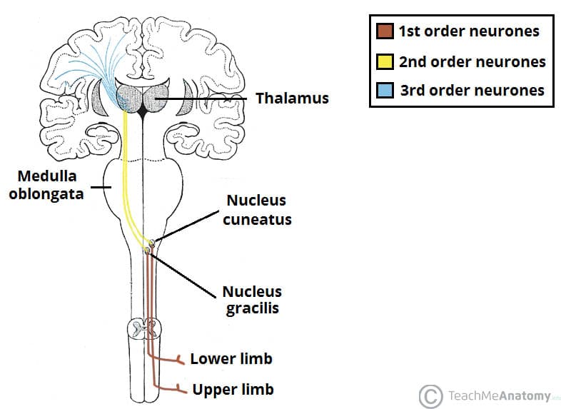
Figure 1 - Dorsal Column Medial Lemniscal Pathway (TeachMeAnatomy)
Clinical Note
A lesion of the DCML on one side causes ipsilateral loss of fine touch, vibration and proprioception below the level of the lesion (since fibers have not yet crossed).
B. Anterolateral (Spinothalamic) Pathway
Sensations Carried
- Pain
- Temperature (crude)
- Crude touch and pressure
Flowchart - Spinothalamic Tract
Peripheral nociceptors / thermoreceptors
        ↓
1st order neuron (DRG cell body)
        ↓
Enters dorsal horn → synapses in REXED LAMINAE I, II, V
        ↓
2nd order neuron CROSSES immediately in anterior commissure
        ↓
Ascends CONTRALATERALLY as spinothalamic tract
(in anterolateral white matter)
        ↓
VPL nucleus of THALAMUS
        ↓
Thalamocortical fibers
        ↓
PRIMARY SOMATOSENSORY CORTEX
Labelled Diagram - Spinothalamic (DCML also shown)
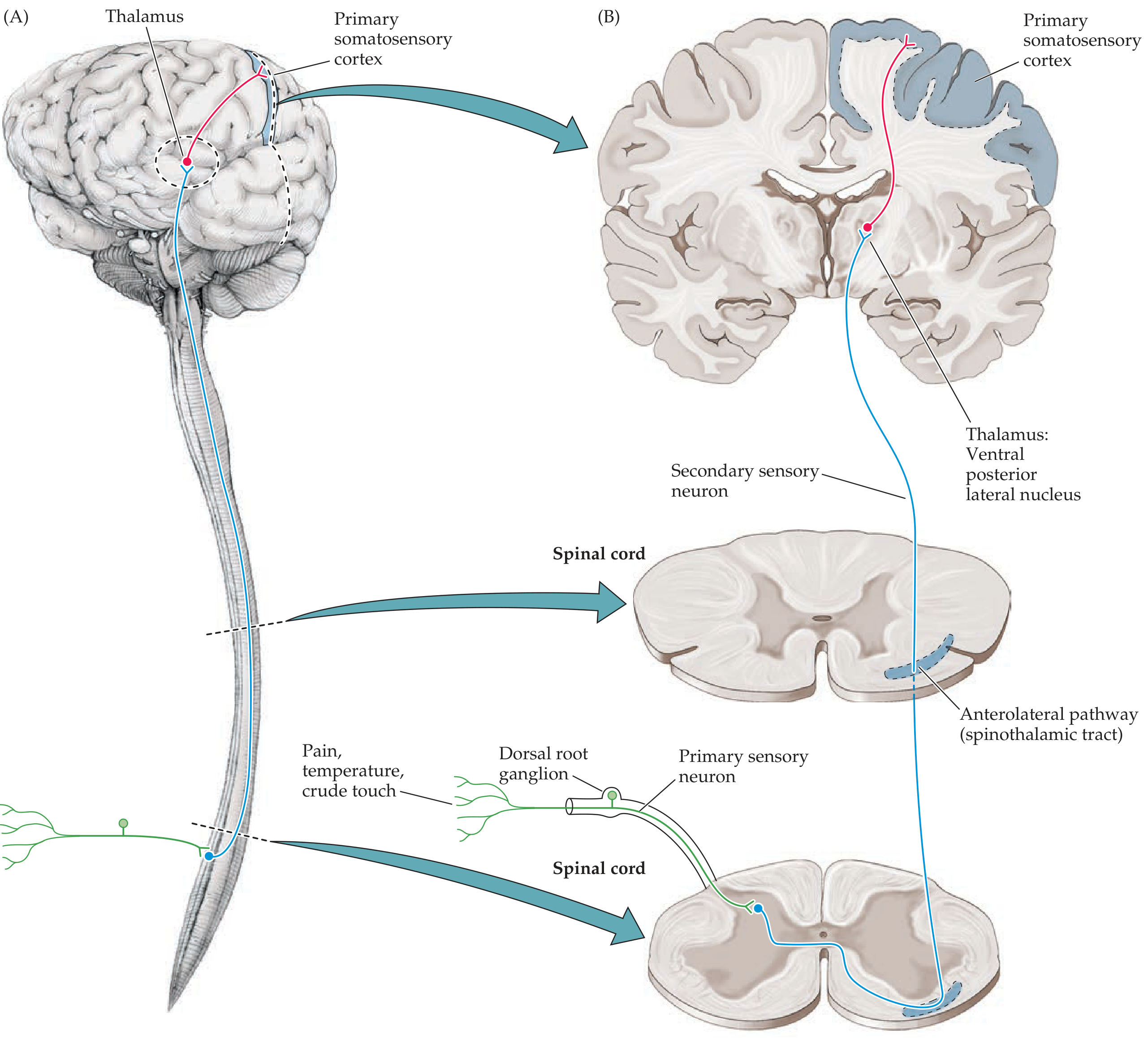
Figure 2 - Spinothalamic (Anterolateral) Sensory Pathway - Neuroanatomy through Clinical Cases
Key Difference between the Two Pathways
| Feature | DCML | Spinothalamic |
|---|---|---|
| Sensations | Fine touch, vibration, proprioception | Pain, temperature, crude touch |
| Decussation level | Medulla | Spinal cord (1-2 levels above entry) |
| 2nd order neuron location | Dorsal column nuclei (medulla) | Dorsal horn of spinal cord |
| Somatotopy in cord | Medial = sacral; lateral = cervical | Lateral = sacral; medial = cervical |
Clinical Relevance: Brown-Séquard Syndrome (Hemisection of cord)
- Ipsilateral loss of fine touch, vibration, proprioception (DCML)
- Contralateral loss of pain and temperature (spinothalamic), 1-2 levels below lesion
2. Corticospinal Tract
Introduction
The corticospinal tract (CST), also called the pyramidal tract, is the most important descending motor pathway. It controls voluntary movement of the limbs and is a two-neuron system: the Upper Motor Neuron (UMN) and the Lower Motor Neuron (LMN).
Origin
Fibers originate approximately from:
- 30% - Primary motor cortex (precentral gyrus, Brodmann area 4)
- 30% - Premotor and supplementary motor areas (area 6)
- 40% - Somatosensory cortex (parietal lobe, areas 3, 1, 2, 5, 7)
Large pyramidal cells in cortical layer 5, including Betz cells (~3% of fibers, largest neurons in the human nervous system, ~60 µm diameter), constitute the most important efferent component.
Course - Flowchart
PRIMARY MOTOR CORTEX (precentral gyrus, area 4)
        ↓
Corona radiata (upper cerebral white matter)
        ↓
POSTERIOR LIMB OF INTERNAL CAPSULE
 (face fibers: anterior; arm: middle; leg: posterior)
        ↓
BASIS PEDUNCULI (cerebral peduncles, midbrain)
 (middle 1/3: CST; medial 1/3: face → arm; lateral 1/3: leg)
        ↓
PONS (dispersed fascicles, ventral pons)
        ↓
MEDULLARY PYRAMIDS (ventral medulla)
        ↓
PYRAMIDAL DECUSSATION (cervicomedullary junction)
 ~85% cross → LATERAL CORTICOSPINAL TRACT (contralateral spinal cord)
 ~15% do NOT cross → VENTRAL CORTICOSPINAL TRACT (ipsilateral)
        ↓
Synapse on INTERNEURONS (mainly) or LMNs in anterior horn
        ↓
SKELETAL MUSCLE (voluntary movement)
Labelled Diagrams
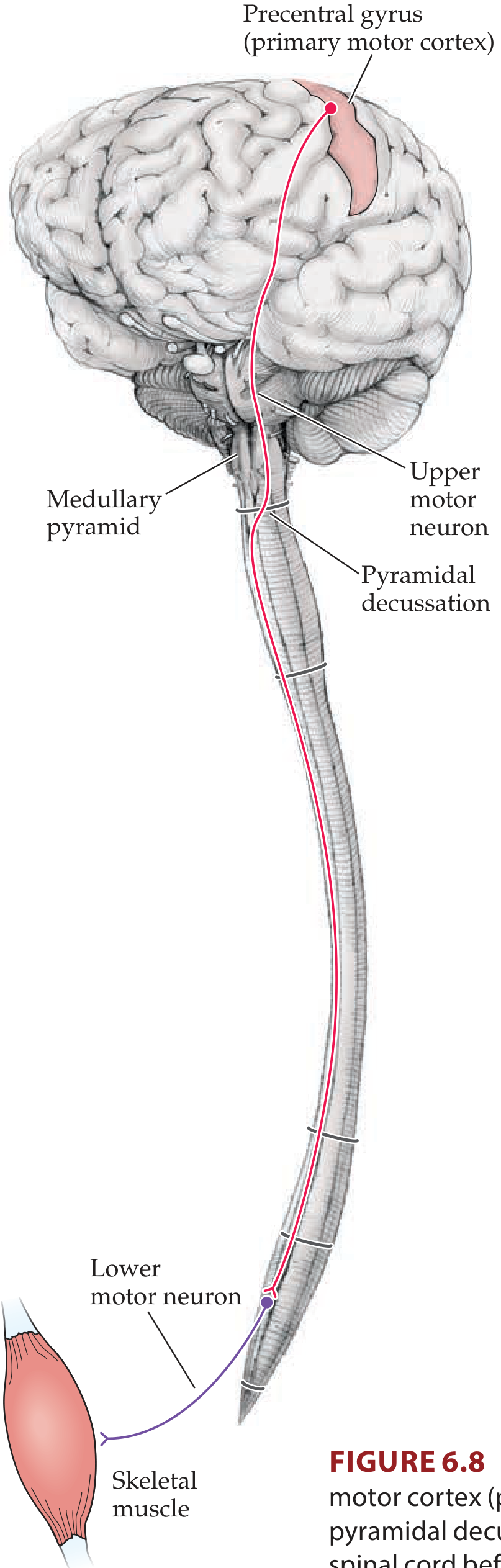
Figure 3 - Lateral Corticospinal Tract (Neuroanatomy through Clinical Cases)
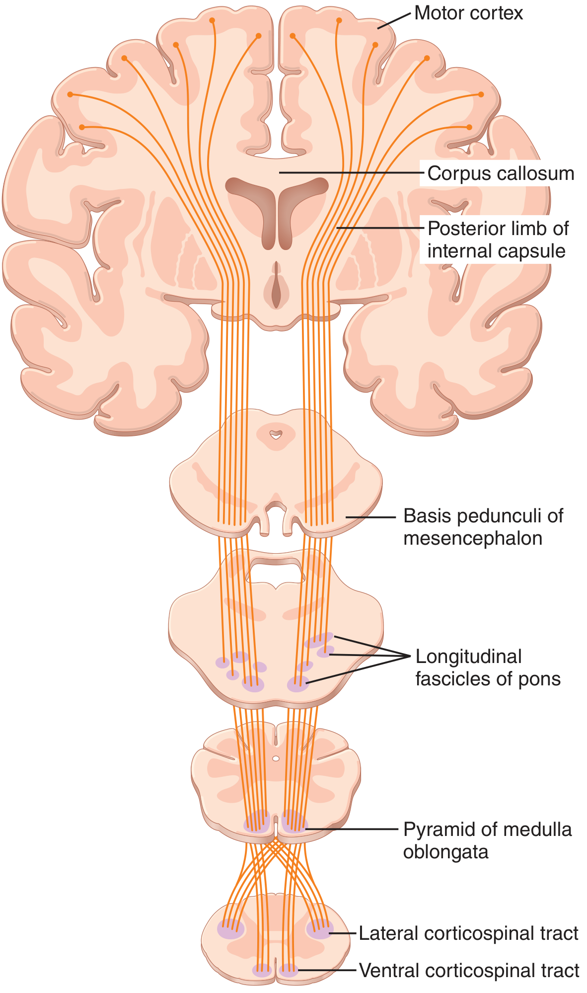
Figure 4 - Corticospinal (Pyramidal) Tract in full (Guyton & Hall)
Key Facts
| Feature | Detail |
|---|---|
| Total fibers per CST | >1 million |
| Large Betz cell fibers | ~34,000 per tract (~3%); 60 µm; conduct at 70 m/s |
| % that cross | ~85% (lateral CST); ~15% ventral CST (many cross later in cord) |
| Main synapse | Intermediary neurons → anterior horn LMNs |
| Somatotopy in lateral column | Upper limb medial; lower limb lateral |
UMN vs. LMN Lesion
| Feature | UMN (above decussation) | LMN (anterior horn/nerve) |
|---|---|---|
| Tone | Increased (spasticity) | Decreased (flaccidity) |
| Reflexes | Hyperreflexia | Hyporeflexia / absent |
| Plantar reflex | Extensor (Babinski +ve) | Flexor or absent |
| Wasting | Minimal (disuse only) | Marked (denervation) |
| Fasciculations | Absent | Present |
3. Functions and Connections of the Thalamus
Introduction
The thalamus is a paired egg-shaped gray matter structure forming the dorsal part of the diencephalon. It sits deep in the cerebral white matter, above the brainstem and behind the basal ganglia. Together with the hypothalamus and epithalamus it forms the diencephalon.
Structure
- Divided by an internal Y-shaped white matter sheet (internal medullary lamina) into multiple nuclear groups
- Surrounded laterally by the reticular nucleus (a thin shell of inhibitory GABAergic neurons)
Major Nuclear Groups
| Nuclear Group | Key Nuclei | Connections / Function |
|---|---|---|
| Anterior | Anterior nucleus | Limbic system (Papez circuit), memory, emotion |
| Medial | Mediodorsal (MD) nucleus | Prefrontal cortex, limbic system; cognition, affect |
| Lateral - dorsal tier | Lateral dorsal, lateral posterior, pulvinar | Association cortex, visual & spatial processing |
| Lateral - ventral tier (relay) | VPL (ventral posterolateral) | Body somatic sensation (DCML + spinothalamic) → postcentral gyrus |
| VPM (ventral posteromedial) | Face sensation (CN V), taste → postcentral gyrus | |
| VA/VL (ventral anterior / lateral) | Basal ganglia + cerebellum → motor cortex | |
| LGN (lateral geniculate nucleus) | Visual cortex (optic radiations) | |
| MGN (medial geniculate nucleus) | Auditory cortex (Heschl's gyrus) | |
| Intralaminar | Centromedian, parafascicular | Widespread cortical activation (arousal), basal ganglia |
| Reticular | Reticular nucleus | Modulates thalamic output (GABAergic inhibition) |
| Posterior | Pulvinar | Visual association, attention |
Connections - Diagram
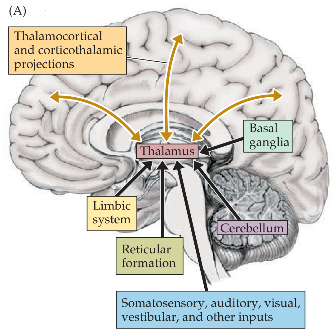
Figure 5 - Thalamic inputs and reciprocal connections (Neuroanatomy through Clinical Cases)
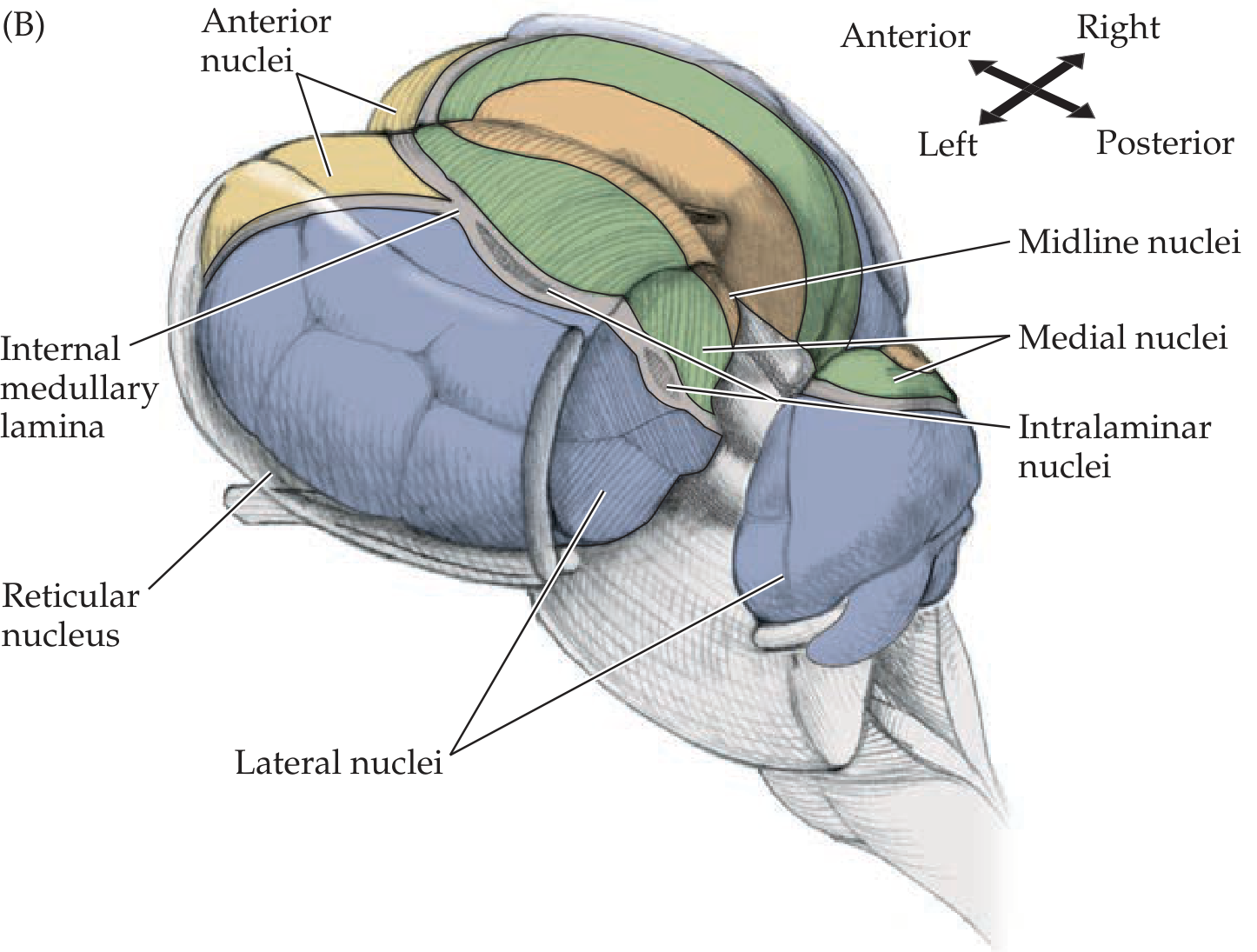
Figure 6 - Major Thalamic Nuclear Divisions (Neuroanatomy through Clinical Cases)
Functions Flowchart
THALAMUS - FUNCTIONS
       |
       ├─ SENSORY RELAY
       │    ├─ VPL → Somatic sensation (body) → Postcentral gyrus
       │    ├─ VPM → Somatic sensation (face) + Taste → Postcentral gyrus
       │    ├─ LGN → Vision → Occipital cortex (area 17)
       │    └─ MGN → Hearing → Temporal cortex (Heschl's gyrus)
       │
       ├─ MOTOR RELAY
       │    └─ VA/VL ← Cerebellum (dentate nucleus) + Basal ganglia
       │            → Motor cortex (area 4 + area 6)
       │
       ├─ LIMBIC / MEMORY
       │    └─ Anterior nucleus ↔ Cingulate gyrus, hippocampus (Papez circuit)
       │
       ├─ AROUSAL / CONSCIOUSNESS
       │    └─ Intralaminar nuclei ← Reticular formation → Widespread cortex
       │
       └─ CORTICAL MODULATION
            └─ Reticular nucleus - gates thalamic output (inhibitory, GABAergic)
Key Points
- All sensory systems (except olfaction) relay in the thalamus before reaching cortex
- Reciprocal connections: every cortical area that receives thalamic input also sends fibers back to the same thalamic nucleus (layer VI of cortex → thalamus)
- Thalamic stroke (VPL infarct): contralateral sensory loss + thalamic pain (Dejerine-Roussy syndrome)
4. Structure, Connections and Functions of the Cerebral Cortex
Introduction
The cerebral cortex is the outermost layer of gray matter of the cerebral hemispheres. It contains >14 billion neurons and 70% of CNS neurons reside here. It is the seat of conscious perception, voluntary movement, language, memory and higher cognitive function.
Gross Structure
- Thrown into folds: gyri (ridges) and sulci (grooves)
- Major sulci divide each hemisphere into 4 lobes: frontal, parietal, temporal, occipital (+ insular cortex)
- Surface area: ~2,500 cm² (most hidden in sulci)
Histological Structure - Layers of Neocortex (6 layers)
| Layer | Name | Major Cell Types | Key Connections |
|---|---|---|---|
| I | Molecular (plexiform) | Few neurons, mostly axons/dendrites | Corticocortical fibers |
| II | External granular | Small pyramidal + granule cells | Short corticocortical |
| III | External pyramidal | Medium pyramidal cells | Corticocortical (long range) |
| IV | Internal granular | Stellate (granule) cells | Main input layer - receives thalamocortical fibers |
| V | Internal pyramidal | Large pyramidal cells (Betz cells in motor cortex) | Main output - corticospinal, corticobulbar, corticostriate |
| VI | Multiform (fusiform) | Spindle-shaped cells | Corticothalamic fibers back to thalamus |
Special Cortical Types
- Granular cortex (koniocortex): thick layer IV, prominent granule cells - sensory areas (postcentral gyrus, primary visual cortex)
- Agranular cortex: thick layer V, no layer IV - motor areas (precentral gyrus)
- Allocortex: 3-layered older cortex (hippocampus, olfactory cortex)
Functional Areas (Brodmann's Areas)
| Area | Brodmann | Lobe | Function |
|---|---|---|---|
| Primary motor cortex | Area 4 | Frontal (precentral gyrus) | Voluntary movement (contralateral body) |
| Premotor cortex | Area 6 | Frontal | Planning of movements |
| Supplementary motor area | Area 6 (medial) | Frontal | Coordination of bilateral movements |
| Broca's area | Areas 44, 45 | Frontal (dominant) | Motor (expressive) speech |
| Primary somatosensory cortex | Areas 3, 1, 2 | Parietal (postcentral gyrus) | Conscious somatic sensation |
| Primary visual cortex | Area 17 | Occipital (calcarine sulcus) | Primary visual perception |
| Visual association cortex | Areas 18, 19 | Occipital | Processing shapes, colors, motion |
| Primary auditory cortex | Areas 41, 42 | Temporal (Heschl's gyrus) | Hearing |
| Wernicke's area | Area 22 | Temporal (dominant) | Auditory comprehension of speech |
| Prefrontal cortex | Areas 9-12, 45-47 | Frontal | Executive function, personality |
Connections
Cortical Connections Flowchart
CEREBRAL CORTEX CONNECTIONS
        |
        ├─ ASSOCIATION FIBERS (within same hemisphere)
        │    ├─ Short (U-fibers): adjacent gyri
        │    └─ Long fasciculi:
        │         ├─ Arcuate fasciculus: frontal ↔ temporal (language)
        │         ├─ Uncinate fasciculus: frontal ↔ temporal (memory/emotion)
        │         ├─ Superior longitudinal fasciculus
        │         └─ Cingulum: cingulate gyrus (limbic)
        │
        ├─ COMMISSURAL FIBERS (between hemispheres)
        │    ├─ Corpus callosum (largest) - connects all lobes
        │    ├─ Anterior commissure - connects temporal lobes
        │    └─ Posterior commissure
        │
        └─ PROJECTION FIBERS (cortex ↔ subcortical structures)
             ├─ Thalamocortical / Corticothalamic (internal capsule)
             ├─ Corticospinal (descending motor)
             ├─ Corticobulbar (to cranial nerve nuclei)
             └─ Corticopontine (to cerebellum via pons)
Key Functions
- Frontal lobe: voluntary motor control, executive function, personality, Broca's speech area (dominant), inhibition of behavior
- Parietal lobe: somatic sensation, spatial awareness, body image; angular gyrus (reading, calculation)
- Temporal lobe: hearing, memory (hippocampus), Wernicke's speech area (dominant), olfaction
- Occipital lobe: vision; dorsal stream (where/how) via parietal, ventral stream (what) via temporal
- Association cortex (70% of neurons): integrates information across modalities; heteromodal association cortex in angular gyrus and prefrontal cortex is responsible for highest cognitive functions
5. Visual Pathway with Lesions
Introduction
The visual pathway carries information from the retina to the primary visual cortex (striate cortex, area 17) in the occipital lobe. Understanding its anatomy allows precise localization of lesions based on the pattern of visual field loss.
Anatomy of the Visual Pathway
Retinal Organization
- Nasal retina receives input from the temporal (lateral) visual field
- Temporal retina receives input from the nasal (medial) visual field
- Macula (central vision): represented disproportionately in the cortex
Course - Flowchart
RETINAL GANGLION CELLS
        ↓
OPTIC NERVE (CN II)
(ipsilateral eye only; carries all visual info from that eye)
        ↓
OPTIC CHIASM (at sella turcica)
 ┌─────────────────────────────────────────┐
 │  Nasal (medial) retinal fibers → CROSS  │
 │  Temporal (lateral) retinal fibers →    │
 │  remain IPSILATERAL                     │
 └─────────────────────────────────────────┘
        ↓
OPTIC TRACT
(carries: ipsilateral temporal + contralateral nasal fibers)
(= all visual info from CONTRALATERAL visual field)
        ↓
LATERAL GENICULATE NUCLEUS (LGN) of thalamus
        ↓
OPTIC RADIATIONS (geniculocalcarine tract)
  ┌────────────────────────────────────────────┐
  │ UPPER fibers → parietal lobe               │
  │   (carry inferior visual field)            │
  │ LOWER fibers → loop through temporal lobe  │
  │   (Meyer's loop - carry superior visual    │
  │   field)                                   │
  └────────────────────────────────────────────┘
        ↓
PRIMARY VISUAL CORTEX (area 17)
Calcarine sulcus, occipital lobe
  Upper bank = inferior visual field
  Lower bank = superior visual field
  Posterior pole = macula (largest representation)
Lesions and Visual Field Defects

Figure 7 - Visual Pathway Lesions and Visual Field Defects (Medbullets/Dominguez)
Lesion Table
| Site | Defect | Cause |
|---|---|---|
| A - Optic nerve (before chiasm) | Monocular blindness (ipsilateral eye) | MS (optic neuritis), ischemia, glaucoma |
| B - Optic chiasm (central) | Bitemporal hemianopia (tunnel vision) | Pituitary adenoma, craniopharyngioma - compresses crossing nasal fibers |
| C / D - Optic tract | Contralateral homonymous hemianopia (incongruous) | Rare: tumor, demyelination |
| E - Meyer's loop (temporal lobe) | Contralateral superior quadrantanopia ("pie in the sky") | Temporal lobe tumor/surgery |
| F - Upper optic radiation (parietal) | Contralateral inferior quadrantanopia ("pie on the floor") | Parietal lobe infarction (MCA superior division) |
| G / H - Entire optic radiation or visual cortex | Contralateral homonymous hemianopia (congruous) | PCA infarct, occipital tumor |
| K - Occipital pole (with PCA occlusion) | Contralateral homonymous hemianopia WITH macular sparing | Posterior cerebral artery infarct (macula has dual blood supply from MCA) |
Key Rule: Pre-chiasmal vs. Post-chiasmal
- Pre-chiasmal lesion (optic nerve): monocular defect in one eye only
- At chiasm: bitemporal heteronymous defect
- Post-chiasmal lesion (optic tract → cortex): contralateral homonymous defect
Congruity
- Optic tract lesions: incongruous (asymmetric defects in both eyes)
- Optic radiation / cortical lesions: congruous (symmetric, identical defects)
6. Organ of Corti
Introduction
The organ of Corti is the sensory transduction apparatus of the inner ear, lying within the cochlea. It converts mechanical vibrations (from sound waves) into electrical nerve impulses transmitted to the CNS via the cochlear division of CN VIII.
Structure of the Cochlea (Context)
The cochlea is a snail-shaped, fluid-filled bony labyrinth with 2.5 turns. In cross-section it has three fluid-filled chambers (scalae):
| Compartment | Fluid | Separated by |
|---|---|---|
| Scala vestibuli | Perilymph | Reissner's membrane (from scala media) |
| Scala media (cochlear duct) | Endolymph | Basilar membrane (from scala tympani) |
| Scala tympani | Perilymph | Basilar membrane |
- Perilymph: high Na⁺, low K⁺ (like ECF)
- Endolymph: high K⁺, low Na⁺ (like ICF) - maintained by stria vascularis in the lateral wall of scala media
Structure of the Organ of Corti
The organ of Corti lies on the basilar membrane and is bathed in endolymph of the scala media.
Components
| Structure | Description |
|---|---|
| Inner hair cells (IHC) | ~3,500; arranged in single row; primary sensory receptors (90-95% of afferent nerve fibers terminate here) |
| Outer hair cells (OHC) | ~12,000; 3-4 rows; electromotility - tune basilar membrane sensitivity |
| Stereocilia | Stiff hairs projecting from hair cells; tips embedded in or touching tectorial membrane |
| Tectorial membrane | Gelatinous membrane overlying the hair cells |
| Basilar membrane | Supports the organ of Corti; narrow & stiff at base (high frequency); wide & floppy at apex (low frequency) |
| Rods of Corti (pillar cells) | Triangular support cells forming the tunnel of Corti |
| Reticular lamina | Rigid plate at the top of hair cells |
| Spiral ganglion | Contains cell bodies of 1st-order cochlear neurons (in modiolus/center of cochlea) |
Labelled Diagram
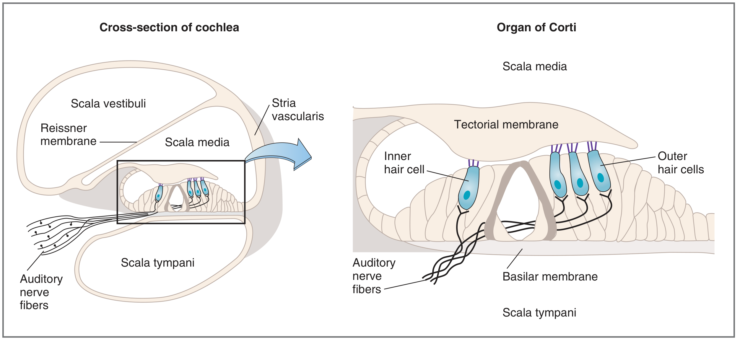
Figure 8 - Structure of the Cochlea and Organ of Corti (Costanzo Physiology)
Mechanism of Auditory Transduction - Flowchart
SOUND WAVES enter external auditory canal
        ↓
TYMPANIC MEMBRANE vibrates
        ↓
OSSICLES (malleus → incus → stapes) transmit vibration
(amplified by lever action + area difference of TM vs. oval window)
        ↓
STAPES pushed into OVAL WINDOW
        ↓
Fluid waves in SCALA VESTIBULI (perilymph)
        ↓
BASILAR MEMBRANE vibrates at specific location
(base = high frequency; apex = low frequency - tonotopy)
        ↓
Organ of Corti moves UP and DOWN
        ↓
SHEARING FORCE between reticular lamina and tectorial membrane
        ↓
STEREOCILIA of hair cells bend
        ↓
TIP LINKS open K⁺/Ca²⁺ mechanosensitive channels
        ↓
K⁺ influx into hair cell (from endolymph) → DEPOLARIZATION
        ↓
Voltage-gated Ca²⁺ channels open at base of hair cell
        ↓
Neurotransmitter (glutamate) released onto cochlear nerve fibers
        ↓
Action potential in SPIRAL GANGLION NEURONS (1st order)
        ↓
Axons form COCHLEAR NERVE (CN VIII) →
        ↓
Cochlear nuclei (dorsal + ventral) in brainstem
        ↓
Superior olivary nucleus (bilateral), inferior colliculus, MGB (medial geniculate body, thalamus)
        ↓
PRIMARY AUDITORY CORTEX (Heschl's gyrus, areas 41, 42 - temporal lobe)
Key Concepts
| Concept | Detail |
|---|---|
| Tonotopy | Base of basilar membrane: high frequency (narrow, stiff); Apex: low frequency (wide, flexible) |
| Inner vs. outer hair cells | IHC = primary sensory transducers; OHC = electromotile (prestin protein), tune sensitivity |
| Endocochlear potential | +80 mV in scala media (from stria vascularis); drives K⁺ into hair cells |
| Tip links | Extracellular filaments between stereocilia tips; gate mechanoelectrical transduction channels |
| Spiral ganglion | Cell bodies of 1st-order afferent neurons in modiolus; ~30,000 axons form cochlear nerve |
| Olivocochlear bundle | Efferent fibers from superior olive → mainly outer hair cells; retrograde control of sensitivity |
Sources
- Neuroanatomy through Clinical Cases, 3rd Edition (Blumenfeld)
- Guyton and Hall Textbook of Medical Physiology (Hall)
- Costanzo Physiology, 7th Edition
- Bradley and Daroff's Neurology in Clinical Practice
- TeachMeAnatomy.info (DCML pathway diagram)
- Medbullets (Visual field defects diagram)
This is a shared conversation. Sign in to Orris to start your own chat.