

IMG_1744.jpg
What is this cell on peripheral smear
analyse_media
Reading File
Finding Sources
Finding Sources
Reading File
Loading Image
The cell being pointed at on the peripheral smear is a Nucleated Red Blood Cell (nRBC) - specifically an Orthochromatic Erythroblast (Normoblast).
Morphological Features (visible in your image)
| Feature | What you see |
|---|---|
| Size | Slightly larger than surrounding RBCs (~8-12 µm) |
| Nucleus | Small, deeply stained, pyknotic (condensed, dark purple-blue), no visible nucleoli |
| Cytoplasm | Eosinophilic/pinkish - similar to surrounding mature RBCs, indicating near-complete hemoglobin synthesis |
| N:C ratio | Low - small nucleus, abundant pink cytoplasm |
| Granules | None |
Compare with the textbook image of erythroblasts/normoblasts:
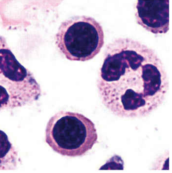
What This Cell Is
The orthochromatic erythroblast (late normoblast) is the last nucleated stage of erythropoiesis. Key facts:
- It has a pyknotic, condensed, inactive nucleus ready for extrusion
- Cytoplasm is predominantly eosinophilic due to high hemoglobin content
- It is no longer capable of mitosis at this stage
- The nucleus is soon ejected; the resulting cell becomes a reticulocyte, then a mature erythrocyte
- Small nuclear fragments left behind after extrusion are called Howell-Jolly bodies
Clinical Significance - Why is an nRBC on a Peripheral Smear?
nRBCs are not normally seen on a peripheral smear in healthy adults. Their presence indicates one of the following:
| Cause | Examples |
|---|---|
| Severe hemolytic anemia | Sickle cell disease, thalassemia major, autoimmune hemolytic anemia |
| Megaloblastic anemia | B12/folate deficiency with ineffective erythropoiesis |
| Hypoxia/stress erythropoiesis | Severe hypoxia, high altitude, cardiopulmonary disease |
| Bone marrow infiltration (leukoerythroblastic reaction) | Myelofibrosis, metastatic carcinoma, leukemia |
| Asplenia | Post-splenectomy (spleen normally clears nRBCs) |
| Neonatal period | Normal finding in newborns |
| Severe sepsis / burns |
Note: In automated CBC analyzers, nRBCs can be miscounted as WBCs, causing falsely elevated WBC counts. The WBC count must be corrected when nRBCs are identified on smear.
Corrected WBC = (Reported WBC × 100) / (100 + number of nRBCs per 100 WBCs)
- Histology: A Text and Atlas with Correlated Cell and Molecular Biology, p. 741-742
This is a shared conversation. Sign in to Orris to start your own chat.