
How hernia happens via deep and superficial ring i dont understand the anatomy and the mechanism explain please with pictures and tell me what is the landmark for them w 2 rings
anatomy illustration of inguinal canal showing deep and superficial inguinal rings and inferior epigastric vessels
inguinal hernia anatomy
inguinal canal deep superficial ring anatomy landmark inferior epigastric vessels authoritative
https://www.ncbi.nlm.nih.gov/books/NBK537156/figure/article-…
- an entrance from inside the abdomen: the deep inguinal ring
- an exit nearer the skin/groin: the superficial inguinal ring
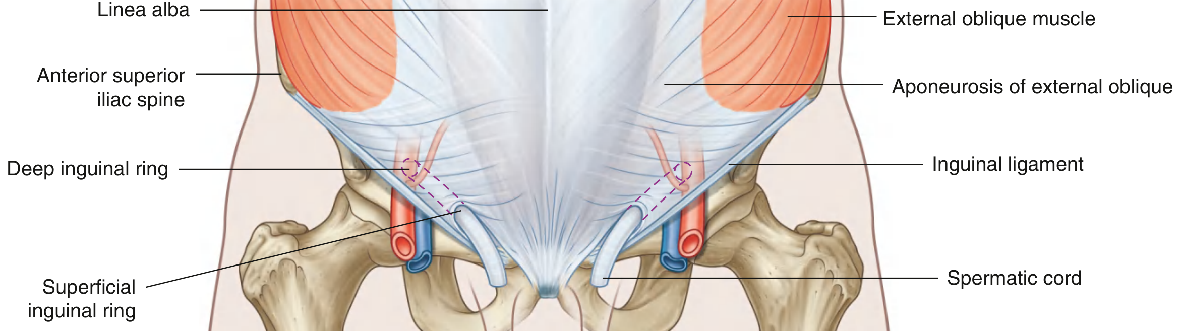
The simple map
Inside abdomen                         Groin / outside

Peritoneum
    |
    |  Deep ring                  Inguinal canal             Superficial ring
    |  (entrance)                 slants down + medially      (exit)
    V
 [   O   ]  ---------------------------------------------->  [  /\  ]
                                                                  |
                                                           spermatic cord
                                                           then scrotum
The two rings: what they actually are
1. Deep inguinal ring = inner entrance
- Located about 1-1.5 cm above the midpoint of the inguinal ligament
- Lateral to the inferior epigastric vessels
Deep ring is lateral to the inferior epigastric vessels.
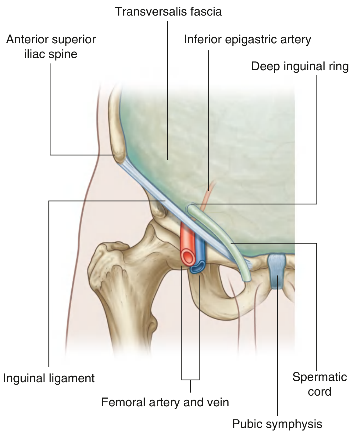
- Midpoint of inguinal ligament = halfway between ASIS and pubic tubercle
- Mid-inguinal point = halfway between ASIS and pubic symphysis
2. Superficial inguinal ring = outer exit
- Just above and lateral to the pubic tubercle
- This is the one near the skin that can be felt clinically, especially by invaginating scrotal skin with a finger.
Superficial ring is above and lateral to the pubic tubercle.
How hernias use this anatomy
A. Indirect inguinal hernia: takes the whole tunnel
Abdomen
   |
   |  enters DEEP ring
   V
  [deep ring] ---> through inguinal canal ---> [superficial ring]
                                                    |
                                             may enter scrotum
Mechanism
One-line memory aid
Indirect = Into the deep ring.
Indirect = lateral to inferior epigastric vessels.
B. Direct inguinal hernia: pushes through the back wall, not through the deep ring
Abdominal pressure
       |
       V
 Weak posterior wall of inguinal canal
       |
       V
  Direct hernia bulges forward
       |
  near/through superficial ring
Hesselbach triangle boundaries
             Rectus abdominis
                  |
                  |  medial boundary
                  |
      Direct hernia bulges here
                  |
inferior epigastric vessels       inguinal ligament
        lateral boundary          inferior boundary
Direct = medial to inferior epigastric vessels.
The comparison that makes it click
| Feature | Indirect inguinal hernia | Direct inguinal hernia |
|---|---|---|
| Starts at deep ring? | Yes | No |
| Goes through full inguinal canal? | Yes | No, pushes through posterior wall |
| Relation to inferior epigastric vessels | Lateral | Medial |
| Can reach scrotum? | Commonly can | Less commonly |
| Usual mechanism | Patent/deep-ring pathway plus pressure | Acquired posterior-wall weakness |
| Memory phrase | “Through the door and tunnel” | “Through the weak wall” |
The single most useful mental image
- Deep ring = the back door into a hallway
- Inguinal canal = the hallway
- Superficial ring = the front door
Clinical surface landmark
Show a labeled diagram of the two inguinal rings
labeled inguinal canal diagram deep inguinal ring superficial inguinal ring anatomy
labeled diagram deep superficial inguinal rings inferior epigastric vessels
inguinal hernia anatomy
Inguinal canal with both rings labeled
Deep ring close-up
ASIS ------------------------------------------- Pubic tubercle
       \___________ inguinal ligament __________/

                     ↑
              DEEP INGUINAL RING
      Just above the inguinal ligament
      LATERAL to inferior epigastric vessels

                \  inguinal canal  /
                 \   down + medial/
                  \               /
                   ↓
         SUPERFICIAL INGUINAL RING
         Just ABOVE and LATERAL to pubic tubercle
Wha is pubic tubercle
pubic tubercle anatomy
pubic tubercle anatomy landmark inguinal ligament authoritative
ASIS (front hip bone) ------------------------ Pubic tubercle
        \_________ inguinal ligament _________/
                                                  ↑
                                     small bony bump you can feel
How to find it on yourself
- Find the center of the lower pelvis, where the two pubic bones meet. This is the pubic symphysis.
- Move your finger a little to the right or left.
- You can feel a firm bony prominence: that is the pubic tubercle.
- The superficial inguinal ring lies just above and lateral to it.
- An inguinal hernia is usually above and medial to it.
- A femoral hernia is usually below and lateral to it.
- The inguinal ligament runs from the front point of the hip bone, the ASIS, to the pubic tubercle. StatPearls description of the inguinal ligament
How the bowel protrudeds from rectus abdomisnis
labeled diagram direct inguinal hernia Hesselbach triangle rectus abdominis inferior epigastric vessels
inguinal hernia anatomy
direct inguinal hernia Hesselbach triangle rectus abdominis mechanism anatomy
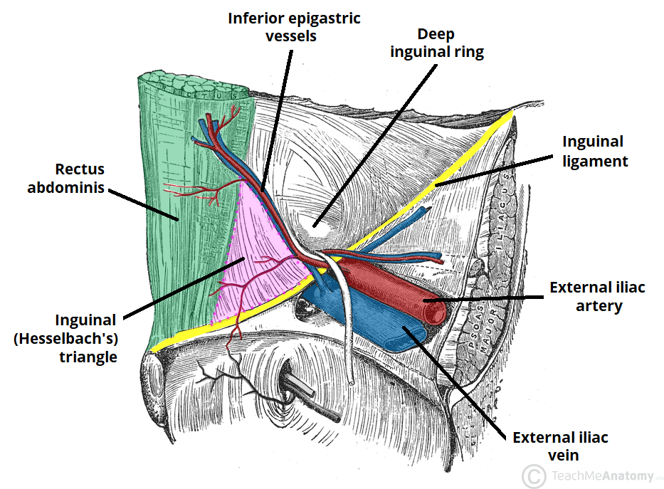
Where exactly does it come through?
                 MIDLINE
                    |
              Rectus abdominis
                    |
                    |\
                    | \       ← weak area:
                    |  \         Hesselbach triangle
                    |   \
                    |    \  Inferior epigastric vessels
                    |     \       (lateral border)
--------------------|------\----------------
          Inguinal ligament  (bottom border)
- Medially: lateral border of the rectus abdominis
- Laterally: inferior epigastric vessels
- Below: inguinal ligament
Mechanism step by step
Inside abdomen                   Abdominal wall                   Outside

Bowel + peritoneum
       |
       | Increased pressure from coughing, straining, etc.
       V
  Weak transversalis fascia
  in Hesselbach triangle
       |
       V
  Hernia sac bulges forward
       |
       V
  Groin lump = direct inguinal hernia
- Medial to the inferior epigastric vessels
- Usually due to acquired weakness of the transversalis fascia
- Usually seen with aging or long-term repetitive raised abdominal pressure, such as chronic cough, constipation/straining, or urinary obstruction
- Usually does not go through the deep inguinal ring
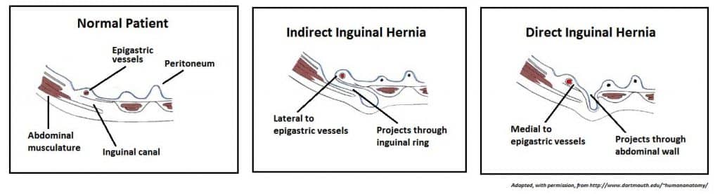
Compare with an indirect hernia
- Direct: bowel pushes through the weak posterior wall beside rectus, within Hesselbach triangle.
- Indirect: bowel enters the deep inguinal ring, which is lateral to the inferior epigastric vessels, then travels through the canal.
Can you show the anatomy of each abdominal layer and how it comes out via a gif maybe?
cross-section layers anterior abdominal wall direct inguinal hernia transversalis fascia Hesselbach triangle labeled
animated gif direct inguinal hernia abdominal wall layers anatomy
inguinal hernia anatomy
~/
mkdir -p "/tmp/workspace/inguinal-hernia-animation/src" "/tmp/workspace/inguinal-hernia-animation/output" && python - <<'PY' from PIL import Image, ImageDraw, ImageFont from pathlib import Path out=Path('/tmp/workspace/inguinal-hernia-animation/output/direct_inguinal_hernia_layers.gif') W,H=1200,760 try: font_b=ImageFont.truetype('/usr/share/fonts/truetype/dejavu/DejaVuSans-Bold.ttf',28) font=ImageFont.truetype('/usr/share/fonts/truetype/dejavu/DejaVuSans.ttf',22) font_s=ImageFont.truetype('/usr/share/fonts/truetype/dejavu/DejaVuSans.ttf',18) font_xs=ImageFont.truetype('/usr/share/fonts/truetype/dejavu/DejaVuSans.ttf',16) except: font_b=font=font_s=font_xs=None # inside (left) to skin (right), simplified cross-section lower abdominal wall layers=[ ('Bowel / abdominal cavity','#f6ce79',135), ('Parietal peritoneum','#9dd5ea',18), ('Transversalis fascia\n(posterior canal wall)','#8fbf9a',44), ('Transversus abdominis','#bc90c9',72), ('Internal oblique','#d8959e',72), ('External oblique aponeurosis','#e6ac64',55), ('Subcutaneous fat','#f4d6a7',65), ('Skin','#d78e71',36), ] xs=[]; x=80 for n,c,w in layers: xs.append((x,x+w,n,c)); x+=w # Hesselbach weakness: only within transversalis, midpoint y 350 frames=[] for step in range(24): im=Image.new('RGB',(W,H),'#fbfbfc'); d=ImageDraw.Draw(im) # title d.text((45,25),'Direct inguinal hernia: bowel pushes through a weak posterior wall',font=font_b,fill='#202735') d.text((45,62),'Simplified side-on slice: inside abdomen is left; skin/groin is right',font=font_s,fill='#4c566a') y0,y1=165,585 # layers, split fascia around defect when step advanced for a,b,name,c in xs: # draw full rectangles except transversalis fascia (index 2) at defect if 'Transversalis' in name and step>2: gap_top=315; gap_bot=440 d.rectangle((a,y0,b,gap_top),fill=c,outline='#555',width=1) d.rectangle((a,gap_bot,b,y1),fill=c,outline='#555',width=1) else: d.rectangle((a,y0,b,y1),fill=c,outline='#555',width=1) # label centered, rotated omitted use multiline tx=(a+b)//2 # draw label in top portion, slight invisible no lines=name.split('\n') yy=615 for line in lines: bb=d.textbbox((0,0),line,font=font_xs); d.text((tx-(bb[2]-bb[0])/2,yy),line,font=font_xs,fill='#222'); yy+=18 # abbreviate use leader legend colored d.rectangle((48,115,940,130), fill='#dedede') # direction d.polygon([(75,135),(105,125),(105,132),(250,132),(250,138),(105,138),(105,145)],fill='#35495e') d.text((265,122),'pressure from inside abdomen',font=font_s,fill='#35495e') # draw bowel loop that progressively bulges in wall in direct route p=max(0,(step-3)/20) # Bowel starts left then expands into defect; anatomical sac carries peritoneum # use bezier via polygon approximation sampled quadratics import math cx=155 + 530*p cy=377 r=80 # sac/bowel shape follows curve, pure ellipse shifted then waist restriction # peritoneal sac outer if p<0.15: bbox=(80,290,245,465) else: # outlined bulge encroaches into layers; cap at external oblique for direct endx=250+420*p # construct joined silhouette with narrow neck at fascia pts=[(120,300),(200,290),(245,315),(250,350),(270,350)] # elongated dome for i in range(21): t=i/20; pts.append((250+(endx-250)*t,350-95*math.sin(math.pi*t))) pts += [(endx+35,377)] for i in range(20,-1,-1): t=i/20; pts.append((250+(endx-250)*t,405+95*math.sin(math.pi*t))) pts += [(270,405),(250,405),(245,440),(200,465),(120,455)] # sac first d.polygon(pts, fill='#b9e6f0', outline='#206b82') # bowel inner ellipse approximately at distal region bx=max(135,endx-35); d.ellipse((bx-52,cy-56,bx+62,cy+56),fill='#f4bd67',outline='#9a5f22',width=3) d.arc((bx-31,cy-32,bx+33,cy+38),20,320,fill='#aa6d25',width=3) d.arc((bx-20,cy-22,bx+20,cy+25),200,120,fill='#aa6d25',width=2) if step>=3: # labels and arrow a,b,_,_=xs[2] d.line((a-5,285,a-5,315),fill='#bd2e2e',width=5) d.line((a-5,440,a-5,470),fill='#bd2e2e',width=5) d.line((a-25,280,a-5,315),fill='#bd2e2e',width=3) d.text((330,245),'Weak spot in transversalis fascia\n(Hesselbach triangle)',font=font_s,fill='#9e2323') d.line((455,295),(a,330),fill='#9e2323',width=2) # fixed landmarks d.text((665,150),'This is DIRECT:',font=font_b,fill='#8d2630') d.text((665,188),'• Does NOT enter deep ring',font=font_s,fill='#303846') d.text((665,216),'• Pushes through posterior wall',font=font_s,fill='#303846') d.text((665,244),'• Medial to inferior epigastric vessels',font=font_s,fill='#303846') # Stage caption if step<3: msg='1. Abdominal pressure acts outward.' elif step<10: msg='2. Peritoneum and bowel press against a weak area.' elif step<18: msg='3. They bulge through the weakened transversalis fascia.' else: msg='4. The bulge moves forward toward the superficial ring and appears in the groin.' d.rounded_rectangle((50,685,1150,735),radius=12,fill='#eaf0f6',outline='#afc2d4') d.text((75,699),msg,font=font_s,fill='#223449') frames.append(im) frames[0].save(out,save_all=True,append_images=frames[1:],duration=[190]*23+[1200],loop=0,optimize=True) # static png first high clarity frames[-1].save('/tmp/workspace/inguinal-hernia-animation/output/direct_inguinal_hernia_layers_final.png') print(out) PY python - <<'PY' from PIL import Image p='/tmp/workspace/inguinal-hernia-animation/output/direct_inguinal_hernia_layers.gif' im=Image.open(p) print(im.size, im.n_frames, 'bytes', __import__('os').path.getsize(p)) PY
python - <<'PY' p='/tmp/workspace/inguinal-hernia-animation/src/make_gif.py' script=r'''from PIL import Image, ImageDraw, ImageFont from pathlib import Path import math out=Path('/tmp/workspace/inguinal-hernia-animation/output/direct_inguinal_hernia_layers.gif') W,H=1200,760 font_b=ImageFont.truetype('/usr/share/fonts/truetype/dejavu/DejaVuSans-Bold.ttf',28) font=ImageFont.truetype('/usr/share/fonts/truetype/dejavu/DejaVuSans.ttf',22) font_s=ImageFont.truetype('/usr/share/fonts/truetype/dejavu/DejaVuSans.ttf',18) font_xs=ImageFont.truetype('/usr/share/fonts/truetype/dejavu/DejaVuSans.ttf',16) layers=[('Bowel / abdominal cavity','#f6ce79',135),('Parietal peritoneum','#9dd5ea',18),('Transversalis fascia\n(posterior canal wall)','#8fbf9a',44),('Transversus abdominis','#bc90c9',72),('Internal oblique','#d8959e',72),('External oblique aponeurosis','#e6ac64',55),('Subcutaneous fat','#f4d6a7',65),('Skin','#d78e71',36)] xs=[]; x=80 for n,c,w in layers: xs.append((x,x+w,n,c)); x+=w frames=[] for step in range(24): im=Image.new('RGB',(W,H),'#fbfbfc'); d=ImageDraw.Draw(im) d.text((45,25),'Direct inguinal hernia: bowel pushes through a weak posterior wall',font=font_b,fill='#202735') d.text((45,62),'Simplified side-on slice: inside abdomen is left; skin/groin is right',font=font_s,fill='#4c566a') y0,y1=165,585 for a,b,name,c in xs: if 'Transversalis' in name and step>2: d.rectangle((a,y0,b,315),fill=c,outline='#555',width=1); d.rectangle((a,440,b,y1),fill=c,outline='#555',width=1) else: d.rectangle((a,y0,b,y1),fill=c,outline='#555',width=1) tx=(a+b)//2; yy=615 for line in name.split('\n'): bb=d.textbbox((0,0),line,font=font_xs); d.text((tx-(bb[2]-bb[0])/2,yy),line,font=font_xs,fill='#222'); yy+=18 d.rectangle((48,115,940,130),fill='#dedede') d.polygon([(75,135),(105,125),(105,132),(250,132),(250,138),(105,138),(105,145)],fill='#35495e') d.text((265,122),'pressure from inside abdomen',font=font_s,fill='#35495e') p=max(0,(step-3)/20) if p<.15: d.ellipse((80,290,245,465),fill='#f4bd67',outline='#9a5f22',width=3) else: endx=250+420*p pts=[(120,300),(200,290),(245,315),(250,350),(270,350)] for i in range(21): t=i/20; pts.append((250+(endx-250)*t,350-95*math.sin(math.pi*t))) pts += [(endx+35,377)] for i in range(20,-1,-1): t=i/20; pts.append((250+(endx-250)*t,405+95*math.sin(math.pi*t))) pts += [(270,405),(250,405),(245,440),(200,465),(120,455)] d.polygon(pts,fill='#b9e6f0',outline='#206b82') bx=max(135,endx-35); d.ellipse((bx-52,321,bx+62,433),fill='#f4bd67',outline='#9a5f22',width=3) d.arc((bx-31,345,bx+33,415),20,320,fill='#aa6d25',width=3) if step>=3: a,b,_,_=xs[2] d.line([(a-5,285),(a-5,315)],fill='#bd2e2e',width=5); d.line([(a-5,440),(a-5,470)],fill='#bd2e2e',width=5) d.text((330,245),'Weak spot in transversalis fascia\n(Hesselbach triangle)',font=font_s,fill='#9e2323'); d.line([(455,295),(a,330)],fill='#9e2323',width=2) d.text((665,150),'This is DIRECT:',font=font_b,fill='#8d2630') d.text((665,188),'• Does NOT enter deep ring',font=font_s,fill='#303846'); d.text((665,216),'• Pushes through posterior wall',font=font_s,fill='#303846'); d.text((665,244),'• Medial to inferior epigastric vessels',font=font_s,fill='#303846') if step<3: msg='1. Abdominal pressure acts outward.' elif step<10: msg='2. Peritoneum and bowel press against a weak area.' elif step<18: msg='3. They bulge through the weakened transversalis fascia.' else: msg='4. The bulge moves forward toward the superficial ring and appears in the groin.' d.rounded_rectangle((50,685,1150,735),radius=12,fill='#eaf0f6',outline='#afc2d4'); d.text((75,699),msg,font=font_s,fill='#223449') frames.append(im) frames[0].save(out,save_all=True,append_images=frames[1:],duration=[190]*23+[1200],loop=0,optimize=True) frames[-1].save('/tmp/workspace/inguinal-hernia-animation/output/direct_inguinal_hernia_layers_final.png') print(out) ''' open(p,'w').write(script) PY python "/tmp/workspace/inguinal-hernia-animation/src/make_gif.py" && python - <<'PY' from PIL import Image import os p='/tmp/workspace/inguinal-hernia-animation/output/direct_inguinal_hernia_layers.gif' im=Image.open(p) print(im.size, im.n_frames, os.path.getsize(p)) PY
python - <<'PY' p='/tmp/workspace/inguinal-hernia-animation/src/make_gif.py' s=open(p).read() s=s.replace("font_b=ImageFont.truetype('/usr/share/fonts/truetype/dejavu/DejaVuSans-Bold.ttf',28)\nfont=ImageFont.truetype('/usr/share/fonts/truetype/dejavu/DejaVuSans.ttf',22)\nfont_s=ImageFont.truetype('/usr/share/fonts/truetype/dejavu/DejaVuSans.ttf',18)\nfont_xs=ImageFont.truetype('/usr/share/fonts/truetype/dejavu/DejaVuSans.ttf',16)","font_b=font=font_s=font_xs=ImageFont.load_default()") open(p,'w').write(s) PY python "/tmp/workspace/inguinal-hernia-animation/src/make_gif.py" && python - <<'PY' from PIL import Image import os p='/tmp/workspace/inguinal-hernia-animation/output/direct_inguinal_hernia_layers.gif' im=Image.open(p) print('GIF:', im.size, im.n_frames, os.path.getsize(p)) PY
direct inguinal hernia layers
GIF File · GIF
Layers, from inside the abdomen to the skin
- Bowel
- Parietal peritoneum: thin inner lining. It becomes the outer lining of the hernia sac.
- Transversalis fascia: the key deep supporting sheet and posterior wall of the inguinal canal. In a direct hernia, this weakens.
- Transversus abdominis
- Internal oblique
- External oblique aponeurosis
- Subcutaneous fat
- Skin