
Gott shunt
Gott shunt cardiac surgery heparin bonded shunt aortic repair
Gott Shunt
What It Is
Why It Was Developed
- Afterload on the left ventricle acutely increases
- All organs distal to the clamp suffer global ischemia (spinal cord, kidneys, bowel, lower limbs)
- Risk of paraplegia and renal failure is directly related to cross-clamp duration
Mechanism
- It is a passive shunt - no pump is required
- Blood flows by a pressure gradient from a proximal source to the distal aorta
- The heparin bonding on the inner surface prevents thrombosis within the shunt without needing systemic heparin
- Typical insertion: proximal end in the left ventricle or proximal descending thoracic aorta, distal end in the distal aorta or femoral artery
Clinical Use
| Feature | Detail |
|---|---|
| Indication | Descending thoracic aortic aneurysm repair, traumatic aortic injury |
| Approach | Left thoracotomy |
| Anticoagulation | Minimal or none systemically (heparin coating does the work) |
| Flow type | Passive (pressure-gradient driven) |
| Advantage over clamp-and-sew | Maintains distal perfusion; reduces paraplegia and renal failure risk |
| Limitation vs. Left Heart Bypass (LHB) | Cannot actively control flow rate; offers less precise hemodynamic support than an active pump |
Comparison With Other Distal Perfusion Strategies
-
Clamp-and-sew - No circulatory support at all. Fastest and simplest but highest risk of paraplegia with longer clamp times.
-
Gott shunt - Passive heparin-bonded conduit. Simplest form of distal perfusion; avoids systemic heparin; no pump needed. Best for short to moderate repairs.
-
Left heart bypass (LHB) / partial bypass - Active centrifugal pump draws blood from the left atrium and returns it to the femoral artery. This is the most commonly used technique today. Allows precise control of proximal and distal pressures. Requires a heparin-coated circuit (dose ~100 units/kg).
-
Full cardiopulmonary bypass (CPB) - Used for the most complex or extensive repairs.
Key Advantages (from the original 1977 Hopkins series, Donahoo, Brawley & Gott)
- Eliminates need for systemic heparin (important in trauma patients with bleeding risk)
- Avoids proximal hypertension during cross-clamping
- Adequate distal organ perfusion without an interposed pump
- Easy to handle; low complication profile
Modern Use
Diagram
Medical diagram of the Gott shunt in descending thoracic aortic surgery. Show a clean anatomical illustration of the heart and aorta from a left lateral view. Label the following structures: heart (left ventricle), ascending aorta, aortic arch, descending thoracic aorta, and femoral artery. Show two aortic cross-clamps on the descending thoracic aorta with an aneurysm between them. Draw the heparin-bonded Gott shunt as a flexible tube bypassing the clamped segment, with proximal end inserted into the left ventricle or proximal descending aorta and distal end going to the distal descending aorta below the clamps. Use arrows to show passive blood flow direction through the shunt. Label the shunt as "Gott Shunt (heparin-bonded passive conduit)". Also label the aneurysm, proximal clamp, distal clamp, and indicate "no systemic heparin required". Use clean medical illustration style with blue for venous structures, red for arterial, and a clear background.
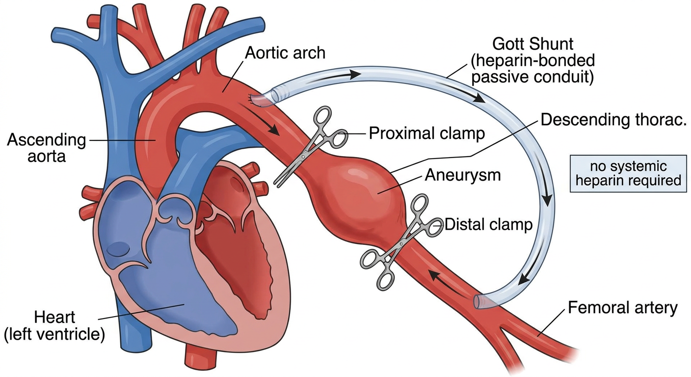
- Proximal clamp - applied to the descending thoracic aorta above the aneurysm
- Distal clamp - applied below the aneurysm to isolate the surgical field
- Gott shunt - the heparin-bonded tube bypasses the clamped segment, running from the left ventricle (or proximal aorta) to the distal aorta
- Passive flow - blood travels down the pressure gradient through the shunt, perfusing the spinal cord, kidneys, and lower body without needing a pump or systemic heparin