
Physiological basis of various liver function tests and it's significance in surgical conditions...give answer for this 20 marks questions in university exam of surgical resident
Reading File
Now let me search the medical library for liver function tests content in parallel:
Finding Sources
Finding Sources
Excellent - I have multiple high-quality sources. Let me read the most relevant surgical textbooks in parallel:
Reading File
Reading File
Reading File
Excellent content from Schwartz's Principles of Surgery and Pye's Surgical Handicraft. Let me read more from Schwartz's (the most detailed surgical source) and also get content on bilirubin metabolism and cholestasis:
Reading File
Finding Sources
Finding Sources
Excellent! Let me get the Child-Pugh and MELD content, and also the remainder of the Schwartz's text:
Reading File
Finding Sources
Finding Sources
Now let me get the Child-Pugh and preoperative hepatic reserve content:
Reading File
Reading File
I now have comprehensive content. Let me get the Child-Pugh table details and also the Pye's remaining content:
Reading File
Reading File
I now have comprehensive, authoritative content from multiple surgical textbooks. Let me compose the complete 20-mark university answer.
Physiological Basis of Liver Function Tests and Their Significance in Surgical Conditions
Introduction
The liver is the largest solid organ in the body, performing over 500 distinct functions including protein synthesis, carbohydrate metabolism, lipid metabolism, detoxification, bile production, and immune surveillance. "Liver function tests" (LFTs) is a panel of serum markers used to evaluate hepatic dysfunction. The term is technically a misnomer - most tests measure cell damage rather than true function. Actual synthetic function is better captured by serum albumin and prothrombin time. The liver possesses enormous functional reserve; significant abnormalities appear only when approximately 80% of parenchyma is destroyed.
LFT abnormalities are best interpreted by classifying them into three categories:
- Hepatocellular injury (transaminases)
- Abnormal synthetic function (albumin, PT/INR)
- Cholestasis (bilirubin, ALP, GGT)
1. Serum Bilirubin
Physiological Basis
Bilirubin is a breakdown product of heme metabolism. The pathway has three phases:
- Prehepatic phase: RBC destruction releases hemoglobin → heme is converted by heme oxygenase to biliverdin → biliverdin reductase converts it to unconjugated (indirect) bilirubin. Unconjugated bilirubin is lipid-soluble and insoluble in water; it is transported to the liver bound to albumin.
- Intrahepatic phase: In hepatocytes, unconjugated bilirubin is conjugated with glucuronic acid by UDP-glucuronosyltransferase, becoming water-soluble conjugated (direct) bilirubin. The rate-limiting step is excretion of conjugated bilirubin from hepatocytes into bile canaliculi.
- Posthepatic phase: Conjugated bilirubin is secreted into bile and passes to the duodenum.
Normal total bilirubin: ~6 mmol/L (upper limit ~20 mmol/L or ~1.2 mg/dL). Jaundice becomes clinically detectable when serum bilirubin rises above 2.5-3 mg/dL.
| Fraction | Character | Elevated in |
|---|---|---|
| Unconjugated (indirect) | Insoluble, albumin-bound | Hemolysis, Gilbert's syndrome, Crigler-Najjar |
| Conjugated (direct) | Water-soluble, urine-positive | Hepatocellular disease, extrahepatic obstruction |
Surgical Significance
- Obstructive jaundice: Elevated direct (conjugated) bilirubin indicates extrahepatic obstruction - choledocholithiasis, cholangiocarcinoma, carcinoma head of pancreas, ampullary carcinoma, post-surgical biliary stricture. Because conjugated bilirubin is water-soluble, it appears in urine (dark urine = bilirubinuria).
- Serial monitoring: Total bilirubin should be repeated every 3-4 days in surgical jaundice. A falling level is a favorable sign; a sudden rise indicates deterioration (Pye's Surgical Handicraft).
- Preoperative risk: Obstructive jaundice increases operative risk significantly - patients are at risk for hepatorenal syndrome, coagulopathy, infection, and poor wound healing. Bilirubin >10 mg/dL substantially raises perioperative mortality.
- Partition into fractions: Although previously used to differentiate hepatic from posthepatic jaundice, the direct van den Bergh reaction measures both mono- and diglucuronides, making fractionation unreliable for this distinction alone. However, elevated indirect fraction strongly suggests prehepatic (hemolytic) cause.
2. Aminotransferases (Transaminases) - AST and ALT
Physiological Basis
Aminotransferases catalyze the transfer of amino groups during gluconeogenesis:
- AST (aspartate aminotransferase / SGOT): Catalyzes transfer of amino group from aspartate to alpha-ketoglutarate → oxaloacetate. Found in liver, cardiac muscle, skeletal muscle, kidney, brain, pancreas, lungs, and RBCs. Less specific for liver.
- ALT (alanine aminotransferase / SGPT): Catalyzes transfer from alanine → pyruvate. Found predominantly in hepatocytes, making it the more liver-specific enzyme. Elevation in ALT is the most useful test to distinguish hepatocellular damage from posthepatic obstruction as a cause of jaundice.
Hepatocellular injury causes membrane disruption → release into circulation. The degree of elevation suggests etiology:
| Elevation | Likely Cause |
|---|---|
| Mild (<3x normal) | NAFLD, chronic viral hepatitis, drug injury |
| Moderate (3-20x) | Acute viral hepatitis, alcoholic hepatitis |
| Severe (>1000 U/L) | Ischemic hepatitis, acetaminophen toxicity, fulminant hepatitis |
| AST:ALT ratio >2:1 | Alcoholic liver disease |
Surgical Significance
- Preoperative assessment: Significantly elevated transaminases (>3x normal) indicate active hepatocellular damage; elective surgery should be deferred.
- Hepatic ischemia detection: Transaminases spike dramatically in hepatic artery thrombosis post-liver transplant, ischemic hepatitis from hypotension/shock (ischemic hepatopathy - "shock liver"), or Pringle maneuver-related ischemic damage during hepatic surgery.
- Post-hepatectomy monitoring: Rising transaminases after liver resection suggest ongoing hepatocellular injury or post-hepatectomy liver failure.
- Blunt abdominal trauma: Elevation of AST/ALT serves as a sensitive marker for hepatic laceration - AST >450 U/L or ALT >250 U/L has high sensitivity for liver injury in trauma.
- Note: Transaminase levels correlate poorly with severity of hepatocellular necrosis - they may not be elevated in cirrhosis or hepatic fibrosis.
3. Alkaline Phosphatase (ALP)
Physiological Basis
ALP is a mixture of isoenzymes derived from multiple tissues - liver, bone, intestinal mucosa, placenta, and kidneys. In the liver, it is expressed by bile duct epithelium. In conditions of biliary obstruction, bile acids disrupt membranes of bile duct cells, increasing ALP synthesis and its release into serum. The half-life of serum ALP is approximately 7 days, so levels may remain elevated for days even after resolution of biliary obstruction.
High ALP is also found in:
- Bone disease: Rickets, osteomalacia, Paget's disease, hyperparathyroidism, bony metastases
- Third trimester of pregnancy (placental isoenzyme)
To confirm hepatic origin, ALP must be interpreted alongside GGT (liver-specific) or by isoenzyme electrophoresis.
Surgical Significance
- Cholestatic pattern: ALP elevation (often >3x) with high bilirubin and GGT = cholestasis. This is the classic pattern in carcinoma head of pancreas, cholangiocarcinoma, primary sclerosing cholangitis (PSC).
- Metastatic liver disease: ALP may be raised from either hepatic involvement OR bone metastases. Distinguished by GGT measurement (GGT elevated with hepatic cause; normal with bony cause) or isoenzyme fractionation.
- Post-transplant biliary complications: Rising ALP post-liver transplant suggests biliary stricture or anastomotic leak.
- PSC: Markedly elevated ALP is a hallmark; PSC has strong associations with inflammatory bowel disease and is a premalignant condition for cholangiocarcinoma - surgical resection or transplant is definitive management.
4. Gamma-Glutamyl Transferase (GGT)
Physiological Basis
GGT is found in hepatocytes and is released from bile duct epithelium. It is an early and sensitive marker of hepatobiliary disease. However, it is non-specific and can be elevated by:
- Alcohol abuse (microsomal enzyme induction)
- Certain medications (phenytoin, phenobarbitone)
- Pancreatic disease, myocardial infarction, renal failure, obstructive pulmonary disease
Surgical Significance
- Confirms hepatic origin of elevated ALP: GGT + ALP both elevated = hepatobiliary cause. ALP elevated + GGT normal = bony cause.
- Alcohol screening: Persistently elevated GGT in the preoperative period suggests chronic alcohol intake - important for anaesthetic planning (altered drug metabolism, risk of alcohol withdrawal post-surgery, thrombocytopenia, coagulopathy).
- Sensitive early marker: GGT rises before other LFTs in early biliary obstruction and in drug-induced liver injury.
5. Serum Albumin
Physiological Basis
The liver produces approximately 10 g of albumin per day. Albumin serves multiple functions: maintaining oncotic pressure, transporting drugs, hormones, and bilirubin. Albumin has a long half-life of 15-20 days, meaning it reflects chronic hepatic synthetic function, not acute failure. Albumin levels are also affected by nutritional status, renal dysfunction (nephrotic syndrome), protein-losing enteropathy, and inflammatory states (negative acute phase protein - falls with inflammation).
Surgical Significance
- Preoperative nutritional marker: Hypoalbuminaemia (<3.5 g/dL) is an independent predictor of surgical complications - poor wound healing, anastomotic dehiscence, increased infection risk, impaired drug binding.
- Chronic liver disease assessment: Albumin is a key component of the Child-Pugh Score for assessing hepatic reserve.
- NOT a marker of acute hepatic failure due to its long half-life - PT/INR is superior in acute settings.
- Cirrhosis: Low albumin + ascites + coagulopathy = decompensated cirrhosis, usually a contraindication to major elective surgery.
6. Prothrombin Time (PT) and INR
Physiological Basis
Most clotting factors (I, II, V, VII, IX, X, XI) are synthesized exclusively in the liver. PT measures the rate of conversion of prothrombin to thrombin (via extrinsic and common pathways). INR (International Normalized Ratio) = Patient PT / Mean normal PT, standardizing results across laboratories. Vitamin K is required for gamma-carboxylation of factors II, VII, IX, and X (Vit K-dependent factors). Therefore, PT/INR can be prolonged in:
- Hepatocellular failure (reduced synthesis)
- Vitamin K deficiency (malabsorption, biliary obstruction - fat-soluble vitamin)
- Anticoagulant therapy (warfarin)
Distinguishing these: IV vitamin K administration corrects PT in Vit K deficiency/biliary obstruction but NOT in hepatocellular failure (no functioning hepatocytes to use the Vit K).
Surgical Significance
- Best acute marker of hepatic synthetic function - short half-life of factor VII (~4-6 hours) makes PT/INR sensitive for acute liver failure.
- Preoperative coagulation assessment: PT >3 seconds above control (INR >1.5) is a contraindication to elective hepatic surgery without correction.
- Biliary obstruction: Prolonged PT in obstructive jaundice = fat malabsorption → Vit K deficiency → correctable with parenteral vitamin K (10 mg IV for 3 days). Failure to correct suggests hepatocellular component.
- Child-Pugh classification: INR is a component - INR >2.3 scores 3 points (worst category).
- Liver failure monitoring: Serial PT/INR is the most sensitive laboratory indicator of progression or recovery in acute liver failure.
7. Composite Scoring Systems - Surgical Decision Making
Child-Pugh Score
The Child-Pugh score incorporates 5 parameters to assess hepatic reserve before surgery:
| Parameter | 1 Point | 2 Points | 3 Points |
|---|---|---|---|
| Bilirubin (mg/dL) | <2 | 2-3 | >3 |
| Albumin (g/dL) | >3.5 | 2.8-3.5 | <2.8 |
| Ascites | Absent | Moderate | Severe |
| Encephalopathy | Absent | Moderate | Severe |
| PT prolonged (sec) / INR | <4 / <1.7 | 4-6 / 1.7-2.3 | >6 / >2.3 |
- Class A (5-6 points): Well-compensated; suitable for major hepatic resection
- Class B (7-9 points): Significant functional compromise; only highly selected patients suitable for resection
- Class C (10-15 points): Decompensated cirrhosis; mortality from surgery is prohibitive - surgery should be avoided; may be considered for transplant listing
MELD Score
MELD (Model for End-Stage Liver Disease) = 3.78 x ln[bilirubin (mg/dL)] + 11.2 x ln[INR] + 9.57 x ln[creatinine (mg/dL)] + 6.43
- Estimates 3-month mortality in patients with chronic liver disease
- Used by UNOS for organ allocation in liver transplantation
- Less useful for predicting post-hepatectomy failure compared to Child-Pugh
- MELD <9 with Child-Pugh A = favorable for liver resection in HCC
ICG Retention Test
Indocyanine green (ICG) is a tricarbocyanine dye exclusively eliminated by the liver without enterohepatic recirculation. ICG retention at 15 minutes (ICG-R15) measures dynamic hepatic function:
- Normal ICG-R15: 1-10%
- ICG-R15 <10% = adequate reserve for major liver resection in cirrhotic patients
- Widely used in Asia (Japan) for hepatic reserve assessment before hepatectomy
- Must be combined with volumetric FLR (future liver remnant) assessment
8. Additional Surgically Relevant Tests
Pseudocholinesterase
Synthesized by the liver cells in a manner parallel to albumin synthesis. Falls in hepatocellular disease - important surgically because:
- Pseudocholinesterase (plasma cholinesterase) is responsible for metabolism of succinylcholine and mivacurium
- In severe liver disease, prolonged neuromuscular blockade can occur with these agents
Serum Ammonia
- Converted in liver via the urea cycle
- Elevated in hepatic failure / porto-systemic shunting
- Clinical significance: Hepatic encephalopathy - ammonia is neurotoxic
- Surgical importance: Portosystemic shunting procedures (e.g. TIPS, surgical shunts) may worsen encephalopathy by bypassing hepatic detoxification
5'-Nucleotidase and Leucine Aminopeptidase
- Elevated in hepatocellular damage
- Used to confirm hepatic origin of elevated ALP when GGT is unavailable
9. Patterns of LFT Abnormality in Common Surgical Conditions
| Condition | Bilirubin | ALT/AST | ALP | GGT | Albumin | PT/INR |
|---|---|---|---|---|---|---|
| Obstructive jaundice (stones/CA) | ↑↑ Direct | Mild ↑ | ↑↑↑ | ↑↑ | Normal | Normal (early) |
| Hepatocellular carcinoma | ↑ Direct | ↑-↑↑ | ↑↑ | ↑↑ | ↓ | Prolonged |
| Cirrhosis (compensated) | Normal-↑ | Mild ↑ | ↑ | ↑ | ↓ | Prolonged |
| Acute hepatic trauma | ↑ | ↑↑↑ | ↑ | ↑ | Normal | Variable |
| Post-hepatectomy failure | ↑↑ | ↑↑↑ | ↑ | ↑ | ↓↓ | ↑↑↑ INR |
| Metastatic liver disease | ↑ Direct | Mild ↑ | ↑↑ | ↑↑ | ↓ | Variable |
Conclusion
A thorough understanding of the physiological basis of each LFT allows the surgeon to:
- Classify the type of liver dysfunction (hepatocellular vs. cholestatic vs. synthetic failure)
- Stratify surgical risk using Child-Pugh, MELD, and ICG retention
- Identify reversible causes (Vit K deficiency, biliary obstruction) before surgery
- Monitor postoperative hepatic recovery
- Time elective surgery appropriately - avoiding operating during active hepatocellular inflammation or decompensated cirrhosis
As emphasized by Pye's Surgical Handicraft, "single readings of any enzyme level are of little help in deciding the nature of hepatic disease - rising or falling values are more useful indicators of the nature and progress of the condition." Serial monitoring integrated with clinical examination remains the gold standard.
References:
- Schwartz's Principles of Surgery, 11th Edition (Hepatocellular Injury, Cholestasis, Jaundice chapters)
- Mulholland and Greenfield's Surgery, 7th Edition (Preoperative Evaluation of Hepatic Reserve, Child-Pugh, MELD, ICG)
- Pye's Surgical Handicraft, 22nd Edition (Liver Function Tests in Surgical Jaundice)
- Bailey and Love's Short Practice of Surgery, 28th Edition
Hepatitis B is another 10 mark University question Hepatorenal syndrome is another 20 mark University question.. biliary fistula is another 10 mark question..give answer for these 3
Finding Sources
Finding Sources
Finding Sources
Now let me read the most relevant sections in parallel:
Reading File
Reading File
Reading File
Good. I need more comprehensive content. Let me get the detailed Hepatitis B virology/serology content and the HRS pathophysiology from Goldman-Cecil and Sleisenger:
Reading File
Reading File
Finding Sources
Excellent content so far. Let me continue reading the HRS pathophysiology and get the biliary fistula content from Goldman-Cecil and Sleisenger:
Reading File
Reading File
Reading File
Excellent. Let me get the HRS diagnostic criteria and treatment section, and HBV serology markers:
Reading File
Reading File
Let me get the HRS treatment section and the HBV treatment content from the clinical chapters:
Reading File
Finding Sources
Finding Sources
I now have comprehensive content from authoritative sources. Let me also fetch the HBV serology image and get the HRS pathophysiology image:
Loading Image
Finding Sources
I now have all the content I need. Let me also quickly get the Robbins content on HBV clinical features and pathology for completeness:
Reading File
I now have comprehensive, authoritative content from the best sources. Let me compile the complete answers.---
QUESTION 1: Hepatitis B (10 Marks)
Introduction
Hepatitis B Virus (HBV) is a member of the Hepadnaviridae family - a group of hepatotropic DNA viruses. It is a globally significant pathogen with over 250 million chronically infected individuals worldwide, causing chronic hepatitis, cirrhosis, and hepatocellular carcinoma (HCC). Its surgical importance lies in both operative risk stratification and the rising prevalence of HBV-related liver disease requiring surgical intervention.
Virology and Replication
HBV is a partially double-stranded DNA virus with four genes:
- S gene - encodes surface antigen (HBsAg) in three forms: large, middle, and small
- C gene - encodes core antigen (HBcAg) and e antigen (HBeAg)
- P gene - encodes DNA polymerase (with reverse transcriptase activity)
- X gene - product not fully characterized; involved in transactivation
Unique to HBV: it replicates via reverse transcriptase (making it susceptible to nucleoside analog antivirals). There are 4 serotypes and 8 genotypes with geographical variation in virulence and treatment response.
Transmission: Blood/body fluids (parenteral, sexual, vertical - mother to neonate). Perinatal transmission is the most epidemiologically important route in endemic regions (SE Asia, Africa).
Serological Markers and Their Significance
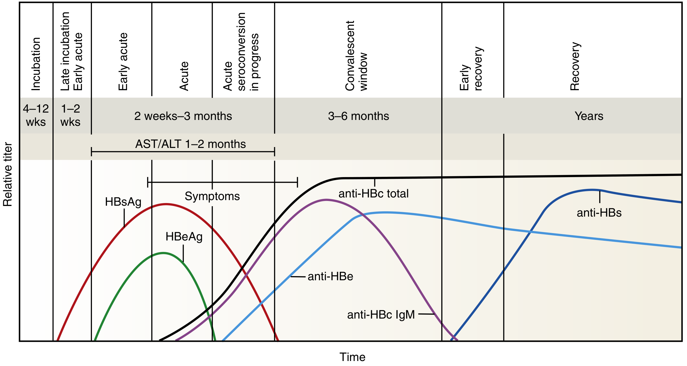
Typical time course for appearance of viral antigens and antibodies in HBV infection (Henry's Clinical Diagnosis, 7th Ed)
| Marker | Significance |
|---|---|
| HBsAg | First marker to appear (4-12 wks incubation). Indicates active infection. Persists >6 months = chronic infection |
| HBeAg | Marker of active viral replication and high infectivity. Persistence = likely progression to chronic hepatitis |
| HBcAg | Not detectable in serum (intracellular only) |
| Anti-HBc IgM | Earliest antibody; diagnostic of acute infection; also appears in reactivation |
| Anti-HBc IgG | Persists for life; marker of past or current infection |
| Anti-HBe | Appears as HBeAg falls; indicates infection is on the wane. Absence in chronic infection implies mutant virus |
| Anti-HBs | Protective antibody; appears last in recovery; basis for vaccination. Absent in chronic infection |
| HBV DNA (PCR) | Gold standard for monitoring viral replication; guides antiviral therapy |
Core Window Period: The gap between disappearance of HBsAg and appearance of anti-HBs, during which only anti-HBc IgM is detectable. This is diagnostically important.
Recommended Test Panels:
- Acute HBV: HBsAg + IgM anti-HBc
- Chronic HBV: HBsAg + IgG anti-HBc + IgG anti-HBs
- Monitoring chronic infection: HBsAg, HBeAg, anti-HBs, anti-HBe, quantitative HBV DNA PCR
Natural History and Clinical Features
| Phase | Features |
|---|---|
| Acute infection | 2/3 are subclinical. Symptomatic: anorexia, fever, RUQ pain, jaundice. Fulminant hepatic failure: 0.1-0.5% |
| Recovery | Most adults clear virus (>90%); HBsAg disappears, anti-HBs appears |
| Chronic infection | Persists in 5-10% of adults; 80-90% of neonates; 5-10% of immunocompromised |
| Chronic hepatitis | Persistent elevation of transaminases; ongoing necroinflammation |
| Cirrhosis | ~20-30% of chronic HBV over 20 years |
| HCC | Risk 100x general population; highest with perinatal acquisition |
Extrahepatic manifestations: glomerulonephritis (membranous), polyarteritis nodosa (immune complex deposition).
Pathology
- Acute HBV: Lobular hepatitis, hepatocyte ballooning, acidophil bodies (apoptotic hepatocytes = Councilman bodies), lymphocytic infiltrate
- Chronic HBV: "Ground glass" hepatocytes (HBsAg accumulation in ER), periportal fibrosis, bridging fibrosis → cirrhosis
- Mechanism of injury: primarily immune-mediated (CD8+ T-cell killing of infected hepatocytes), not direct viral cytopathic effect
Treatment
| Strategy | Agent/Approach |
|---|---|
| Acute HBV | Supportive (self-limited in most adults) |
| Chronic HBV - antivirals | Entecavir (1st line) or Tenofovir (TDF or TAF) - potent, low resistance profile |
| Chronic HBV - immunotherapy | Pegylated interferon-alfa (finite course, higher side effects) |
| Prevention | HBV vaccine (recombinant HBsAg); 3-dose series; 95% efficacy; Universal infant immunization |
| Post-exposure prophylaxis | HBIG + HBV vaccine |
| Pre-transplant | Antiviral suppression before liver transplant reduces graft reinfection |
| Post-transplant | HBIG + Entecavir/Tenofovir combination prevents recurrence in the graft |
Surgical Relevance
- Preoperative screening: All surgical patients should be screened for HBsAg - active HBV may decompensate with surgical stress
- Universal precautions: High infectivity of HBV (viable on dry surfaces for >7 days, HBsAg-positive blood is highly infectious); needlestick transmission ~30%
- Liver resection in HBV: HBV-related HCC is a common indication for hepatectomy; viral suppression with antiviral therapy pre- and post-operatively is mandatory to prevent post-hepatectomy liver failure and HCC recurrence
- Liver transplantation: HBV-related cirrhosis/HCC is a major indication for LT; post-transplant HBIg + antivirals prevent graft reinfection
- Post-transplant reactivation: Immunosuppression after any organ transplant or chemotherapy can reactivate occult HBV (anti-HBc positive, HBsAg negative); screen all candidates and use prophylactic antivirals
- Coagulopathy and hepatic reserve: Chronic HBV with cirrhosis - assess Child-Pugh/MELD before any elective surgery
QUESTION 2: Hepatorenal Syndrome (20 Marks)
Definition
Hepatorenal syndrome (HRS) is a functional renal failure occurring in the setting of severe acute or chronic liver disease (cirrhosis with ascites), in the absence of any identifiable intrinsic renal pathology. It represents the extreme end of hemodynamic derangement in cirrhosis. Kidneys are histologically normal in HRS - function may be restored by liver transplantation, correction of portal hypertension, or in some cases medical therapy. The term was first used in 1939 to describe AKI following biliary tract surgery.
Epidemiology
- Acute renal dysfunction occurs in 15-25% of hospitalized cirrhotic patients
- HRS accounts for 10-30% of these cases
- Annual frequency in cirrhotic patients with ascites: ~8% (up to 40% in some reports)
- HRS develops in ~30% of cirrhotic patients admitted with SBP, 25% with severe alcoholic hepatitis, and 10% requiring serial large-volume paracentesis
Pathophysiology
HRS results from three interrelated mechanisms:
1. Splanchnic Arterial Vasodilation
Portal hypertension and gut bacterial translocation lead to release of endogenous vasodilators (primarily nitric oxide (NO), also carbon monoxide, prostacyclin, glucagon, adrenomedullin) in the splanchnic circulation. This causes:
- Splanchnic and systemic vasodilation
- Decreased effective arterial blood volume
- Decreased mean arterial pressure (MAP)
In early stages, compensatory increases in heart rate and cardiac output maintain a hyperdynamic circulation.
2. Compensatory Renal Vasoconstriction
As liver disease progresses and vasodilatation worsens, compensatory mechanisms are activated:
- Stimulation of the sympathetic nervous system (SNS)
- Activation of the renin-angiotensin-aldosterone system (RAAS)
- Non-osmotic release of arginine vasopressin (ADH)
- Local intrarenal effects: altered endothelins, prostaglandins, kallikreins
These lead to intense renal arterial vasoconstriction, opening of intrarenal arteriovenous communications, reduced renal blood flow, decreased GFR, and sodium and water retention (ascites, hyponatremia). The balance between systemic vasodilation and renal vasoconstriction is ultimately lost.
3. Cardiac Dysfunction (Cirrhotic Cardiomyopathy)
Impaired cardiac contractility and diastolic dysfunction in advanced cirrhosis (cirrhotic cardiomyopathy) further reduces cardiac output and contributes to renal hypoperfusion.
Summary Cascade:
Cirrhosis → Portal hypertension → Splanchnic vasodilation → ↓ Effective circulating volume → RAAS + SNS + ADH activation → Renal vasoconstriction → ↓ GFR → Renal failure
Classification (ICA 2015 Updated Criteria)
| Type | Old Nomenclature | New Nomenclature | Features |
|---|---|---|---|
| Type 1 | HRS-1 | AKI-HRS | Stage 2 or 3 AKI; rapid deterioration (creatinine doubles in <2 wks); precipitated by SBP, sepsis, acute alcoholic hepatitis, GI bleeding; median survival <2 wks without treatment |
| Type 2 | HRS-2 | CKD-HRS | Moderate, slowly progressive renal failure; closely linked to refractory ascites; RAAS activation predominates; median survival 3-6 months |
Diagnostic Criteria (International Club of Ascites, 2015)
All of the following must be present:
- Cirrhosis with ascites
- AKI - serum creatinine rise ≥0.3 mg/dL in 48 hrs OR ≥50% rise from baseline within 7 days
- No response after at least 2 days of diuretic withdrawal AND albumin volume expansion (1 g/kg/day, max 100 g/day)
- Absence of shock
- No recent nephrotoxic agents (NSAIDs, ACEIs, ARBs, aminoglycosides)
- No intrinsic renal disease - proteinuria <500 mg/day, no microhematuria, normal renal ultrasound
Key diagnostic features:
- Oliguria (urine output <500 mL/day)
- Urine sodium <10 mmol/L (avid sodium retention)
- Urine osmolality > plasma osmolality
- Urine red cells <50/HPF
- Normal renal ultrasound
Important caveat: In cirrhotic patients, profound muscle wasting and reduced urea synthesis can mask rising creatinine - even small increments in creatinine deserve attention.
Precipitating Factors
| Factor | Mechanism |
|---|---|
| Spontaneous bacterial peritonitis (SBP) | Bacterial translocation → cytokine surge → renal vasoconstriction |
| GI bleeding | Volume depletion |
| Large-volume paracentesis without albumin | Circulatory dysfunction |
| Aggressive diuresis | Volume depletion |
| Nephrotoxic drugs (NSAIDs, ACEIs, aminoglycosides) | Direct renal insult |
| Sepsis/bacteremia | Systemic inflammatory response |
| Severe alcoholic hepatitis | Acute-on-chronic liver failure |
Differential Diagnosis
| Condition | Features distinguishing from HRS |
|---|---|
| Pre-renal AKI | Responds to fluid resuscitation |
| Acute tubular necrosis (ATN) | Granular casts in urine, urine Na >20 mmol/L, no response to vasoconstrictors |
| Glomerulonephritis | Proteinuria >500 mg/day, haematuria |
| Drug-induced nephrotoxicity | Temporal association with nephrotoxic drug |
| Obstructive uropathy | Abnormal renal ultrasound |
Prevention
- Avoid excessive diuresis
- Give albumin (1.5 g/kg on day 1, then 1 g/kg on day 3) with IV antibiotics in SBP - reduces HRS development by 40% and significantly reduces mortality (Sort et al. NEJM 1999)
- Avoid nephrotoxic drugs in patients with ascites
- Treat infections promptly (SBP prophylaxis with norfloxacin/ciprofloxacin in high-risk patients)
- Administer albumin (6-8 g per litre drained) after large-volume paracentesis (>5 L)
- Pentoxifylline (400 mg TDS) in severe alcoholic hepatitis - reduces incidence of HRS
Treatment
General Measures
- Discontinue all nephrotoxic agents (ACEIs, ARBs, NSAIDs, diuretics)
- Treat precipitating infections with antibiotics
- IV albumin: bolus 1 g/kg/day on presentation (max 100 g), then 20-60 g/day to maintain CVP 10-15 cm H₂O
Vasopressor Therapy (in combination with albumin)
The rationale is to counteract splanchnic vasodilation, increase MAP, and improve renal perfusion.
Option 1 - Terlipressin (Preferred where available):
- Vasopressin V1 receptor agonist → splanchnic vasoconstriction
- Start: 1 mg IV every 4 hours; increase to 2 mg every 4 hrs if creatinine does not improve by 25% at day 3
- Most evidence-based option; reverses HRS in 40-50% of cases
- Duration: maximum 2 weeks, until HRS reverses or liver transplant performed
Option 2 - Midodrine + Octreotide + Albumin:
- Midodrine (alpha-1 agonist, oral): 2.5-5 mg TDS, up to max 15 mg TDS
- Octreotide (inhibits glucagon, reduces splanchnic vasodilation): 100-200 µg SC TDS OR 25 µg/hr IV infusion
- Titrate midodrine to achieve MAP increase of ≥15 mmHg
Option 3 - Norepinephrine (ICU setting):
- 0.1-0.7 µg/kg/min IV infusion
- Increase by 0.05 µg/kg/min every 4 hrs to achieve MAP increase of ≥10 mmHg
Renal Replacement Therapy (RRT)
- Used as bridge to liver transplantation
- Does not improve outcome unless liver recovers or transplant is performed
- Not recommended for non-transplant candidates given dismal prognosis
TIPS (Transjugular Intrahepatic Portosystemic Shunt)
- Reduces portal hypertension → reduces splanchnic vasodilation
- Useful in type 2 HRS / refractory ascites
- Contraindicated in Child-Pugh C with severe hepatic dysfunction, active sepsis, encephalopathy
Liver Transplantation (Definitive Treatment)
- The only curative option for HRS
- Post-transplant, renal function recovers in most patients (since HRS is functional, not structural)
- Consider simultaneous liver-kidney transplant if HRS has persisted >4-6 weeks (risk of irreversible renal damage)
Prognosis
- Type 1 HRS (AKI-HRS): median survival without treatment <2 weeks; 3-month mortality >80%
- Type 2 HRS (CKD-HRS): median survival 3-6 months
- Successful reversal with vasoconstrictors improves short-term survival and serves as bridge to transplant
- Post-liver transplant: most HRS reverses; 5-year survival similar to non-HRS transplant recipients
QUESTION 3: Biliary Fistula (10 Marks)
Definition
A biliary fistula is an abnormal communication between the biliary system and an adjacent organ, body cavity, or skin surface, resulting in the diversion of bile from its normal pathway. It may be:
- Internal: between biliary tree and a hollow viscus (e.g., duodenum, colon, stomach)
- External: between biliary tree and the skin surface (biliary cutaneous fistula)
Aetiology and Classification
A. Internal Biliary Fistula (Entero-biliary/Cholecystoenteric)
| Type | Communication | Cause |
|---|---|---|
| Cholecystoduodenal | Gallbladder → duodenum (most common ~60%) | Chronic cholecystitis, gallstone erosion |
| Cholecystocolic | Gallbladder → colon | Severe cholecystitis |
| Cholecystogastric | Gallbladder → stomach | Cholecystitis with adhesions |
| Choledochoduodenal | CBD → duodenum | Choledocholithiasis, duodenal ulcer |
| Mirizzi syndrome | External compression of CBD by impacted stone in cystic duct | Type II Mirizzi = erosion into CBD |
Gallstone ileus is a dreaded complication of cholecystoduodenal fistula - a large gallstone passes into the intestinal lumen, travels to the terminal ileum, and causes small bowel obstruction (Rigler's triad on X-ray: pneumobilia + small bowel obstruction + ectopic gallstone).
B. External Biliary Fistula (Post-operative/Post-traumatic)
Causes:
- Iatrogenic bile duct injury - commonest cause. Incidence <0.5% after laparoscopic cholecystectomy, slightly less with open cholecystectomy. Incidence has increased since the advent of laparoscopic cholecystectomy
- CBD exploration (T-tube leak/dislodgement)
- Liver trauma/resection - one-third of liver trauma complications are biliary
- Spontaneous: from long-standing untreated biliary disease, perforated hydatid cyst, hepatic abscess
- Post-liver transplant anastomotic leak
C. Physiological Effects of Biliary Fistula
When bile is diverted from entering the small intestine (biliary fistula), bile acid synthesis rises markedly (up to 20-fold) due to loss of negative feedback from the enterohepatic circulation. Normal hepatic bile acid secretion is 12-18 g/day; maximum synthesis capacity is only 4-6 g/day. Therefore:
- Intestinal bile acid concentration falls below critical micellar concentration
- Malabsorption of fat-soluble vitamins (A, D, E, K) occurs
- Fat malabsorption (especially long-chain saturated fatty acids)
- Vitamin K deficiency → coagulopathy (prolonged PT/INR)
- Vitamin D deficiency → osteomalacia (long-standing cases)
- Net result: negative bile acid balance despite maximal synthesis
Clinical Features
External Biliary Fistula (Post-operative):
- Usually presents within 1 week of surgery (Goldman-Cecil)
- Abdominal pain (90%), tenderness (80%), fever (75%), nausea/vomiting (50%), jaundice (40%)
- Bilious output from surgical drains is diagnostic
- Clinically detectable ascites is rare
- Biochemical testing is nonspecific: variable elevation in LFTs and WBC
Internal Biliary Fistula:
- May be asymptomatic (discovered incidentally)
- Cholangitis - recurrent episodes (bacteria colonize biliary tree from bowel)
- Pneumobilia on plain X-ray/CT (air in biliary tree from communication with bowel)
- Gallstone ileus (see above)
- Diarrhea (bile acids in colon)
- Steatorrhea (fat malabsorption)
Post-traumatic Biliary Fistula:
- Biloma (contained bile collection): may present as RUQ pain, fever, rising bilirubin
- Bile peritonitis: systemic inflammatory response, peritonism, sepsis
Investigations
| Test | Finding/Use |
|---|---|
| Ultrasound | Biloma, pericholecystic fluid, pneumobilia, dilated ducts |
| CT abdomen | Best for biloma, fistula tract, and associated pathology |
| ERCP | Most sensitive for detecting biliary fistula; allows simultaneous therapy (stent/sphincterotomy) |
| HIDA (hepatobiliary iminodiacetic acid) scan | Demonstrates bile leak dynamically; shows site of extravasation |
| Percutaneous transhepatic cholangiography (PTC) | Used when ERCP fails; especially for hilar lesions |
| MRCP | Non-invasive mapping of biliary anatomy before surgical repair |
| Plain X-ray | Pneumobilia (air in biliary tree = hallmark of internal fistula) |
Management
External Biliary Fistula
Conservative/Endoscopic (majority of cases):
- Small leaks (<300 mL/day): Percutaneous drainage + ERCP with sphincterotomy and biliary stent placement → most close spontaneously
- Sphincterotomy reduces resistance to bile flow → bile preferentially passes into duodenum rather than leaking
- Larger/persistent leaks: Percutaneous drainage + ERCP with stent + sphincterotomy
- Bile peritonitis: Percutaneous/laparoscopic washout and drainage; open surgery rarely indicated
- PTBD (Percutaneous transhepatic biliary drainage): When ERCP fails or for proximal lesions
Surgical Repair (indicated when endoscopic management fails or major ductal transection):
- Hepaticojejunostomy (Roux-en-Y): Gold standard for bile duct injury repair; creates bilio-enteric anastomosis
- Timing: If bile duct injury recognized intraoperatively → immediate repair. If delayed → wait 6-8 weeks for inflammation to settle
Nutritional support:
- Correct fat-soluble vitamin deficiencies (Vitamin K IV/IM, Vitamins A, D, E supplementation)
- Medium-chain triglycerides (MCTs) are better absorbed (don't require bile micelles)
- Total parenteral nutrition if severe malabsorption
Internal Biliary Fistula
Cholecystoduodenal/enteric fistula:
- Surgical management: cholecystectomy + closure of fistulous opening + primary repair or bowel resection of affected segment
- Gallstone ileus: Enterolithotomy (stone extraction) ± cholecystectomy with fistula repair (one or two-stage depending on patient condition)
Classification by Stump Closure
The Csendes classification for Mirizzi syndrome (Type I-IV) guides management:
- Type I (external compression): cholecystectomy
- Type II-III (partial CBD erosion): bile duct repair
- Type IV (complete CBD obliteration): Roux-en-Y hepaticojejunostomy
Prognosis
- Most postoperative bile leaks (small fistulae) resolve with endoscopic management and percutaneous drainage
- Major bile duct injuries without proper repair progress to biliary stricture → secondary biliary cirrhosis → liver failure
- Mortality of untreated bile peritonitis: high
- After successful Roux-en-Y hepaticojejunostomy: excellent long-term outcomes if performed at specialized centres
References:
- Goldman-Cecil Medicine (Biliary Fistula, Cholestatic Liver Disease)
- Sleisenger & Fordtran's GI and Liver Disease (HRS Pathophysiology and Treatment; Biliary Fistula)
- Fischer's Mastery of Surgery, 8th Edition (Biloma, Biliary Fistula, Bile Leak)
- Current Surgical Therapy, 14th Edition (Hepatorenal Syndrome)
- Henry's Clinical Diagnosis by Laboratory Methods (Hepatitis B serology)
- Robbins & Cotran Pathologic Basis of Disease (HBV pathology and natural history)
- Jawetz Medical Microbiology, 28th Edition (HBV virology)
ERCP 10 mark University question answer
Finding Sources
Finding Sources
Finding Sources
Multiple excellent surgical sources. Let me read Bailey & Love, Harrison's, and the dedicated endoscopy textbook in parallel:
Reading File
Reading File
Reading File
Let me now get the dedicated ERCP chapter content from Bailey & Love (the biliary surgery chapter) and Mulholland's surgical section:
Reading File
Reading File
Good. Now let me get the detailed ERCP indications/complications content and also the Pye's and Maingot's sections:
Reading File
Finding Sources
Now let me get the detailed ERCP therapeutic procedures and complications from Schwartz's and the dedicated endoscopy textbook:
Reading File
Reading File
Excellent - I have comprehensive content from multiple surgical sources. Let me fetch one ERCP image showing the procedure:
Loading Image
Reading File
MRCP 10 mark University question answer
I'll answer both the pending ERCP question and the new MRCP question together. Let me first gather the MRCP content from the medical library.
Finding Sources
Finding Sources
No need of answer for ERCP..only give answer for 10 mark question MRCP
Finding Sources
Finding Sources
Excellent sources found. Let me read the most relevant surgical textbooks in parallel:
Reading File
Reading File
Reading File
Excellent content. Let me now get the Yamada Gastroenterology dedicated MRCP chapter and Grainger & Allison radiology textbook content, plus fetch the normal MRCP image:
Reading File
Reading File
Loading Image
I have comprehensive, authoritative content from multiple surgical, radiological, and gastroenterology textbooks. Now let me write the complete answer.
Magnetic Resonance Cholangiopancreatography (MRCP) - 10 Mark University Answer
Introduction
Magnetic resonance cholangiopancreatography (MRCP) is a non-invasive, non-ionizing imaging modality introduced in the early 1990s that uses magnetic resonance imaging (MRI) to produce high-resolution images of the biliary tree, gallbladder, and pancreatic duct - without contrast injection into the ducts, ionizing radiation, or endoscopic access. It has largely replaced diagnostic ERCP in the evaluation of hepatobiliary and pancreatic pathology, and has substantially replaced diagnostic percutaneous transhepatic cholangiography (PTC) as well.
Physical Principles and Technical Basis
MRCP exploits the intrinsic water content of bile and pancreatic secretions as a natural contrast medium. The technique relies on:
- Heavily T2-weighted fast spin-echo sequences (HASTE/RARE): Stationary or slow-moving fluid (bile, pancreatic juice) in the ductal system appears as bright (hyperintense) signal, while background solid tissues (liver parenchyma, fat, moving blood) are suppressed to near-zero signal. This produces an "endoscopy-like" roadmap of the ductal anatomy.
- No exogenous contrast required for standard MRCP (unlike CT-IVC). Bile itself is the intrinsic contrast agent.
- Two main acquisition techniques:
- Single-slab (thick-collimation 30-70 mm): Rapid acquisition in 2-3 seconds; ERCP-like overview images in oblique coronal plane; can be acquired in multiple rotational angles
- Multi-slice thin-collimation (3-4 mm slices): Better for small intraductal stones and fine ductal detail; used alongside the thick-slab for completeness
- 3D MIP (Maximum Intensity Projection) reconstruction: Post-processing of thin-slice images creates 3D maps of the biliary tree and pancreatic duct
Normal MRCP image - showing common bile duct (CBD) and pancreatic duct (PD):
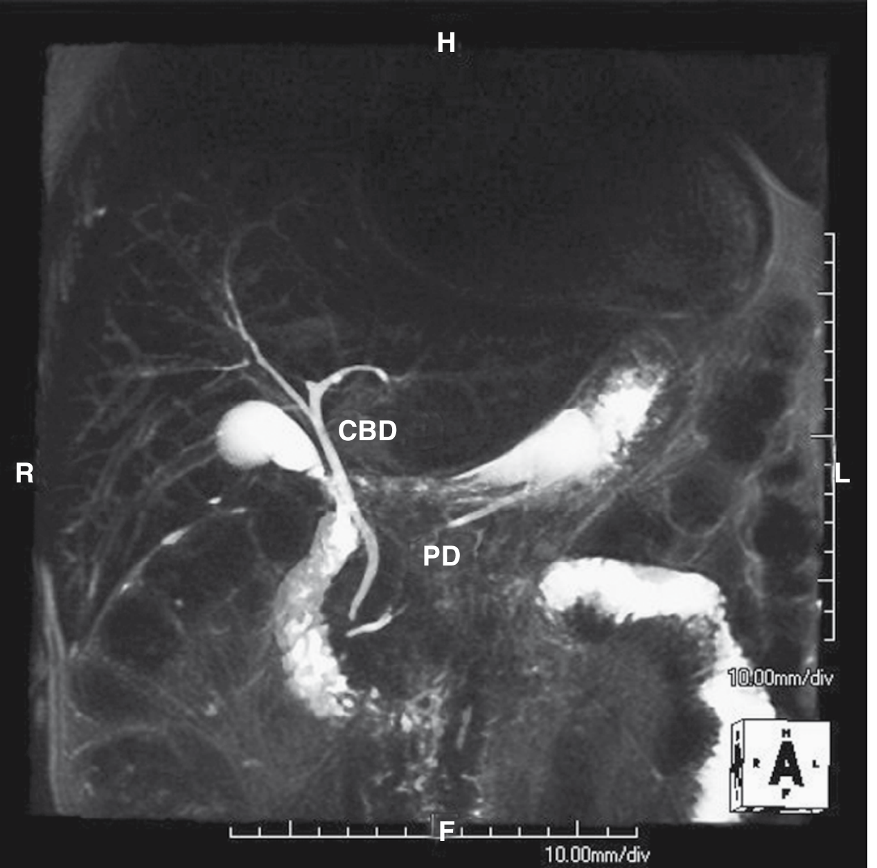
Normal MRCP: The CBD and pancreatic duct (PD) are clearly delineated as bright structures. Gallbladder is also seen as a bright round structure (Sabiston Surgery, 21st Ed)
Hepatobiliary Contrast-Enhanced MRCP
More recently, hepatobiliary MR contrast agents (gadobenate dimeglumine, gadoxetate disodium/Eovist) have been combined with MRCP:
- These agents shorten T1 relaxation and are excreted via hepatocytes into bile (biliary phase begins 10-30 minutes post-injection)
- T1-weighted sequences provide functional information in addition to anatomical detail
- Particularly useful for detecting biliary leaks, variant anatomy, and small liver metastases in the hepatobiliary phase
- Requires near-normal hepatocyte function for adequate biliary excretion
Indications for MRCP
| Category | Specific Indications |
|---|---|
| Choledocholithiasis | Suspected CBD stones in intermediate-risk patients before laparoscopic cholecystectomy (sensitivity >90%, specificity ~99%); defines need for ERCP |
| Biliary strictures | Malignant vs. benign strictures; Bismuth classification of hilar cholangiocarcinoma (Klatskin tumour); PSC |
| Obstructive jaundice | Non-invasive delineation of level and cause of obstruction |
| Pancreatic pathology | Pancreatic ductal carcinoma (double duct sign); chronic pancreatitis (duct dilation, strictures, stones, side branch changes); IPMN; pancreatic divisum |
| Congenital anomalies | Choledochal cysts (Todani classification); biliary atresia evaluation; anomalous pancreaticobiliary junction |
| Post-surgical assessment | Biliary anastomotic strictures after hepaticojejunostomy; post-cholecystectomy bile leaks; altered anatomy (Roux-en-Y gastric bypass, Billroth II) where ERCP is technically difficult/impossible |
| Failed or incomplete ERCP | When cannulation fails (5-10% of ERCP cases); MRCP provides roadmap for PTC or re-attempt |
| Pre-operative planning | Before liver resection for biliary tumours; hilar cholangiocarcinoma staging; mapping biliary anatomy before complex hepatectomy |
| Primary sclerosing cholangitis (PSC) | Classical "beads on a string" appearance of multifocal strictures alternating with dilated segments in both intra- and extrahepatic ducts |
MRCP Findings in Common Surgical Conditions
| Condition | MRCP Finding |
|---|---|
| CBD stone (choledocholithiasis) | Low-signal (dark) filling defect within the hyperintense bile duct, usually with upstream duct dilatation; target sign on axial images |
| Carcinoma head of pancreas | Abrupt cut-off of both CBD and pancreatic duct = "Double duct sign" (pathognomonic of malignant obstruction at the head); proximal duct dilation |
| Cholangiocarcinoma (Klatskin) | Stricture at biliary confluence; upstream segmental or bilobar intrahepatic duct dilation; Bismuth type I-IV classified by extent of involvement |
| Benign biliary stricture | Smooth, gradual tapering; typically short segment |
| Chronic pancreatitis | Main pancreatic duct dilation (>3mm), beading, irregularity; intraductal stones (signal voids); side branch dilation; communicating pseudocysts |
| Choledochal cyst | Cystic dilation of CBD and/or intrahepatic ducts; Todani classification |
| PSC | "Beading" - multifocal strictures and dilations throughout intrahepatic and extrahepatic ducts |
| Mirizzi syndrome | Extrinsic compression/stricture of CHD at level of cystic duct by impacted gallstone |
| Post-surgical bile leak | Extravasation of high-signal fluid outside the duct; with hepatobiliary contrast - pooling of excreted agent at site of leak |
Advantages of MRCP
- Non-invasive - no endoscopic access, no radiation, no ductal contrast injection
- No ionizing radiation - safe in pregnancy (ERCP in pregnancy carries fetal radiation risk)
- No sedation or anaesthesia required in most cases
- No risk of post-procedure pancreatitis (the most feared ERCP complication)
- Images both sides of a complete obstruction - ERCP can only show ducts distal to total obstruction; MRCP shows both the proximal and distal biliary tree
- Can be combined with conventional MRI - evaluates periductal structures, hepatic parenchyma, vascular anatomy, lymph nodes in one examination (ERCP cannot)
- Useful in altered anatomy - Roux-en-Y, Billroth II, hepaticojejunostomy - where ERCP access is impossible or technically very difficult
- No operator-dependent failure - unlike ERCP which fails in 5-10% due to unsuccessful cannulation
- Excellent sensitivity/specificity - for choledocholithiasis: sensitivity >90%, specificity ~99%
Limitations and Disadvantages of MRCP
| Limitation | Detail |
|---|---|
| No therapeutic capability | Cannot perform sphincterotomy, stone extraction, stent placement, biopsy - purely diagnostic |
| Motion artefacts | Longer acquisition times make it prone to respiratory and peristaltic motion artefacts (less of an issue with breath-hold sequences) |
| Small stones may be missed | Stones <3 mm, especially at the ampulla, can be missed; source thin-section images must always be reviewed as MIP post-processing can obscure small stones |
| False positives - clip artefacts | Surgical clips cause local signal voids that can mimic or obscure stones |
| Bile flow voids | Turbulent bile flow can mimic filling defects; these are centrally placed with less defined margins than true stones |
| Intraductal gas/blood | Can cause signal voids mimicking stones (pneumobilia after biliary-enteric anastomosis, after sphincterotomy) |
| Concentrated/inspissated bile | May produce little signal on long-TE sequences and obscure ducts |
| Claustrophobia and patient cooperation | Cannot be used in claustrophobic patients or those unable to lie still; not suitable for ventilated ICU patients without modification |
| Cost and availability | More expensive and less universally available than ERCP in resource-limited settings |
| No functional assessment | Standard MRCP does not show dynamics of bile flow (hepatobiliary contrast agents partially address this) |
| Pacemakers/metallic implants | Contraindicated in patients with non-MRI-compatible implants |
MRCP vs ERCP - Comparison
| Feature | MRCP | ERCP |
|---|---|---|
| Invasiveness | Non-invasive | Invasive (endoscope + fluoroscopy) |
| Radiation | None | Fluoroscopic radiation |
| Sedation | Usually not required | Required (conscious sedation/GA) |
| Therapeutic | No | Yes (sphincterotomy, stenting, stone extraction, biopsy) |
| Post-procedure pancreatitis | Nil | 3-5% |
| Perforation risk | Nil | 0.3-1% |
| Bleeding risk | Nil | 1-2% (after sphincterotomy) |
| Cholangitis risk | Nil | 1-3% |
| Mortality | 0% | 0.1-1% |
| Both sides of obstruction | Yes | No (only distal side) |
| Periductal assessment | Yes (combined MRI) | No |
| Altered anatomy | Excellent | Very difficult/impossible |
| Stone sensitivity | >90% | >95% |
| Success rate | ~100% | 90-95% |
Current consensus: MRCP is the first-line diagnostic investigation for suspected biliary/pancreatic pathology. ERCP is reserved for therapeutic interventions once pathology is confirmed on MRCP. "MRCP first, ERCP only if therapy needed."
MRCP in Surgical Decision Making
-
Choledocholithiasis workup before cholecystectomy:
- Low risk patients: proceed to laparoscopic cholecystectomy with intraoperative cholangiogram
- Intermediate risk: MRCP first - if CBD stone confirmed, pre-operative ERCP + stone clearance then cholecystectomy; if negative, proceed directly to cholecystectomy
- High risk (cholangitis, dilated CBD >8 mm): proceed to ERCP directly
-
Suspected cholangiocarcinoma: MRCP + MRI defines Bismuth classification, vascular involvement, and resectability - guides decision between resection, transplant, or palliation
-
Pre-hepatectomy biliary mapping: MRCP defines variant biliary anatomy (right posterior sectoral duct draining into CHD, trifurcation) to prevent inadvertent ductal injury during liver resection
-
Post-cholecystectomy bile leak/injury: MRCP characterizes the level and type of bile duct injury (Strasberg classification) before surgical repair planning
-
Acute biliary pancreatitis: MRCP detects persistent CBD stone to determine if ERCP is needed
Conclusion
MRCP has transformed the investigation of hepatobiliary and pancreatic diseases. Its combination of excellent diagnostic accuracy, complete safety, non-invasiveness, and ability to assess periductal structures makes it the first-line imaging modality for evaluating the biliary and pancreatic ductal systems. In surgical practice, MRCP provides the biliary "road map" before any operative or endoscopic intervention, reduces unnecessary diagnostic ERCP, and is irreplaceable in patients with altered anatomy or after failed ERCP.
References:
- Bailey and Love's Short Practice of Surgery, 28th Edition
- Sabiston Textbook of Surgery, 21st Edition (MRCP, Choledocholithiasis)
- Mulholland & Greenfield's Surgery, 7th Edition
- Yamada's Textbook of Gastroenterology, 7th Edition (MRCP of Biliary System)
- Grainger & Allison's Diagnostic Radiology (Biliary Imaging)
This is a shared conversation. Sign in to Orris to start your own chat.