
Ca stomach in detail
"gastric cancer"[MeSH Terms] AND "treatment"[MeSH Terms]
gastric cancer carcinoma stomach endoscopy gross pathology linitis plastica
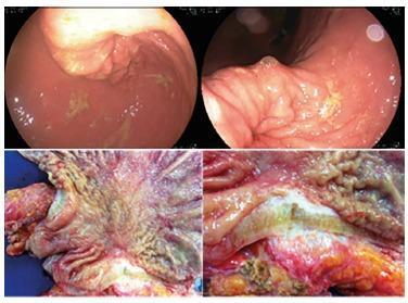
This composite figure illustrates the endoscopic and surgical presentation of metastatic breast cancer to the stomach, manifesting as linitis plastica. The top panels show upper endoscopy views of the gastric mucosa, characterized by thickened, non-distensible folds, mucosal irregularities, and pale, flesh-toned nodules. The lower panels display gross surgical pathology of resected stomach tissue. These images demonstrate significant mural thickening and rigidity, typical of the 'leather bottle' appearance seen in linitis plastica. The cut surfaces reveal a dense, fibrous, whitish-yellow infiltrating process that replaces the normal gastric layers, alongside areas of mucosal ulceration and erythematous inflammation. This visual comparison highlights the correlation between live endoscopic findings of malignancy and the resultant architectural distortion and transmural fibrosis observed in surgical specimens.
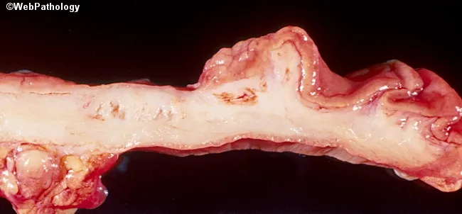
This is a gross pathology photograph of a gastric wall segment from a surgical resection, illustrating diffuse tumor infiltration consistent with signet ring cell carcinoma of the stomach. Imaging modality: gross pathology photography; specimen is a stomach/gastric wall section oriented longitudinally to reveal the mural involvement. The gastric wall is markedly thickened and firm, with a pale, tan to gray appearance and irregular, cobblestoned surface; there is minimal discreet tumor mass, reflecting a diffuse infiltrative growth pattern. The lesion corresponds to the diffuse-type gastric adenocarcinoma (signet ring cell) and demonstrates linitis plastica–like remodeling of the gastric wall, with infiltration extending from mucosa through submucosa and muscularis propria. Histologic subtype: signet ring cell carcinoma characterized by mucin-filled cells with peripherally displaced round nuclei, often discohesive, producing diffuse spread rather than a mass-forming lesion. Clinically, this pattern is associated with aggressive behavior, early transmural spread, and poor prognosis. Diagnostic significance: confirms gastric malignancy type as diffuse signet ring cell carcinoma; supports staging workup, prognosis assessment, and therapeutic planning (gastrectomy with lymphadenectomy and systemic therapy). Potential clinical use cases include education on diffuse gastric cancer morphology, gross-pathology correlation, and radiologic-pathologic correlation for linitis plastica presentations.
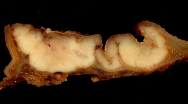
Gross pathology photograph of a gastric wall longitudinal section demonstrating infiltrative adenocarcinoma with diffuse wall thickening. The specimen shows the stomach cut along its greater curvature, with the duodenal margin at the left, allowing assessment of tumor extension across the pyloric region. The tumor invades deeply, producing marked, irregular luminal distortion and a diffuse, leathery thickening consistent with linitis plastica. The mucosa appears variably pale and fibrotic, with firm, white-tan tumor tissue intermingled with submucosal fat and serosa. Histologically, the described subtype is signet ring cell carcinoma, characterized by tumor cells with intracytoplasmic mucin pushing the nucleus to the periphery, resulting in cells with a signet-ring appearance; this pattern accounts for the diffuse infiltration and stiffness. The lesion lacks clear circumscribed mass and instead permeates through wall layers, explaining the poor prognosis associated with diffuse-type gastric cancer. Clinically relevant implications include aggressive behavior, early transmural spread, and potential involvement of regional lymphatics. This image is useful for education on gross morphology of diffuse gastric adenocarcinoma, signet-ring cytology, and to illustrate differential considerations such as intestinal-type carcinoma, lymphoma, or inflammatory processes. Documentation notes: image courtesy of Ed Uthman, MD, Houston, Texas. Clinical note: correlates with gross pathology findings and surgical planning.
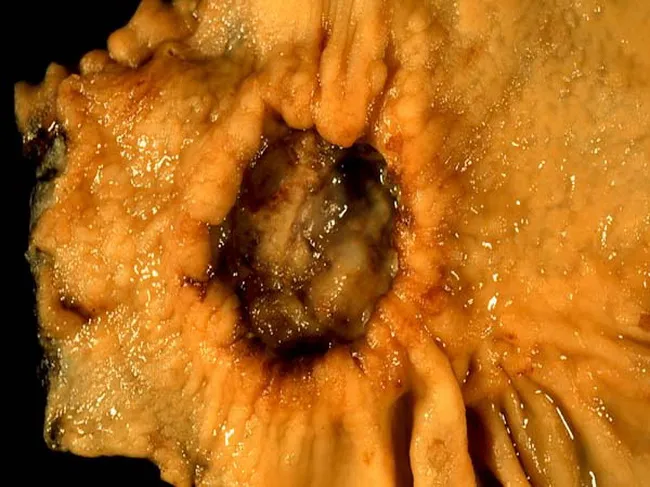
This gross pathology photograph documents a subtotal gastrectomy specimen from a 40-year-old female with abdominal pain. The gastric wall at the anterior aspect exhibits marked thickening and rigidity with a central ulcer crater on the lesser curvature. The lesion is firm and tan-yellow with irregular, serpiginous margins and surrounding diffusely involved mucosa. The pyloric margin is oriented to the left, consistent with distal stomach resection. The thickened wall and ulceration are characteristic of diffuse-type gastric carcinoma, particularly signet ring cell carcinoma, which classically produces diffuse infiltration (linitis plastica pattern) and loss of normal gastric rugal folds. While histology confirms signet-ring cells with intracellular mucin and nuclear displacement, gross assessment emphasizes wall stiffening and circumferential spread rather than a discrete mass. The clinical history notes a biopsy-proven signet ring cell carcinoma, and this resection aims to relieve obstruction and achieve cytoreduction. In diffuse gastric cancer, involvement of the prepyloric region and lesser curvature correlates with pyloric obstruction risk and poor prognosis; subtotal gastrectomy provides palliation and potential survival benefit in selected cases. This image is educational for surgical pathology, illustrating tumor localization, wall thickening, ulceration, and gross patterns of diffuse gastric malignancy, and can support differential diagnoses such as linitis plastica vs focal mucosal ulcerating carcinoma in teaching cases.
early gastric cancer Japanese classification endoscopy
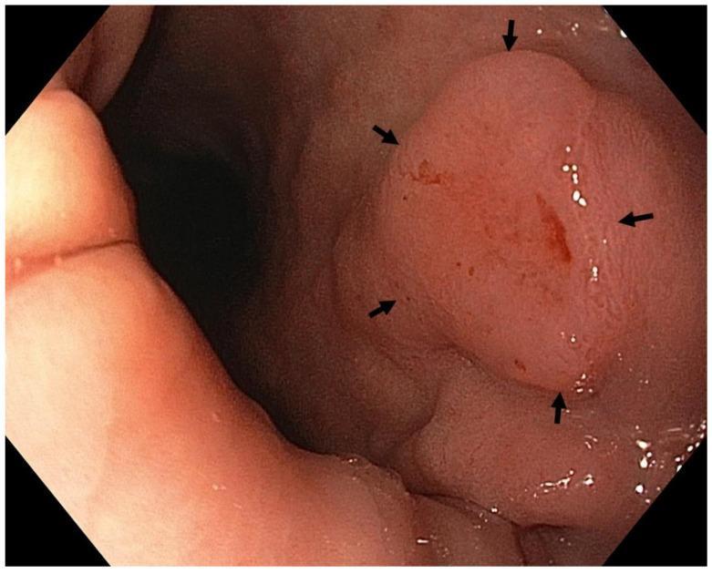
This High-Definition White Light Endoscopy (WLE) image displays a prominent gastric lesion located in the antrum. The content demonstrates clinical findings of early gastric cancer, specifically a moderately differentiated intramucosal adenocarcinoma categorized as Paris classification 0-IIb. The primary lesion is an elevated, rounded, pinkish mass with relatively well-defined margins indicated by black arrows. The surface of the mass exhibits subtle irregularities and focal areas of erythema, including small red streaks and spots suggestive of spontaneous oozing or hypervascularity. The surrounding gastric mucosa shows signs of autoimmune gastritis with atrophy, characterized by a paler pink hue and a smoother texture compared to the neoplastic lesion. This diagnostic image serves as an educational example for identifying precancerous and early neoplastic changes in the stomach, highlighting the transition from atrophic gastritis to intestinal-type adenocarcinoma.
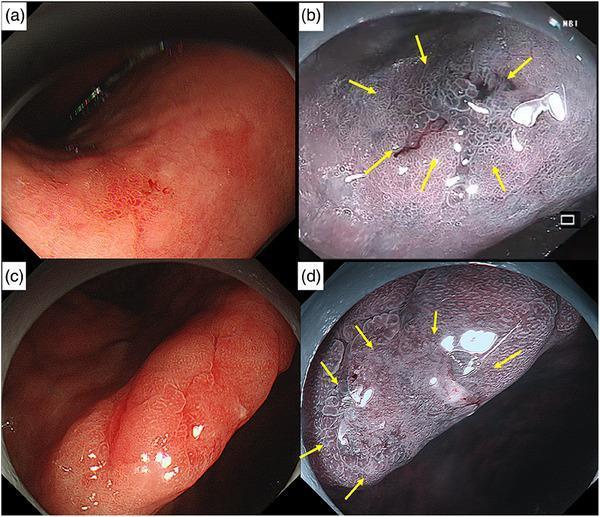
This set of diagnostic images showcases early gastric cancer (EGC) using endoscopy in the stomach. Panels (a) and (c) display conventional white light imaging (WLI), revealing small (7–8 mm), reddish, superficial depressed lesions (Paris classification 0-IIc) located at the lesser curvature of the lower corpus and the incisura angularis, respectively. Panels (b) and (d) utilize Narrow Band Imaging (NBI) and magnified NBI to enhance visualization of the pathology. These images clearly demonstrate an irregular mucosal surface pattern and irregular microvascular architecture, both delimited by a sharp demarcation line (indicated by yellow arrows) that separates the malignant tissue from the surrounding atrophic gastric mucosa. The findings are characteristic of well-differentiated early gastric adenocarcinoma limited to the lamina propria. These visuals serve as educational examples of utilizing advanced endoscopic modalities to identify subtle neoplastic changes in the setting of chronic atrophic gastritis and Helicobacter pylori infection.
Carcinoma of the Stomach - Comprehensive Review
1. Epidemiology
| Region | Incidence (per 100,000/year) |
|---|---|
| USA | ~10 |
| UK | ~15 |
| Eastern Europe | ~40 |
| Japan | ~70 |
| Some areas of China | >70 |
- Men are affected more than women; incidence rises with age.
- Overall cure rates in the West are only 5-10%, though Japan achieves far better outcomes due to population screening.
- In the West, distal gastric cancer incidence is falling ~1% per year, but proximal (cardia/GOJ) cancer is rising - associated with obesity and higher socioeconomic status rather than H. pylori. - Bailey and Love's Surgery, 28th ed.
2. Aetiology and Risk Factors
- H. pylori - strongly associated with carcinoma of the body and distal stomach (intestinal type); NOT a risk factor for proximal/cardia cancers.
- Pernicious anaemia and gastric atrophy
- Gastric adenomatous polyps
- Intestinal metaplasia
- Post-gastrectomy stomach (Billroth II / Polya gastrectomy / gastroenterostomy) - ~4x average risk, related to bile reflux
- High salt intake
- Deficiency of antioxidants (vitamin C, E, beta-carotene)
- Exposure to N-nitroso compounds
- Cigarette smoking
- Industrial dust ingestion
- CDH1 (E-cadherin) mutations - associated with hereditary diffuse gastric cancer
- Mutations in BRCA2, TP53, ARID1A, RHOA
- Lynch syndrome (MSI-H tumours)
3. Pathology
A. Histological Type - Laurén Classification
| Feature | Intestinal Type | Diffuse Type |
|---|---|---|
| Morphology | Gland-forming, tubular | Discohesive cells, signet-ring cells |
| Growth pattern | Polypoid or ulcerating | Infiltrative (no mass lesion) |
| Origin | Areas of intestinal metaplasia | De novo |
| H. pylori association | Yes | Less so |
| CDH1 mutation | Rare | Common |
| Prognosis | Better | Worse |
| Spread | Less aggressive early spread | Submucosal/subserosal spread, early transmural |
B. Japanese Classification - Early Gastric Cancer (EGC)
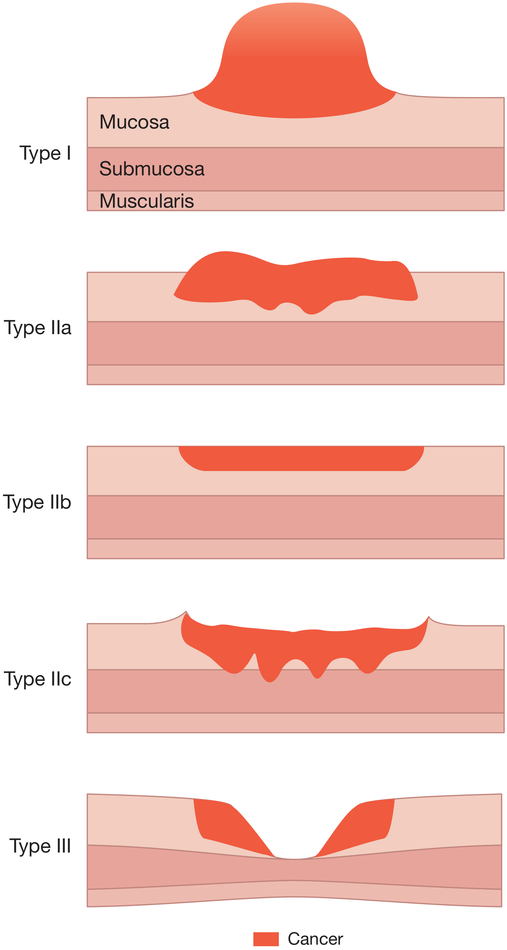
| Type | Description |
|---|---|
| Type I | Protruding |
| Type IIa | Superficial elevated |
| Type IIb | Superficial flat |
| Type IIc | Superficial depressed |
| Type III | Excavated/ulcerated |
C. Borrmann Classification - Advanced Gastric Cancer
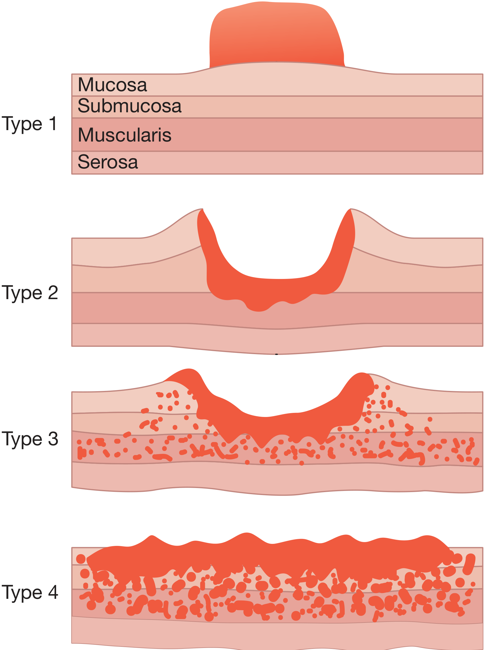
| Type | Appearance | Prognosis |
|---|---|---|
| Type 1 | Polypoid | Best |
| Type 2 | Ulcerating with sharp margins | Good |
| Type 3 | Ulcerating + infiltrating | Intermediate |
| Type 4 | Diffuse infiltrating (linitis plastica) | Worst |
4. Molecular Subtypes (TCGA Classification)
- EBV-positive - PIK3CA mutations, CDKN2A silencing; associated with better immunotherapy responses
- Microsatellite Unstable (MSI-H) - hypermutation, MLH1 silencing; best prognosis; responds to immunotherapy (pembrolizumab)
- Genomically Stable (GS) - CDH1/RHOA mutations; corresponds to diffuse/signet-ring type; worst prognosis
- Chromosomal Instability (CIN) - TP53 mutations, receptor tyrosine kinase amplification; intestinal type; HER2 amplification common here
5. Modes of Spread
Direct spread
- Tumour penetrates muscularis → serosa → adjacent organs (pancreas, colon, liver)
Lymphatic spread
- Via permeation and emboli to regional lymph node stations (Japanese classification assigns numbers to each)
- Troisier's sign = tumour in left supraclavicular node (Virchow's node)
- Unlike breast cancer, nodal involvement does NOT necessarily imply systemic dissemination
Blood-borne spread
- First to the liver, then lung and bone
- Uncommon in the absence of nodal disease
Transperitoneal spread
- Once tumour reaches serosa - implies incurability
- Krukenberg tumour = transcoelomic spread to ovaries
- Sister Mary Joseph's nodule = umbilical metastasis
- Blumer's shelf = rectal shelf deposit
Special features of diffuse type
- Spreads via submucosal and subserosal lymphatic plexus, penetrating the gastric wall early - Bailey and Love's Surgery, 28th ed.
6. Clinical Features
Symptoms of early gastric cancer:
- No specific features - indistinguishable from benign dyspepsia
- High index of suspicion is required (alarm features: unexplained weight loss, dysphagia, vomiting, anaemia >55 years)
Symptoms of advanced cancer:
- Early satiety, bloating, distension
- Vomiting (pyloric obstruction → gastric outlet obstruction with hypochloraemic hypokalaemic alkalosis, though less pronounced than duodenal ulcer obstruction)
- Iron deficiency anaemia (occult bleeding)
- Dysphagia (GOJ/cardia tumours)
- Profound weight loss, cachexia
Paraneoplastic/non-metastatic signs:
- Trousseau's sign - migratory thrombophlebitis
- Deep vein thrombosis
- Acanthosis nigricans
Signs of advanced/metastatic disease:
- Epigastric mass (palpable tumour or liver)
- Virchow's node (Troisier's sign)
- Sister Mary Joseph's nodule (umbilical)
- Blumer's shelf (rectal)
- Krukenberg tumour (ovarian)
- Ascites
7. Investigations
Endoscopy
- Gold standard for diagnosis - allows direct visualization and biopsy
- Multiple biopsies needed (at least 8-10 from suspicious lesions)
- Endoscopic ultrasound (EUS) - best for local T and N staging
Imaging
- CT chest/abdomen/pelvis - distant metastases, lymph nodes, ascites
- PET-CT - occult metastases
- Staging laparoscopy - mandatory before planned surgery; biopsy of suspicious lesions; peritoneal washings (positive cytology = M1 disease)
- Barium meal (historical) - "rat's tail" stricture at cardia, filling defect in body
Laboratory
- FBC (anaemia), LFTs, tumour markers (CEA, CA 19-9, CA 72-4 - not specific but useful for monitoring)
- HER2 testing (IHC or FISH) - guides use of trastuzumab
- MSI/MMR testing - guides immunotherapy eligibility
8. TNM Staging (UICC 8th Edition)
| Stage | Criteria |
|---|---|
| Tis | Carcinoma in situ - no lamina propria invasion |
| T1a | Invades lamina propria or muscularis mucosae |
| T1b | Invades submucosa |
| T2 | Invades muscularis propria |
| T3 | Invades subserosa |
| T4a | Perforates serosa (visceral peritoneum) |
| T4b | Invades adjacent structures |
| N0 | No nodal metastasis |
| N1 | 1-2 regional nodes |
| N2 | 3-6 regional nodes |
| N3a | 7-15 regional nodes |
| N3b | ≥16 regional nodes |
| M1 | Distant metastasis (including positive peritoneal cytology) |
9. Treatment
A. Endoscopic Resection (EGC only)
- Endoscopic Mucosal Resection (EMR) or Endoscopic Submucosal Dissection (ESD)
- Suitable for T1a lesions <2 cm, well-differentiated, no ulceration, no lymphovascular invasion
- Very high cure rates in Japan
B. Neoadjuvant (Perioperative) Chemotherapy
- Indicated for all operable patients - Level 1 evidence of improved survival
- FLOT regimen (fluorouracil, leucovorin, oxaliplatin, docetaxel) is now standard of care, replacing ECF
- FLOT4 trial: FLOT vs ECF - median survival 50 months vs 35 months (significant advantage for FLOT)
- MRC-MAGIC trial established perioperative chemotherapy principle (ECF regimen)
- Note: <50% of patients receive the full postoperative component in practice
C. Surgery - Curative Intent
- Resection margins must be >5 cm clear of tumour; frozen section if in doubt
- Minimum D2 lymphadenectomy is standard at specialist centres
- Spleen and pancreas preservation wherever possible
| Operation | Indication |
|---|---|
| Total gastrectomy | Proximal and mid-body tumours |
| Subtotal (distal) gastrectomy | Antral and distal tumours |
| Proximal gastrectomy | Cardia tumours (controversial) |
- D1 = perigastric nodes (station 1-6)
- D2 = D1 + nodes along major arterial trunks (stations 7-11)
- D2 is the standard; associated with improved locoregional control
- Station 10 (splenic hilum) nodes typically spared when spleen is conserved
D. Palliative Surgery
- Gastrectomy for palliation of obstruction or bleeding
- REGATTA trial: palliative gastrectomy + chemotherapy vs chemotherapy alone showed NO survival benefit; chemotherapy alone is preferred for metastatic disease
E. Palliative Chemotherapy (Advanced/Metastatic Disease)
- First-line platinum + fluoropyrimidine backbone (FOLFOX, XELOX/CAPOX)
- HER2-positive tumours (~15-20%): add trastuzumab (Herceptin) to chemotherapy - ToGA trial showed survival benefit
- Trastuzumab deruxtecan (T-DXd): antibody-drug conjugate for HER2-overexpressing tumours after prior therapy
- MSI-H/dMMR tumours: Pembrolizumab (anti-PD-1) approved for first-line treatment; also nivolumab (CHECKMATE trials)
- PD-L1 CPS (combined positive score) guides immunotherapy eligibility
F. Radiotherapy
- Adjuvant chemoradiation (INT-0116/MacDonald regimen) - used mainly in the USA for R0 resection
- Less commonly used in the UK/Europe where perioperative chemotherapy is preferred
G. Peritoneal Disease - HIPEC
- CRS (cytoreductive surgery) + HIPEC (hyperthermic intraperitoneal chemotherapy) - considered for patients with limited peritoneal disease (PCI <6) who respond to systemic therapy, ideally in a trial setting
- GASTRICHIP trial ongoing; GYMSSA trial showed median survival 11.3 vs 4.3 months (CRS+HIPEC vs systemic alone)
- Sabiston Textbook of Surgery, 21st ed.
10. Complications of Surgery
- Anastomotic leak (oesophagojejunal or duodenal stump)
- Secondary haemorrhage from exposed vessels
- Biliary peritonitis (duodenal stump leak)
- Long-term:
- Dumping syndrome (early and late)
- Diarrhoea, bile reflux gastritis
- Vitamin B12 deficiency (loss of parietal cell mass) - requires lifelong B12 replacement
- Nutritional deficiencies (iron, fat-soluble vitamins)
- Afferent loop syndrome (after Billroth II)
11. Prognosis
| Stage | Approximate 5-Year Survival |
|---|---|
| Early gastric cancer (T1) | ~90% |
| Stage I | 60-80% |
| Stage II | 30-50% |
| Stage III | 10-30% |
| Stage IV (metastatic) | <5% |
12. Special Situations
Hereditary Diffuse Gastric Cancer (HDGC)
- Autosomal dominant CDH1 mutation
- Lifetime risk of diffuse gastric cancer ~70-80%
- Prophylactic total gastrectomy recommended for confirmed mutation carriers
Gastric Stump Cancer
- Occurs 15-30 years after Billroth II or other drainage procedures
- 4x increased risk compared to general population
- Related to bile reflux and intestinal metaplasia at the anastomotic site
- Regular endoscopic surveillance recommended from 15 years post-surgery
GOJ/Siewert Classification
- Type I: Distal oesophageal (adenocarcinoma arising from Barrett's oesophagus)
- Type II: True cardia tumour (epicentre 1 cm above to 2 cm below GOJ)
- Type III: Subcardia gastric tumour (2-5 cm below GOJ)
Create a treatment algorithm flowchart for gastric cancer management
A detailed clinical treatment algorithm flowchart for gastric cancer management. The flowchart starts at the top with "GASTRIC CANCER DIAGNOSIS (Endoscopy + Biopsy + CT Staging)" and branches downward through the following decision points and pathways: First decision diamond: "Staging Workup Complete: CT Chest/Abdomen/Pelvis + EUS + Staging Laparoscopy" Second decision diamond: "Metastatic / Unresectable Disease?" with YES branch going right and NO branch going down. YES branch (Metastatic pathway - shown in red/orange): - Box: "Test: HER2 status (IHC/FISH) + MSI/MMR status + PD-L1 CPS" - Three sub-branches: 1. "HER2+ (IHC 3+ or 2+/FISH+)" → "Trastuzumab + Platinum + Fluoropyrimidine (TOGA regimen)" 2. "MSI-H / dMMR" → "Pembrolizumab + Chemotherapy (1st line) or Pembrolizumab monotherapy" 3. "HER2- / MSI-stable" → "FOLFOX or XELOX (Platinum + Fluoropyrimidine)" - All three merge → "Best Supportive Care / Palliative care" - Note: "2nd line: Ramucirumab ± Paclitaxel; T-DXd for HER2+ refractory" NO branch (Potentially Resectable - shown in blue/green): Decision diamond: "Early Gastric Cancer (T1a, well-diff, <2cm, no LVI)?" - YES → "Endoscopic Resection: EMR or ESD" → "Surveillance Endoscopy" - NO → Continue down Next box: "Medically Fit for Surgery?" - NO → "Palliative Chemotherapy / BSC" - YES → Continue Next box: "Neoadjuvant Chemotherapy (FLOT × 4 cycles): Fluorouracil + Leucovorin + Oxaliplatin + Docetaxel" → "Restage: CT + EUS" Decision diamond: "Resectable after neoadjuvant?" - NO → "Palliative Chemotherapy" - YES → Continue Next box: "SURGERY" Two branches: - "Distal/Antral Tumour" → "Subtotal (Distal) Gastrectomy + D2 Lymphadenectomy" - "Proximal/Body Tumour or Diffuse" → "Total Gastrectomy + D2 Lymphadenectomy + Roux-en-Y Reconstruction" Both merge → Decision diamond: "Resection Margin Status?" - "R0 (clear)" → "Adjuvant FLOT × 4 cycles → Surveillance" - "R1/R2 (involved)" → "Consider adjuvant chemoradiation or further chemotherapy" Bottom of flowchart: "SURVEILLANCE: Clinical review + CT every 6 months × 3 years, then annually. Vitamin B12 replacement (post-gastrectomy)" Use a clean professional medical infographic style with color-coded pathways: blue for resectable/surgical pathway, red/orange for metastatic pathway, green for early cancer/endoscopic pathway. Diamond shapes for decision points, rounded rectangles for actions. Clear arrows connecting all boxes. White background with bold readable text. Title at top: "GASTRIC CANCER MANAGEMENT ALGORITHM" with subtitle "Based on FLOT4, TOGA, KEYNOTE trials"
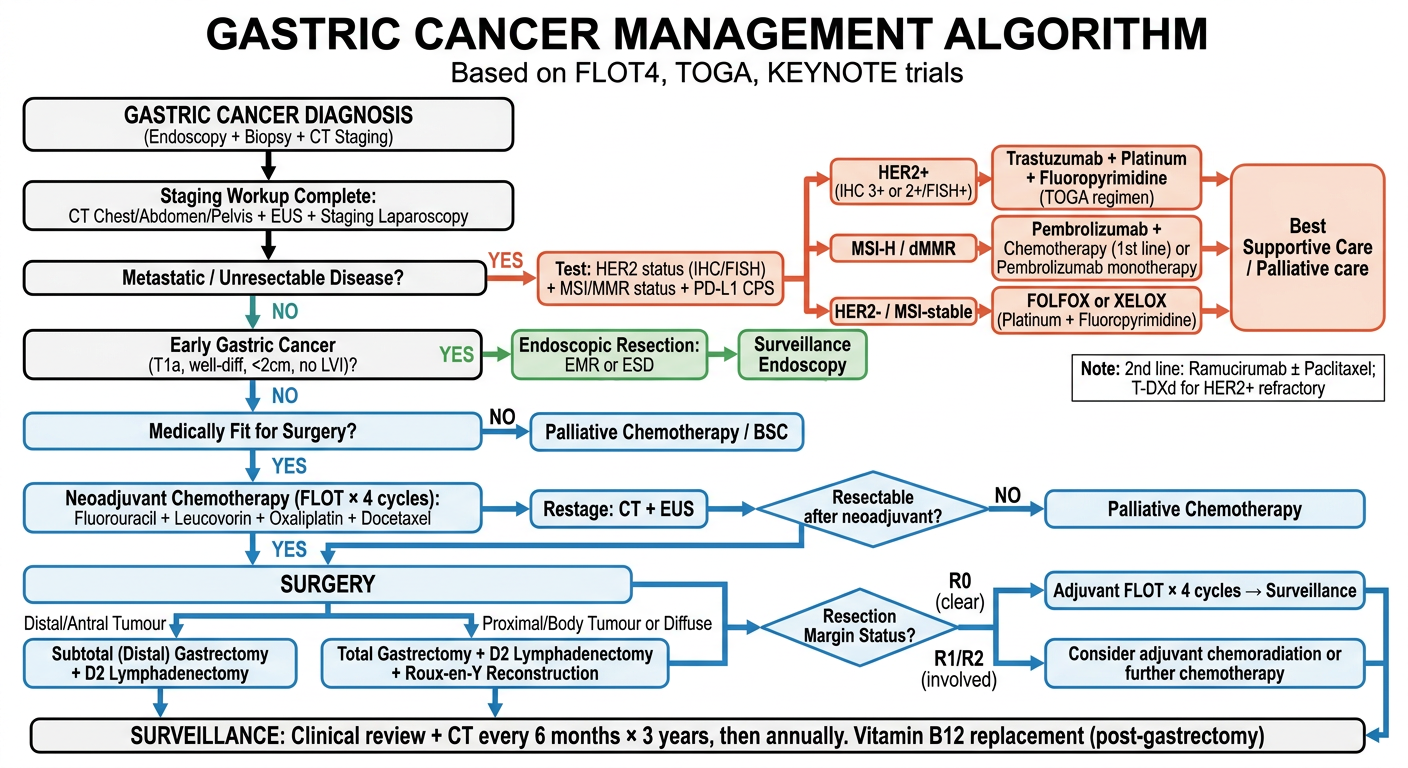
Resectable / Surgical Pathway (Blue)
- Early gastric cancer (T1a, well-differentiated, <2 cm, no LVI): Endoscopic resection (EMR/ESD) + surveillance
- Resectable disease: Neoadjuvant FLOT x4 cycles → Surgery → Adjuvant FLOT x4 cycles
- Distal/antral tumours: Subtotal gastrectomy + D2 lymphadenectomy
- Proximal/body/diffuse: Total gastrectomy + D2 + Roux-en-Y reconstruction
- R0 → adjuvant chemotherapy + surveillance
- R1/R2 → chemoradiation considered
Metastatic / Palliative Pathway (Red/Orange)
- Biomarker testing first: HER2 (IHC/FISH), MSI/MMR, PD-L1 CPS
- HER2+: Trastuzumab + platinum + fluoropyrimidine (TOGA regimen)
- MSI-H/dMMR: Pembrolizumab + chemotherapy (KEYNOTE-590/811)
- HER2−, MSI-stable: FOLFOX or XELOX
- 2nd line: Ramucirumab ± paclitaxel; T-DXd for HER2+ refractory disease
Post-gastrectomy
- Surveillance CT every 6 months x3 years
- Vitamin B12 replacement lifelong (loss of parietal cell mass)