
https://youtu.be/g5B6WOOY1E0?si=7QEN3UISfIu7OMKa /generatebestnotes on the link given above for 1st year mbbs student act as a professor and /teachme each and every point in /deepdive don't miss even a single point explain with the help of images Provide USMLE type questions on this topic with /3levels /doughtkiller.
Reading File
First, let me analyse the YouTube video to understand what topic it covers.
analyse_media
The video covers Ear Anatomy - External, Middle, and Inner Ear (Special Senses). Now let me search the medical library for comprehensive content on this topic in parallel.
Finding Sources
Finding Sources
Searching the Web
ear anatomy external middle inner ear physiology hearing balance 1st year MBBS labeled diagrams
Finding Sources
Finding Sources
Loading Image
Excellent content found. Now let me read the key textbook sections in parallel for deep content.
Reading File
Reading File
Loading Image
Reading File
Reading File
Loading Image
Reading File
Loading Image
Searching the Web
auricle pinna anatomy landmarks helix antihelix tragus diagram labeled USMLE
Loading Image
Loading Image
Finding Sources
Reading File
Reading File
I now have rich, comprehensive content from Gray's Anatomy for Students, Guyton & Hall Medical Physiology, and supporting images. Let me compile the complete masterclass notes.
🎓 PROFESSOR'S MASTERCLASS NOTES
Ear Anatomy & Physiology of Hearing and Balance
Based on: Taim Talks Med | For 1st Year MBBS
Watch the original video here:
🗺️ THE BIG PICTURE FIRST
The ear is a precision sensory organ with two entirely distinct jobs:
- Hearing - convert sound waves (air vibrations) into electrical nerve impulses
- Balance - detect the position and movement of your head in space
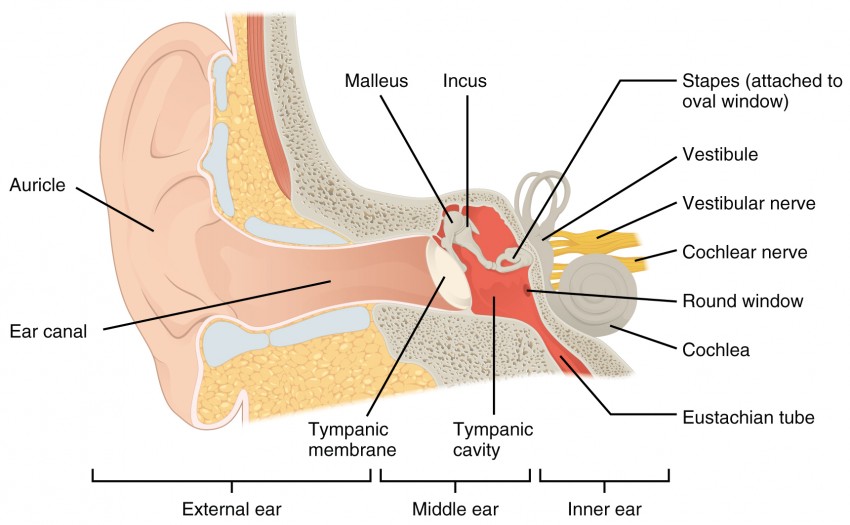
It is divided into three anatomical regions:
| Region | Boundary | Main Function |
|---|---|---|
| External Ear | Outer world → Tympanic membrane | Collect & funnel sound |
| Middle Ear | Tympanic membrane → Oval window | Amplify & transmit vibrations |
| Inner Ear | Oval window inward | Transduce vibration into nerve signals; balance |
Here is the complete overview:
PART 1: THE EXTERNAL EAR (Auris Externa)
1A. The Auricle / Pinna
The auricle (pinna) is the visible, projecting part of the ear made of elastic cartilage covered by thin skin. Its job is to collect sound waves and funnel them into the ear canal. It has no cartilage in the earlobe (lobule) - that's why earrings don't hurt the way they would elsewhere.
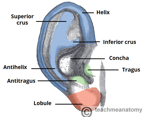
Key Landmarks of the Auricle (MUST MEMORIZE):
| Landmark | What It Is | Memory Hook |
|---|---|---|
| Helix | The curved outer rim | "H for Hood" - outermost rim |
| Antihelix | The inner curved ridge parallel to helix | "Anti" = opposite/inner |
| Superior & Inferior crura | Two branches of antihelix forming a Y | The "fork" of antihelix |
| Concha | The deep central hollow (bowl) | "Concha" = shell in Latin; funnels sound |
| Tragus | Small projection over ear canal opening | "Trag" = goat (has hairs in old age) |
| Antitragus | Small projection opposite tragus | Opposite to tragus |
| Intertragic notch | Notch between tragus & antitragus | The gap between them |
| Lobule | The earlobe at the bottom | No cartilage - has fat |
| Triangular fossa | Depression between superior & inferior crura | Triangle-shaped pit |
| Scapha | Narrow groove between helix & antihelix | "Boat"-shaped groove |
🔑 Professor's Tip: In otoscopy, you pull the auricle upward, backward, and outward in adults (downward in children) to straighten the ear canal.
1B. External Acoustic Meatus (Ear Canal)
The ear canal runs from the concha to the tympanic membrane - approximately 2.5 cm long.
Two Segments:
- Outer 1/3 = Cartilaginous - contains ceruminous glands (makes earwax) and hair follicles
- Inner 2/3 = Bony - very sensitive, directly drills into the temporal bone
Cerumen (Earwax): Made by modified sweat glands (ceruminous glands). It has antibacterial properties and prevents insects and debris from reaching the eardrum.
⚠️ Clinical Tip: The bony part is exquisitely sensitive. Instruments in the bony ear canal are very painful. A cotton swab pushed in too far can impact cerumen against the eardrum.
1C. The Tympanic Membrane (Eardrum)
The tympanic membrane is the boundary between the external and middle ear. It is held in position by a fibrocartilaginous ring called the annulus (tympanic ring), seated in the tympanic sulcus.
Structure: Three layers -
- Outer layer: Stratified squamous epithelium (continuous with skin of canal)
- Middle layer: Fibrous tissue (gives it stiffness)
- Inner layer: Mucous membrane (continuous with middle ear)
Two Zones:
| Zone | Location | Properties |
|---|---|---|
| Pars tensa | Lower ~90% | Taut; has all three layers; vibrates for sound |
| Pars flaccida (Shrapnell's membrane) | Superior ~10% | Lax; only 2 layers; clinically important - cholesteatoma forms here |
Key Landmarks (visible on otoscopy):
- Umbo - the central depression where the handle of the malleus attaches from behind
- Malleolar stria - bright streak from umbo upward (outline of the malleus handle beneath)
- Malleolar prominence - small white elevation at the top of the malleolar stria
- Cone of Light - triangular bright reflection in the anteroinferior quadrant (5 o'clock right ear, 7 o'clock left ear)
🔦 Otoscopy Tip: Loss of the "cone of light" in a patient with ear pain and fever = acute otitis media until proven otherwise.
Sensory Innervation of the External Ear (Classic Exam Topic!)
This is tested constantly because it explains referred otalgia (ear pain from non-ear causes):
| Nerve | Region Supplied | Parent Nerve |
|---|---|---|
| Auriculotemporal nerve | Anterosuperior auricle, anterior canal, anterior TM | CN V₃ (mandibular) |
| Great auricular nerve | Most of the auricle (lateral and medial surfaces) | C2, C3 (cervical plexus) |
| Lesser occipital nerve | Superior/posterior auricle | C2 (cervical plexus) |
| Arnold's nerve (auricular branch of vagus) | Posterior wall and floor of canal, posterior TM | CN X (vagus) |
🎯 Arnold's Reflex: Stimulating the posterior ear canal (e.g., with a syringe) can trigger coughing - because Arnold's nerve (CN X branch) supplies the ear AND the cough reflex. Dentists and ENT surgeons must know this!
🎯 Referred Otalgia (Ear pain without ear disease): A patient complains of ear pain but the ear looks completely normal. Think: dental abscess (CN V₃), tonsillitis (CN IX = Jacobson's nerve in TM), laryngeal cancer (CN X = Arnold's nerve), TMJ disease (CN V₃).
PART 2: THE MIDDLE EAR (Auris Media / Tympanic Cavity)
The middle ear is an air-filled cavity within the petrous part of the temporal bone. Think of it as a small rectangular room with 6 walls.
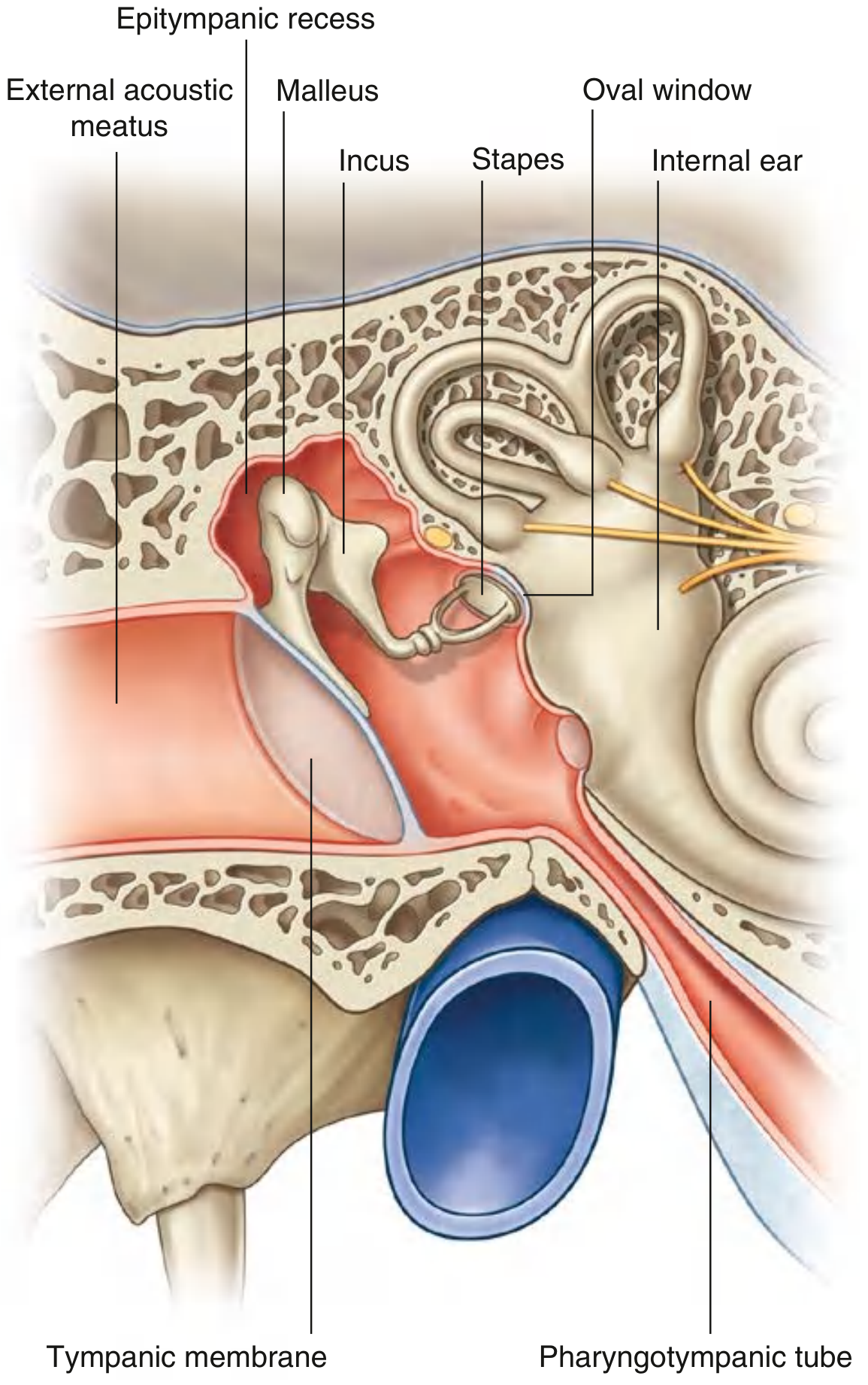
2A. The Six Walls of the Tympanic Cavity
| Wall | Also Called | Key Content / Relations |
|---|---|---|
| Roof (superior) | Tegmental wall | Tegmen tympani (thin bone) - separates middle ear from middle cranial fossa |
| Floor (inferior) | Jugular wall | Thin bone over internal jugular vein; tympanic branch of CN IX enters here |
| Lateral wall | Membranous wall | Mostly the tympanic membrane |
| Medial wall | Labyrinthine wall | Oval window, round window, promontory (cochlear turns below it) |
| Anterior wall | Carotid wall | Opening of Eustachian (pharyngotympanic) tube; canal for tensor tympani |
| Posterior wall | Mastoid wall | Aditus (opening to mastoid antrum); pyramidal eminence for stapedius muscle |
🔑 The middle ear has two regions: the mesotympanum (main part beside the TM) and the epitympanic recess (attic) - the space above the TM that contains the head of malleus and body of incus.
2B. The Auditory Ossicles (The Three Smallest Bones in the Body)
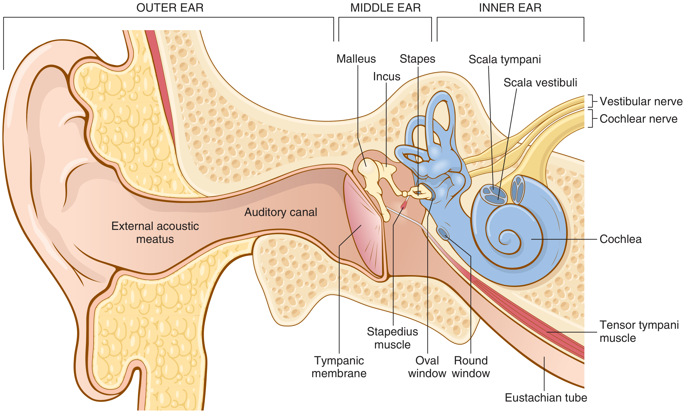
The ossicles form a chain from the eardrum to the oval window:
MALLEUS (Hammer)
- Parts: Head (sits in epitympanic recess), Neck, Handle (manubrium - embedded in TM), Lateral process, Anterior process
- The handle is attached to the center of the TM at the umbo
- Held taut by the tensor tympani muscle (innervated by CN V₃ - medial pterygoid nerve)
INCUS (Anvil)
- Parts: Body, Short limb (points backward to mastoid), Long limb (descends to connect with stapes), Lenticular process (tip)
- Connects malleus to stapes
STAPES (Stirrup) - the smallest bone in the human body
- Parts: Head, Neck, Two crura (anterior and posterior), Footplate (base)
- The footplate sits in the oval window, sealed by the annular ligament
- Moves like a piston to push perilymph in the cochlea
- Controlled by the stapedius muscle (smallest skeletal muscle; innervated by CN VII - facial nerve)
The Two Muscles of the Middle Ear
| Muscle | Bone Moved | Nerve | Function |
|---|---|---|---|
| Tensor tympani | Malleus (pulls handle medially) | CN V₃ (medial pterygoid n.) | Tenses TM; limits vibration amplitude |
| Stapedius | Stapes (pulls backward) | CN VII (facial nerve) | Acoustic reflex - protects inner ear from loud sounds |
🎯 Acoustic Reflex: A loud sound → stapedius contracts bilaterally → stiffens ossicular chain → reduces transmission of low-frequency loud sounds. This PROTECTS the cochlea. Testing the acoustic reflex with a tympanometer tests the integrity of CN VII (stapedius reflex is absent in Bell's palsy).
🎯 Hyperacusis (sounds seem painfully loud) = stapedius paralysis in Bell's palsy (CN VII damage). Patient hears own chewing as unbearably loud.
2C. Impedance Matching - The Physics of Why Ossicles Matter
Sound travels in air in the external ear but must enter fluid (perilymph) in the cochlea. The problem: fluid has much greater inertia (resistance to movement) than air. Without amplification, 99.9% of sound energy would be reflected!
The ossicular system solves this through two mechanisms:
-
Area ratio: Tympanic membrane area ≈ 55 mm² vs. stapes footplate ≈ 3.2 mm² → 17:1 ratio - the same force applied over a smaller area = greater pressure
-
Lever ratio: The ossicular lever system gives an additional 1.3x force amplification (the malleus handle is longer than the long process of the incus)
Total amplification = 17 × 1.3 = ~22 times more force in cochlear fluid than in air
This is called impedance matching and is 50-75% efficient for 300-3000 Hz - exactly the frequency range of human speech! (Source: Guyton and Hall, p.656)
💡 Without ossicles, hearing drops by 15-20 dB - equivalent to going from a normal speaking voice to barely perceptible whispering.
2D. The Pharyngotympanic (Eustachian) Tube
This tube connects the middle ear to the nasopharynx.
- Length: ~36 mm
- Composition: 1/3 bony (near middle ear) + 2/3 cartilaginous (near nasopharynx)
- Opens during: Swallowing, yawning, sneezing (levator and tensor veli palatini muscles open it)
- Function: Equalizes pressure on both sides of the TM
⚠️ Otitis Media: In children, the Eustachian tube is shorter, more horizontal, and flabbier → bacteria from the nasopharynx (e.g., S. pneumoniae, H. influenzae) travel up more easily → middle ear infection. In adults, the tube is longer and more angled (45°), making infection less common.
⚠️ Aeroplane ear (barotrauma): Rapid descent → cabin pressure rises → TM pushed inward → pain. Yawning or swallowing (Valsalva maneuver) opens the tube and equalizes pressure.
PART 3: THE INNER EAR (Auris Interna / Labyrinth)
The inner ear is the most complex part - a fluid-filled cavity system carved into the petrous temporal bone. It contains:
- The cochlea (for hearing)
- The vestibular apparatus (for balance)

3A. The Bony Labyrinth vs. Membranous Labyrinth
This is a classic MBBS exam concept:
| Bony Labyrinth | Membranous Labyrinth | |
|---|---|---|
| What is it? | Cavity in petrous temporal bone | Set of tubes/sacs inside the bony labyrinth |
| Fluid inside | Perilymph (high Na⁺, like ECF) | Endolymph (high K⁺, like ICF) |
| Divisions | Cochlea, Vestibule, 3 Semicircular Canals | Cochlear duct, Utricle, Saccule, 3 Semicircular Ducts |
🧠 Mnemonic: "Endo = Inside = like ICF = high K⁺" | "Peri = Peripheral/outside = like ECF = high Na⁺"
3B. The Cochlea - The Organ of Hearing
The cochlea is a snail-shaped spiral making 2¾ turns around a central bony axis called the modiolus.
Three fluid-filled compartments (scalae):
| Compartment | Fluid | Location |
|---|---|---|
| Scala vestibuli | Perilymph | Upper chamber; starts at oval window |
| Scala tympani | Perilymph | Lower chamber; ends at round window |
| Scala media (Cochlear duct) | Endolymph | Middle; contains Organ of Corti |
Scala vestibuli and scala tympani communicate at the apex through a small opening called the helicotrema.
Walls of the Scala Media:
- Roof: Reissner's membrane (vestibular membrane)
- Floor: Basilar membrane (contains Organ of Corti)
- Lateral wall: Stria vascularis (produces endolymph, maintains +80mV endocochlear potential)
3C. The Organ of Corti - The End Organ of Hearing
Sitting on the basilar membrane, the Organ of Corti contains the actual sound receptor cells.
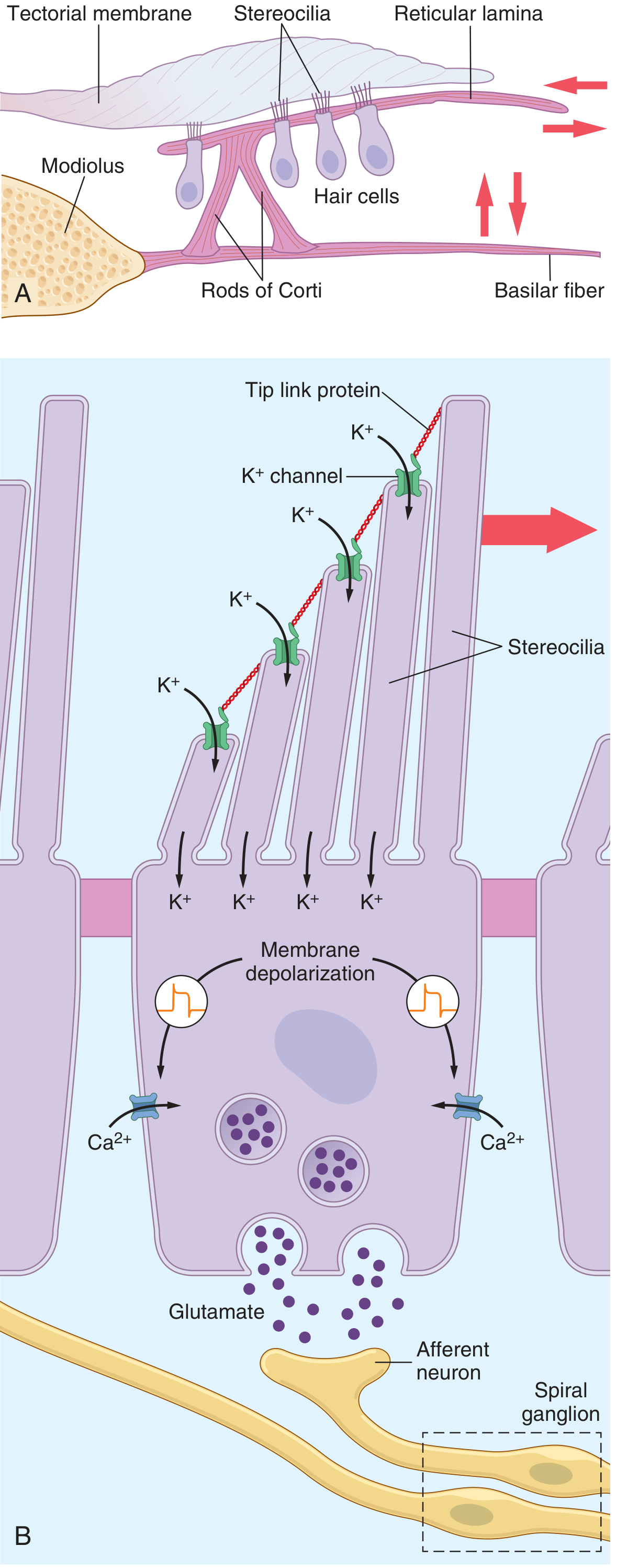
Cell types in the Organ of Corti:
- Inner hair cells (IHCs): ~3,500 cells in a single row; these are the true auditory receptors (~90% of auditory nerve fibers connect here)
- Outer hair cells (OHCs): ~12,000 cells in 3-4 rows; function in amplification and "tuning" (electromotility); if damaged → significant hearing loss
- Supporting cells: Pillar cells (rods of Corti), Deiters' cells (phalangeal cells), Hensen's cells
The Tectorial Membrane: A gelatinous membrane that lies on top of the hair cells. The stereocilia of outer hair cells are embedded in it. When the basilar membrane moves, the stereocilia shear against the tectorial membrane.
3D. Tonotopy - The Frequency Map of the Cochlea
The basilar membrane is not uniform - it acts as a frequency analyzer:
| Location | Width | Stiffness | Best Frequency |
|---|---|---|---|
| Base (near oval window) | Narrow | Stiff | High frequencies (20,000 Hz) |
| Apex (helicotrema) | Wide | Floppy | Low frequencies (20 Hz) |
🎵 Mnemonic: "BASE = High-pitched BASS guitar sounds sharp" | "APEX = Low-pitched deep bass"
This tonotopic organization is preserved all the way from the cochlea to the auditory cortex (Heschl's gyrus, superior temporal lobe).
3E. Hair Cell Transduction - How Sound Becomes Electricity
This is the most fundamental mechanism in auditory physiology:
(Source: Guyton and Hall Textbook of Medical Physiology, p.659)
Step-by-step:
- Sound wave → stapes piston movement → oval window vibrates
- Perilymph wave travels up scala vestibuli → through helicotrema → down scala tympani → bulges round window outward (round window acts as pressure relief valve)
- This fluid movement causes basilar membrane to vibrate (traveling wave)
- Basilar membrane vibration → reticular lamina rocks → stereocilia of hair cells shear against tectorial membrane
- Stereocilia bend toward taller stereocilia → tip-link proteins pull open K⁺ channels
- K⁺ rushes INTO hair cell (from endolymph which has high K⁺ and is at +80mV) → depolarization
- Depolarization opens voltage-gated Ca²⁺ channels
- Ca²⁺ influx → vesicles release glutamate onto cochlear nerve fibers
- Glutamate → action potential → CN VIII (vestibulocochlear nerve) → brain
🔑 When stereocilia bend AWAY from taller stereocilia → K⁺ channels CLOSE → hyperpolarization → no signal
3F. The Auditory Pathway (Central)
(Source: Guyton and Hall, p.662)
Sound signal travels:
Cochlea (Spiral ganglion of Corti) → CN VIII → Cochlear nuclei (dorsal and ventral, medulla) → Cross to contralateral side (trapezoid body) → Superior olivary nucleus → Lateral lemniscus → Inferior colliculus (midbrain) → Medial geniculate nucleus (thalamus) → Auditory radiation → Primary auditory cortex (Heschl's gyrus, Superior temporal gyrus, Brodmann areas 41 & 42)
🎯 Key fact: Auditory signals cross at MULTIPLE levels (trapezoid body, commissures of lateral lemnisci, inferior collicular commissure). A unilateral lesion above the cochlear nuclei does NOT cause total unilateral deafness because BOTH ears are represented in BOTH hemispheres.
3G. The Vestibular Apparatus - Balance
(Source: Gray's Anatomy for Students)
Five structures handle balance:
- 2 otolith organs (in the vestibule): Utricle + Saccule
- 3 semicircular ducts (in bony semicircular canals): Anterior (Superior), Posterior, Lateral
| Structure | Detects | Receptor |
|---|---|---|
| Utricle | Linear acceleration - horizontal plane; sideways head tilts | Macula utriculi |
| Saccule | Linear acceleration - vertical plane (up/down, forward/back) | Macula sacculi |
| 3 Semicircular canals | Rotational (angular) acceleration in any direction | Crista ampullaris |
How the Maculae Work (Otolith Organs):
- Maculae contain hair cells with stereocilia embedded in a gelatinous otolithic membrane
- On top of this membrane sit otoliths/otoconia (calcium carbonate crystals - tiny "stones")
- When your head tilts, gravity pulls the otoliths → shearing force on stereocilia → signal
🎯 BPPV (Benign Paroxysmal Positional Vertigo): Otoconia (ear crystals) fall out of the utricle and into the posterior semicircular canal. Rolling over in bed triggers massive vertigo. Treatment: Epley maneuver (repositions the crystals).
How the Semicircular Canals Work:
- Each canal has an enlarged end called the ampulla containing the crista ampullaris
- Hair cells of the crista are embedded in a gelatinous mass called the cupula
- When you rotate, the endolymph lags behind (inertia) → deflects the cupula → bends stereocilia → signal
- Three canals in three planes → detect rotation in any direction
PART 4: CLINICAL CORRELATIONS (Doubt Killer 🎯)
Types of Hearing Loss
| Feature | Conductive Hearing Loss | Sensorineural Hearing Loss |
|---|---|---|
| Site of problem | External or Middle ear | Inner ear (cochlea) or CN VIII |
| Weber test | Lateralizes to WORSE ear | Lateralizes to BETTER ear |
| Rinne test | BC > AC (abnormal) | AC > BC (normal ratio, both reduced) |
| Causes | Cerumen impaction, otitis media, otosclerosis, perforated TM | Noise-induced, presbycusis, ototoxic drugs (aminoglycosides, cisplatin), Menière's disease |
Otosclerosis: Abnormal bone growth FIXES the stapes footplate → conductive hearing loss. Treated with stapedectomy (replace stapes with prosthesis).
Key Clinical Syndromes to Know
| Condition | Key Feature | Mechanism |
|---|---|---|
| Acute Otitis Media | Ear pain, fever, bulging red TM, loss of cone of light | Eustachian tube dysfunction; bacterial ascent |
| Otitis Media with Effusion (Glue Ear) | Dull, retracted TM; fluid level; conductive hearing loss | Chronic Eustachian dysfunction; treated with grommets |
| Cholesteatoma | Retraction pocket in pars flaccida; keratin debris; bone erosion | Can erode ossicles, facial canal, labyrinth; requires surgery |
| Menière's Disease | Triad: Fluctuating hearing loss + tinnitus + episodic vertigo | Endolymphatic hydrops (excess endolymph) |
| Presbycusis | Gradual high-frequency sensorineural hearing loss in elderly | Cochlear hair cell degeneration, starting at base |
| BPPV | Episodic vertigo with positional change | Otoconia displacement into semicircular canal |
🏆 USMLE-STYLE QUESTIONS - THREE LEVELS
⭐ LEVEL 1: Foundation (Step 1 - Straight Recall)
Q1. A 25-year-old medical student is studying ear anatomy. The stapes footplate sits in which of the following structures?
- A) Round window
- B) Oval window
- C) Helicotrema
- D) Foramen rotundum
- E) Aditus ad antrum
Answer: B - Oval windowExplanation: The footplate (base) of the stapes is seated in the oval window, secured by the annular ligament. The round window is covered by the secondary tympanic membrane and acts as a pressure release valve. The helicotrema is the communication between scala vestibuli and scala tympani at the cochlear apex.
Q2. During an ENT procedure, stimulation of the posterior wall of the external auditory canal triggers a cough. This reflex is mediated by which cranial nerve?
- A) CN V₃
- B) CN VII
- C) CN IX
- D) CN X
- E) CN XI
Answer: D - CN X (Vagus nerve)Explanation: Arnold's nerve (auricular branch of CN X) supplies the posterior wall and floor of the external auditory canal. Stimulation triggers the cough reflex (Arnold's reflex). This is why patients sometimes cough during ear syringing.
Q3. The tensor tympani muscle is innervated by which nerve?
- A) CN VII (facial)
- B) CN VIII
- C) CN V₃ (medial pterygoid nerve)
- D) CN IX
- E) CN X
Answer: C - CN V₃ (mandibular division via medial pterygoid nerve)Explanation: Tensor tympani = CN V₃. Stapedius = CN VII. This is a classic paired fact. Tensor tympani tenses the tympanic membrane by pulling the malleus handle medially.
Q4. Which fluid fills the scala media (cochlear duct)?
- A) Perilymph - high Na⁺
- B) CSF
- C) Endolymph - high K⁺
- D) Plasma
- E) Perilymph - high K⁺
Answer: C - Endolymph (high K⁺)Explanation: The scala media contains endolymph (high K⁺, like ICF - produced by the stria vascularis). Scala vestibuli and scala tympani contain perilymph (high Na⁺, like ECF). This ionic difference is critical for hair cell depolarization - K⁺ flows INTO hair cells FROM endolymph when stereocilia channels open.
Q5. The primary auditory cortex is located in which lobe?
- A) Frontal lobe
- B) Parietal lobe
- C) Occipital lobe
- D) Temporal lobe
- E) Insula
Answer: D - Temporal lobeExplanation: Primary auditory cortex (Heschl's gyrus, Brodmann areas 41 & 42) is in the superior temporal gyrus of the temporal lobe. Tonotopic organization is preserved here - high frequencies are processed anterolaterally, low frequencies posteromedially.
⭐⭐ LEVEL 2: Intermediate (Clinical Application)
Q6. A 45-year-old woman presents with sudden right-sided facial paralysis (Bell's palsy). She also reports that sounds seem unbearably loud on the right side (hyperacusis). Which structure's dysfunction explains the hyperacusis?
- A) Tensor tympani
- B) Stapedius
- C) External auditory canal
- D) Tympanic membrane
- E) Malleus
Answer: B - StapediusExplanation: The stapedius is innervated by CN VII. In Bell's palsy, CN VII is damaged → stapedius paralysis → the acoustic reflex is lost on that side → the ossicular chain is no longer dampened during loud sounds → sounds seem painfully amplified (hyperacusis). Testing the stapedius reflex (acoustic reflex on tympanometry) is used to localize facial nerve lesions.
Q7. A 6-year-old boy is brought by his mother for recurrent bilateral ear infections. She notes he has had 5 episodes in the past year. The physician explains that children are more prone to otitis media than adults due to anatomical differences. Which of the following best describes the relevant anatomical difference?
- A) Children lack ceruminous glands
- B) Children have a larger tympanic membrane
- C) Children's Eustachian tubes are shorter, more horizontal, and flabbier
- D) Children's ossicles are not yet ossified
- E) Children lack stapedius muscle function until age 7
Answer: CExplanation: In children, the Eustachian tube is approximately 1/2 the length of an adult's, more horizontally oriented (nearly horizontal vs. 45° in adults), and has weaker cartilaginous support. This allows nasopharyngeal bacteria to ascend more easily into the middle ear. By age 7-8, the tube gradually assumes adult proportions, which is why otitis media frequency declines with age.
Q8. A 65-year-old carpenter who worked without ear protection for 40 years complains of gradual hearing loss. Audiometry shows a 4000 Hz notch (worse hearing at 4 kHz). Where in the cochlea is the maximal damage?
- A) Apex (helicotrema region)
- B) Base (near the oval window)
- C) Modiolus
- D) Stria vascularis throughout
- E) Middle turns equally
Answer: B - Base of cochleaExplanation: The cochlea is tonotopically organized. The base is narrow and stiff → responds to HIGH frequencies (4000-20,000 Hz). Noise-induced hearing loss (NIHL) typically first destroys outer hair cells at the basal turn → explains the characteristic 4 kHz notch on audiogram. 4 kHz is particularly vulnerable because of resonance properties of the ear canal.
Q9. A 55-year-old woman has sudden onset severe rotational vertigo, nausea, tinnitus in the right ear, and a feeling of right ear fullness. Episodes last 20-60 minutes and recur over months. Pure tone audiogram shows low-frequency sensorineural hearing loss in the right ear. What is the most likely pathophysiology?
- A) Displaced otoconia in posterior semicircular canal
- B) Viral infection of vestibular nerve
- C) Acoustic neuroma
- D) Endolymphatic hydrops (excess endolymph)
- E) Otosclerosis
Answer: D - Endolymphatic hydrops (Menière's disease)Explanation: The classic triad of Menière's disease: episodic vertigo + ipsilateral sensorineural hearing loss (low frequencies early) + tinnitus, often with aural fullness. Pathophysiology = endolymphatic hydrops (distension of membranous labyrinth due to impaired reabsorption of endolymph). Compare: BPPV (brief seconds-long vertigo with positional change, no hearing loss); vestibular neuritis (single prolonged episode, no hearing loss); acoustic neuroma (slowly progressive unilateral hearing loss, no vertigo early).
Q10. After a traumatic temporal bone fracture, a patient has Weber test lateralizing to the RIGHT, and Rinne test on the left shows BC > AC. On the right, Rinne shows AC > BC but both are reduced. Which side has conductive hearing loss and which has sensorineural?
- A) Bilateral conductive hearing loss
- B) Right: conductive; Left: sensorineural
- C) Left: conductive; Right: sensorineural
- D) Bilateral sensorineural
- E) Left: sensorineural; Right: normal
Answer: C - Left conductive, Right sensorineuralExplanation:
- Weber lateralizes to the WORSE ear in conductive loss → right side can't be the conductive side; left conductive loss explains Weber going RIGHT (sound lateralizes to affected conductive side)
- Rinne on left: BC > AC → conductive hearing loss (bone conduction bypasses the blocked pathway)
- Rinne on right: AC > BC but both reduced → sensorineural (normal Rinne pattern but overall quiet)
⭐⭐⭐ LEVEL 3: Advanced (Complex Reasoning / Doubt Killer)
Q11. A researcher applies a toxin that selectively blocks the K⁺ channels on the apical surface of cochlear outer hair cells. What would be the IMMEDIATE effect on sound transduction?
- A) Hyperpolarization of hair cells → increased glutamate release
- B) Depolarization block - hair cells would not respond to further mechanical stimulation
- C) Increased Ca²⁺ influx → excessive glutamate → excitotoxicity
- D) Loss of the endocochlear potential
- E) Rupture of Reissner's membrane
Answer: B - Depolarization block / failure of mechanotransductionExplanation: Sound transduction depends on K⁺ flowing FROM endolymph INTO hair cell through tip-link activated channels, causing depolarization. If channels are blocked → no K⁺ entry → no receptor potential → no Ca²⁺ influx → no glutamate release → no action potential in CN VIII → SILENCE. This is the mechanism by which aminoglycosides (gentamicin) damage hair cells - they enter through these mechanotransduction channels.
Q12. A 40-year-old woman undergoes surgery for cholesteatoma. Post-operatively, she has dry eyes on the right side, absent corneal reflex, right facial paralysis, and hyperacusis. She also has loss of taste over the anterior 2/3 of the right tongue. Which segment of the facial nerve is most likely damaged?
- A) Within the parotid gland
- B) At the stylomastoid foramen
- C) Chorda tympani branch in the infratemporal fossa
- D) In the middle ear (horizontal/tympanic segment)
- E) At the internal acoustic meatus
Answer: D - Tympanic (horizontal) segment of CN VII in the middle earExplanation: Let's work backward through CN VII branches from proximal to distal:
- Greater petrosal nerve (from geniculate ganglion) → tear glands; absent → dry eye + loss of corneal reflex lacrimation component
- Nerve to stapedius (in mastoid segment) → hyperacusis
- Chorda tympani (leaves facial nerve WITHIN middle ear, travels through it) → taste anterior 2/3 tongue + submandibular gland secretion All these branches, plus motor to face, are affected → the lesion must be PROXIMAL to all these branches, i.e., the tympanic segment where the facial nerve runs along the medial wall of the middle ear. Cholesteatoma can erode the Fallopian canal and damage the facial nerve here.
Q13. A pure tone audiogram shows BILATERAL high-frequency sensorineural hearing loss with normal middle ear function (normal tympanograms, normal acoustic reflexes). The patient is a 70-year-old who uses a hearing aid. A dip at 4 kHz is also noted. Which combination of diagnoses best explains these findings?
- A) Bilateral otosclerosis
- B) Bilateral Menière's disease
- C) Presbycusis + superimposed noise-induced hearing loss
- D) Bilateral acoustic neuromas (NF2)
- E) Ototoxicity from aminoglycosides
Answer: C - Presbycusis + noise-induced hearing lossExplanation:
- Presbycusis: Age-related sensorineural hearing loss, bilateral, symmetric, gradual, predominantly high-frequency (basal cochlea degeneration), common in 70-year-olds
- The 4 kHz NOTCH specifically points to NOISE-INDUCED HEARING LOSS (NIHL) superimposed on presbycusis
- Normal tympanograms + normal acoustic reflexes = middle ear is intact = NOT conductive
- Bilateral acoustic neuromas (NF2) is possible but rare, would show retrocochlear pattern on ABR
- Aminoglycoside ototoxicity would show basal-to-apical progression but no specific 4 kHz notch
Q14. In a patient with a left-sided lesion of the lateral lemniscus, what pattern of hearing deficit would you expect?
- A) Complete left-sided deafness
- B) Complete right-sided deafness
- C) Mild bilateral hearing loss, worse in the right ear
- D) No hearing loss (cortical compensation)
- E) Bilateral severe deafness
Answer: C - Mild bilateral hearing loss, worse in the right ear (contralateral)Explanation: The auditory pathway crosses primarily (but not exclusively) at the trapezoid body/superior olivary nucleus level. By the time signals reach the lateral lemniscus, BOTH ears are represented - but the contralateral ear's signals predominate in each lateral lemniscus. Therefore, a LEFT lateral lemniscus lesion affects mainly RIGHT ear signals (contralateral) → mild bilateral loss, worse on the right. This is why unilateral lesions above the cochlear nuclei rarely cause TOTAL deafness in one ear.
Q15. (Integrative Case) A 28-year-old medical officer gets a right-side temporal bone fracture in a road traffic accident. He presents with:
- Right-sided sensorineural hearing loss
- Right-sided vertigo and imbalance
- Intact right facial movement
- No CSF otorrhea
What type of temporal bone fracture pattern best explains this presentation, and which structures are damaged?
- A) Right transverse fracture; cochlea and vestibular labyrinth damaged; CN VII intact
- B) Right longitudinal fracture; ossicular chain disrupted; CN VII damaged
- C) Right transverse fracture (through labyrinth); cochlea and semicircular canals damaged; CN VII spared because it runs anteriorly
- D) Left longitudinal fracture with crossed effects
- E) Right transverse fracture; stapes dislocated; middle ear effusion
Answer: CDetailed Explanation (Doubt Killer):Temporal bone fractures - two patterns:
Feature Longitudinal (80%) Transverse (20%) Fracture line Along petrous pyramid (parallel to EAC) Across petrous pyramid (perpendicular to EAC) Hearing loss Conductive (ossicular disruption, TM tear) Sensorineural (cochlea/CN VIII) CN VII 20% incidence 50% incidence (but spared here) Vertigo Mild Severe (labyrinth destruction) CSF otorrhea Common (TM torn) Rare (TM intact) This patient has: sensorineural hearing loss + severe vertigo + intact facial nerve + no CSF otorrhea → classic TRANSVERSE fracture through the labyrinthine capsule, destroying cochlea and vestibular end organs. CN VII may be spared if the fracture doesn't cross the Fallopian canal.
📋 RAPID REVIEW SUMMARY TABLE
| Topic | Key Point to Remember |
|---|---|
| Pinna cartilage | Elastic cartilage; lobule has NO cartilage |
| Arnold's nerve | CN X → posterior EAC → cough reflex |
| TM cone of light | Anteroinferior; 5 o'clock (R), 7 o'clock (L) |
| Pars flaccida | Site of cholesteatoma formation |
| Tensor tympani | CN V₃ - tenses TM |
| Stapedius | CN VII - acoustic reflex, protects cochlea |
| Impedance matching | ×22 pressure amplification; 17:1 area ratio + 1.3 lever ratio |
| Ossicles order | Malleus → Incus → Stapes (MIS) |
| Endolymph | High K⁺, +80 mV; in scala media + membranous labyrinth |
| Tonotopy | Base = high frequency; Apex = low frequency |
| IHC vs OHC | IHC = main receptor (90% fibers); OHC = amplification |
| Hair cell transduction | K⁺ entry → depolarization → Ca²⁺ → glutamate → CN VIII |
| Utricle | Linear acceleration, horizontal + head tilt |
| Saccule | Linear acceleration, vertical |
| Semicircular canals | Rotational (angular) acceleration |
| BPPV | Otoconia in posterior canal; Epley maneuver |
| Menière's | Endolymphatic hydrops; triad: hearing loss + vertigo + tinnitus |
| Conductive loss | Weber → bad ear; Rinne BC > AC |
| Sensorineural loss | Weber → good ear; Rinne AC > BC (both reduced) |
| Auditory cortex | Heschl's gyrus, superior temporal lobe, BA 41/42 |
Sources: Gray's Anatomy for Students (p.1098-1108), Guyton and Hall Textbook of Medical Physiology (p.655-664), TeachMeAnatomy
This is a shared conversation. Sign in to Orris to start your own chat.