
Blephritis
I'll look this up in the medical library for you right away.
Searching sources...
Reading File
Finding Sources
Searching PubMed
blepharitis treatment management
Good hits. Now I'll read the key ophthalmology sources in parallel.
Reading File
Reading File
Reading File
Reading File
Reading File
Reading File
Now let me get the full Kanski treatment section:
Reading File
I now have comprehensive information to give a thorough answer. Let me fetch the key clinical image as well.
Loading Image
Loading Image
Here is a comprehensive overview of blepharitis drawn from Kanski's Clinical Ophthalmology (10th ed.), the Wills Eye Manual, and Goldman-Cecil Medicine.
Blepharitis
Blepharitis is a common, usually bilateral, chronic inflammation of the eyelid margins. It is one of the most frequent causes of ocular discomfort and irritation seen in clinical practice. A permanent cure is rarely achievable, but symptom control is usually possible.
Classification
Blepharitis is divided into anterior and posterior forms; mixed disease (overlap of both) is the most common presentation.
Anterior Blepharitis
Affects the skin and lash follicles at the lid margin. Two subtypes:
| Subtype | Key Features | Associated Condition |
|---|---|---|
| Staphylococcal | Hard scales/crusts, collarettes at lash bases, ulceration, lash loss, madarosis, trichiasis | Atopic dermatitis |
| Seborrhoeic | Soft, greasy scales, less lash loss, foamy secretions | Seborrhoeic dermatitis (scalp, nasolabial folds) |
Posterior Blepharitis (Meibomian Gland Dysfunction - MGD)
- Caused by dysfunction of the meibomian glands within the tarsal plate
- Bacterial lipases generate free fatty acids from meibum, raising its melting point and causing inspissation (plugging) of gland orifices
- Results in unstable tear film, increased evaporation, and ocular surface irritation
- Strongly associated with acne rosacea
Aetiology and Pathophysiology
- Staphylococcus aureus is the most common infectious agent overall
- An abnormal cell-mediated immune response to S. aureus cell wall components may cause the peripheral corneal infiltrates seen in some patients
- Demodex mites (D. folliculorum longus in anterior; D. folliculorum brevis in posterior) play a causative role in many chronic cases
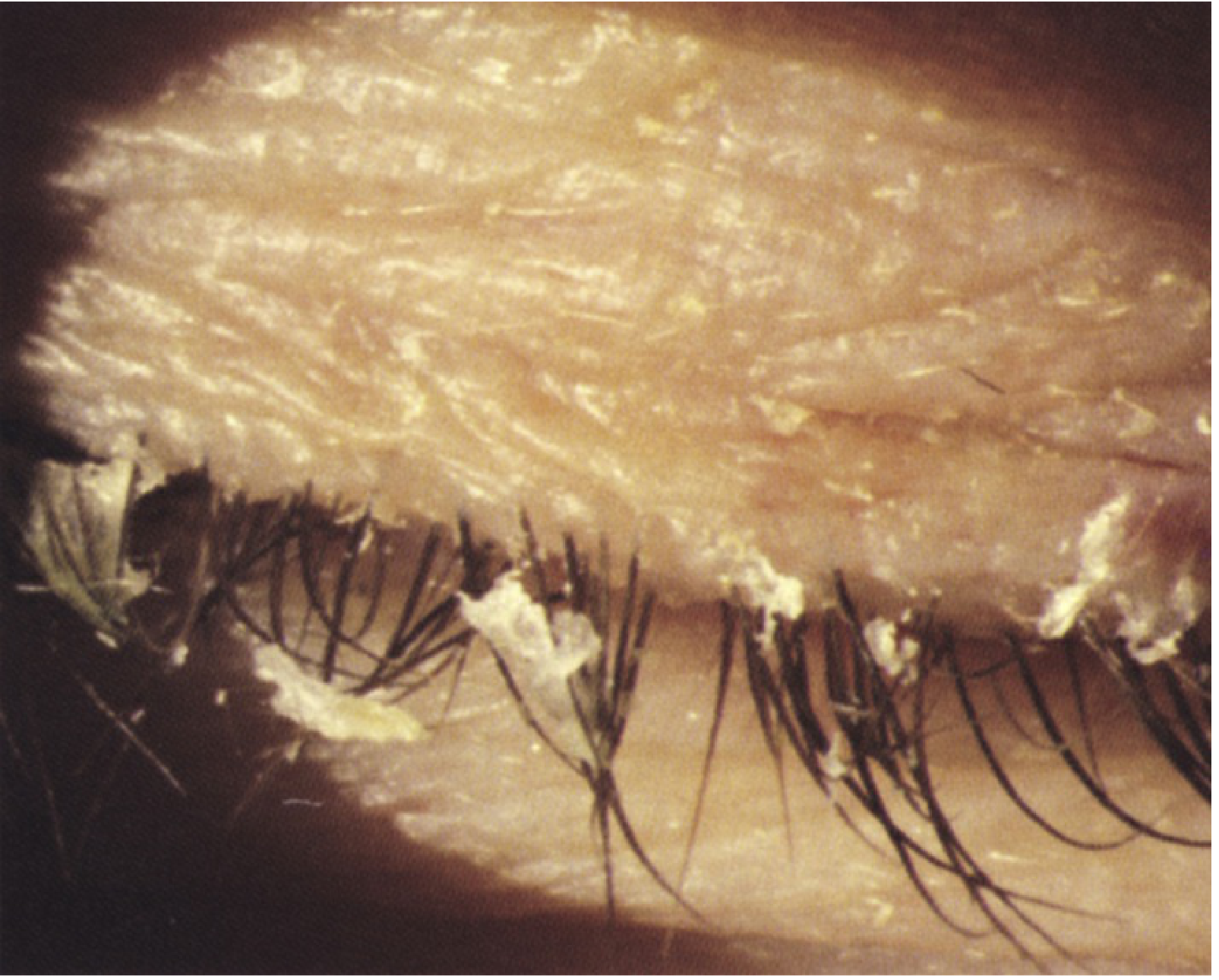
- Collarettes (cylindrical sleeves of keratin/mite eggs at lash bases) are considered pathognomonic of Demodex infestation
- Microscopic examination of an epilated lash confirms the diagnosis
Symptoms
- Burning, grittiness, itching, foreign body sensation
- Mild photophobia
- Crusting around the eyes worse in the mornings (in contrast to dry eye, which worsens later in the day)
- Tearing, mild mucous discharge
- Contact lens intolerance
- Remissions and exacerbations are typical
Signs
Blepharitis with collarettes (Wills Eye Manual, Fig. 5.8.1):
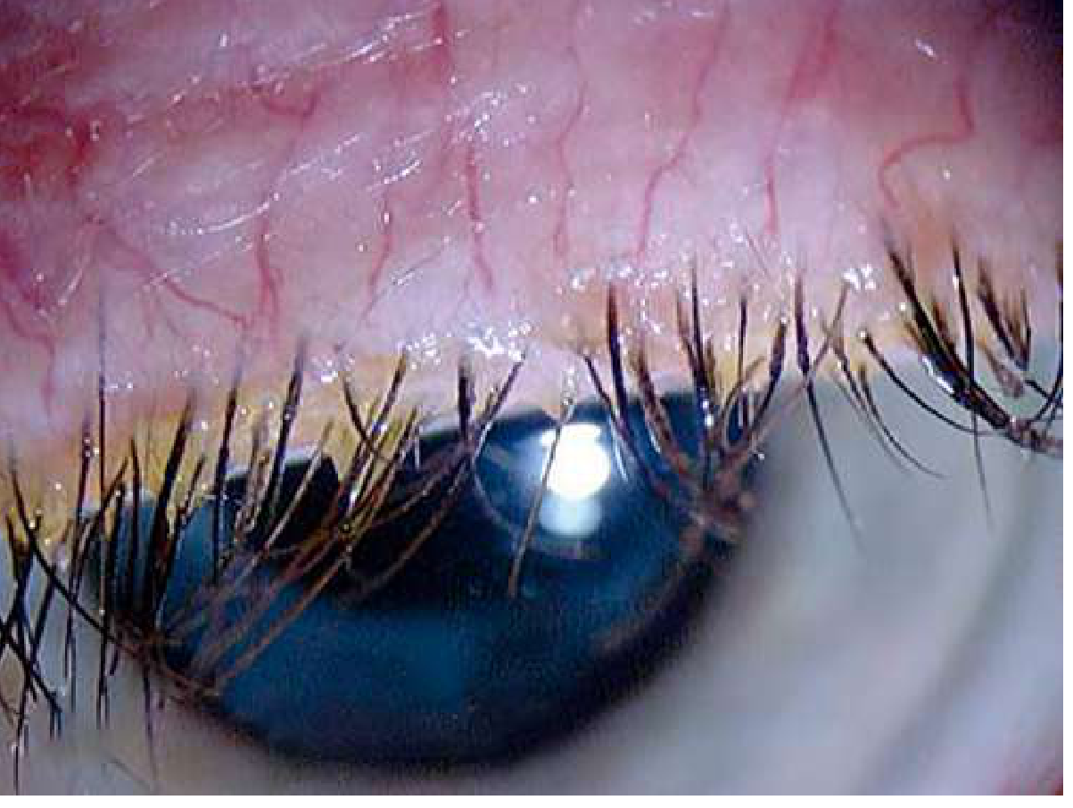
Blepharitis - inflamed, erythematous eyelid margin with visible telangiectasias:
Critical Signs
- Crusty, red, thickened eyelid margins with prominent blood vessels
- Inspissated (plugged) meibomian gland orifices
- Collarettes or cylindrical sleeves around lashes
- Scales at lash bases (hard in staphylococcal; soft/greasy in seborrhoeic)
Other Signs
- Conjunctival injection, papillary conjunctivitis
- Superficial punctate keratopathy (SPK)
- Corneal infiltrates, marginal keratitis, phlyctenules
- Corneal vascularization/pannus (in severe or chronic disease)
- Foaming at lid margins (posterior/MGD)
- Hordeolum (stye) or chalazion formation
- Lash loss (madarosis), misdirected lashes (trichiasis)
Complications / Secondary Changes
- Blepharoconjunctivitis (conjunctival and corneal co-inflammation)
- Salzmann nodular degeneration
- Bacterial keratitis (especially in contact lens wearers)
- Phlyctenular eye disease
- Dry eye syndrome (unstable tear film)
Differential Diagnosis
- Pediculosis (Phthirus pubis infestation of lashes)
- Demodicosis (look specifically for cylindrical sleeves)
- Ocular rosacea (telangiectasias of lid margin, facial flushing)
- Sebaceous carcinoma - important: intractable, unilateral, or asymmetric blepharitis (upper vs. lower lid asymmetry) should raise suspicion and warrants workup
Treatment
There is limited high-quality evidence for any single treatment protocol. The following stepwise approach is standard:
1. Lid Hygiene (First-line for all forms)
- Apply warm compresses for 5-10 minutes b.i.d. to q.i.d. to soften crusts and melt inspissated meibum
- Follow with lid margin scrubbing using a clean wet cloth or commercial eyelid scrub pads
- Note: Baby shampoo is NOT recommended by Kanski; it destabilizes the tear film and can cause contact dermatitis
- For significant MGD: manually express meibomian glands by rolling a finger over the lid margin
2. Dry Eye / Tear Support
- Preservative-free artificial tears 4-8 times/day if associated dry eye is present
3. Topical Antibiotics (Moderate-severe anterior disease)
- Erythromycin ointment, azithromycin gel-drop, sodium fusidic acid, bacitracin, or chloramphenicol applied to the lid margin at bedtime (q.h.s.)
4. Oral Antibiotics (Significant posterior/MGD or refractory cases)
- Doxycycline: 50-100 mg twice daily for 1 week, then daily for 6-24 weeks
- Lymecycline: 408 mg daily for up to 3 months
- Azithromycin: 500 mg/day x 3 days for 3 cycles at 1-week intervals
- Tetracyclines reduce bacterial colonization and inhibit staphylococcal lipase production
- Contraindicated in pregnancy, breastfeeding, and children ≤8 years - use erythromycin 200 mg b.i.d. in these groups
5. Anti-inflammatory / Immunomodulatory Agents
- Cyclosporine 0.05% or 0.09% eye drops b.i.d.
- Lifitegrast 5% drops b.i.d.
- Low-dose topical steroids (prednisolone 0.1% or fluorometholone 0.1%) for phlyctenular disease or corneal infiltrates
- Omega-3 fatty acid oral supplementation
6. Demodex-specific Treatment
- Suspected when collarettes are present and standard treatment has failed
- Tea tree oil eyelid scrubs or eyelid cleansing agent with hypochlorous acid once or twice daily for minimum 6 weeks
7. Advanced/Interventional (Refractory MGD)
- Thermal treatment with meibomian gland expression
- Intense pulsed light (IPL) laser therapy
- Microblepharoexfoliation
- Probing of meibomian glands
Special Variants
Angular Blepharitis
- Erythema, scaling, maceration, and fissuring at the lateral and/or medial canthi
- Caused by Moraxella lacunata or S. aureus
- Treatment: topical chloramphenicol, bacitracin, or erythromycin
Childhood Blepharokeratoconjunctivitis (BKC)
- More severe in Asian and Middle Eastern children; onset around age 6
- Presents with recurrent anterior/posterior blepharitis, styes, chalazia, eye rubbing, photophobia
- May be mistaken for allergic eye disease
- Treatment: lid hygiene + topical antibiotic ointment; erythromycin syrup 125 mg/day for 4-6 weeks for systemic coverage; low-dose topical steroids as needed
Follow-Up
- Review at 2-4 weeks depending on severity
- Lid scrubs and warm compresses can be reduced to once daily as symptoms improve, but often must be maintained indefinitely to prevent relapse
- Advise patients that the condition is chronic and a permanent cure is unlikely
Sources: Kanski's Clinical Ophthalmology: A Systematic Approach, 10th ed. | Wills Eye Manual, 8th ed. | Goldman-Cecil Medicine, International Edition
This is a shared conversation. Sign in to Orris to start your own chat.