
Devlopment of the face and defect anatomy mbbs 1st year
Reading File
Finding Sources
Finding Sources
Finding Sources
Reading File
Reading File
This is a broad MBBS 1st-year Embryology topic (Development of the Face + associated defects) that would take many pages to cover fully. Here's the map of what it includes, a brief overview, and then I'll ask you which part to go deep on.
Topic Breakdown
- Facial primordia formation - stomodeum, frontonasal prominence, paired maxillary and mandibular prominences (4th week)
- Development of the nose - nasal placodes, medial and lateral nasal prominences, formation of external nose
- Development of the upper lip and palate - fusion of medial nasal prominences (intermaxillary segment) and maxillary prominences; primary and secondary palate formation
- Pharyngeal (branchial) arch contribution - 1st arch (mandibular) derivatives forming lower face/jaw
- Molecular control - sonic hedgehog, neural crest cell migration, growth factors
- Congenital defects:
- Cleft lip (unilateral/bilateral, complete/incomplete)
- Cleft palate (primary vs secondary palate clefts, complete/incomplete)
- Median cleft lip, oblique facial clefts
- Macrostomia, microstomia
- First arch syndromes (Treacher Collins syndrome, Pierre Robin sequence)
Brief Overview
The face develops between the 4th and 10th weeks of gestation from five primordia surrounding the stomodeum (primitive mouth): one frontonasal prominence (unpaired, forms forehead, nose bridge, and via nasal prominences the sides of the nose and philtrum of the upper lip) and paired maxillary and mandibular prominences (from the 1st pharyngeal arch). Nasal placodes on the frontonasal prominence sink to form nasal pits, flanked by medial and lateral nasal prominences. The medial nasal prominences fuse in the midline with each other and with the maxillary prominences to form the intermaxillary segment - this gives rise to the philtrum of the upper lip, four incisor teeth, and the primary palate. The maxillary prominences give rise to the lateral parts of the upper lip and the secondary palate (from palatal shelves that fuse in the midline and with the nasal septum). The mandibular prominences fuse early in the midline to form the lower jaw and lip.
Failure of fusion at these various junctions produces the classic defects: failure of the maxillary prominence to fuse with the intermaxillary segment causes cleft lip (lateral to philtrum, along the line of the incisive fossa); failure of palatal shelves to fuse with each other and the nasal septum causes cleft palate. These can occur together or separately since primary and secondary palate formation are distinct events, which is why classification is based on the incisive foramen as the landmark. Cleft lip/palate affects roughly 1 in 700-1000 births with male predominance for cleft lip and female predominance for isolated cleft palate.
Asking for Preferences
Which part of Development of the Face would you like covered in detail?
This is a broad MBBS 1st-year Embryology topic (Development of the Face + associated defects) that would take many pages to cover fully. Here's the map of what it includes, a brief overview, and then I'll ask you which part to go deep on.
Topic Breakdown
- Facial primordia formation - stomodeum, frontonasal prominence, paired maxillary and mandibular prominences (4th week)
- Development of the nose - nasal placodes, medial and lateral nasal prominences, formation of external nose
- Development of the upper lip and palate - fusion of medial nasal prominences (intermaxillary segment) and maxillary prominences; primary and secondary palate formation
- Pharyngeal (branchial) arch contribution - 1st arch (mandibular) derivatives forming lower face/jaw
- Molecular control - sonic hedgehog, neural crest cell migration, growth factors
- Congenital defects:
- Cleft lip (unilateral/bilateral, complete/incomplete)
- Cleft palate (primary vs secondary palate clefts, complete/incomplete)
- Median cleft lip, oblique facial clefts
- Macrostomia, microstomia
- First arch syndromes (Treacher Collins syndrome, Pierre Robin sequence)
Brief Overview
The face develops between the 4th and 10th weeks of gestation from five primordia surrounding the stomodeum (primitive mouth): one frontonasal prominence (unpaired, forms forehead, nose bridge, and via nasal prominences the sides of the nose and philtrum of the upper lip) and paired maxillary and mandibular prominences (from the 1st pharyngeal arch). Nasal placodes on the frontonasal prominence sink to form nasal pits, flanked by medial and lateral nasal prominences. The medial nasal prominences fuse in the midline with each other and with the maxillary prominences to form the intermaxillary segment - this gives rise to the philtrum of the upper lip, four incisor teeth, and the primary palate. The maxillary prominences give rise to the lateral parts of the upper lip and the secondary palate (from palatal shelves that fuse in the midline and with the nasal septum). The mandibular prominences fuse early in the midline to form the lower jaw and lip.
Failure of fusion at these various junctions produces the classic defects: failure of the maxillary prominence to fuse with the intermaxillary segment causes cleft lip (lateral to philtrum, along the line of the incisive fossa); failure of palatal shelves to fuse with each other and the nasal septum causes cleft palate. These can occur together or separately since primary and secondary palate formation are distinct events, which is why classification is based on the incisive foramen as the landmark. Cleft lip/palate affects roughly 1 in 700-1000 births with male predominance for cleft lip and female predominance for isolated cleft palate.
Devlopment of the face and defect anatomy mbbs 1st year with proper heading and diagram
Reading File
Reading File
Reading File
Searching Images
development of face embryology frontonasal prominence facial primordia
Reading File
Reading File
Reading File
Searching Images
five facial prominences frontonasal maxillary mandibular embryo development week
Development of the Face and Facial Defects — MBBS 1st Year Embryology
1. Timeline and the Five Facial Primordia
Facial development begins early in the 4th week of intrauterine life and is essentially complete by the end of the 10th week. It occurs around the stomodeum (the primitive mouth, a depression lined by ectoderm where the oral cavity will form).
Facial development is induced by the forebrain (via sonic hedgehog signaling gradients), the frontonasal ectodermal zone, and the developing eye. Five swellings (primordia), made of neural-crest-derived mesenchyme, appear around the stomodeum:
- One frontonasal prominence (unpaired) - superior to the stomodeum, formed by proliferation of mesenchyme ventral to the forebrain
- Two maxillary prominences (paired) - lateral to the stomodeum, derivatives of the first pharyngeal arch
- Two mandibular prominences (paired) - caudal to the stomodeum, also derivatives of the first pharyngeal arch; they fuse early (by end of week 4) to form the lower jaw and lower lip
(The Developing Human Clinically Oriented Embryology, p. 484)
2. Development of the Nose
Bilateral thickenings of surface ectoderm called nasal (olfactory) placodes appear on the frontonasal prominence around day 28. Mesenchyme proliferates around the margins of each placode, forming horseshoe-shaped elevations:
- Medial nasal prominences (inner limb)
- Lateral nasal prominences (outer limb)
The placodes sink to form nasal pits, which deepen to become the primitive nostrils and later communicate with the developing pharynx via the nasal sacs (oronasal membrane ruptures to form the primitive choanae).
- The lateral nasal prominences form the alae (sides) of the nose
- The medial nasal prominences merge with each other in the midline to form the intermaxillary segment, which gives rise to the bridge and tip of the nose, the philtrum of the upper lip, the premaxillary part of the maxilla, and the primary palate
3. Development of the Upper Lip
The upper lip forms from three components:
- Two maxillary prominences (lateral parts of the upper lip and cheek)
- The fused medial nasal prominences / intermaxillary segment (philtrum, the central part)
Mesenchyme from the maxillary prominences proliferates medially, merging with the medial nasal prominences along a groove (nasolacrimal groove separates lateral nasal from maxillary prominence, which is later canalized to form the nasolacrimal duct). Normal fusion smooths out the grooves between prominences.
4. Development of the Palate
The palate develops from two primordia:
| Component | Origin | Structure formed |
|---|---|---|
| Primary palate | Intermaxillary segment (fused medial nasal prominences) | Small, triangular part of hard palate anterior to the incisive fossa (premaxillary part) |
| Secondary palate | Two lateral palatine processes ("palatal shelves") from the maxillary prominences | Hard palate (posterior to incisive fossa) and the entire soft palate/uvula |
The palatal shelves are initially vertical alongside the tongue; as the mandible grows and the tongue drops, they reorient horizontally, fuse with each other in the midline, and fuse anteriorly with the primary palate and superiorly with the nasal septum. The incisive fossa (in the region of the incisive foramen) is the key anatomical landmark separating primary from secondary palate derivatives and is used to classify cleft defects.
5. Molecular Control (brief)
Neural crest cells migrating from the neuroectoderm form nearly all facial mesenchyme. Key signaling molecules include sonic hedgehog (SHH), BMPs, FGF8, and transcription factors like MSX1 and PAX genes. TCOF1 (treacle protein, ribosomal RNA biogenesis) and IRF6 are clinically important genes discussed below.
Congenital Defects of the Face
A. Cleft Lip and Cleft Palate
Clefts of the lip and palate are the most common craniofacial birth defects (roughly 1 in 1000 births), and can be detected antenatally by ultrasound.
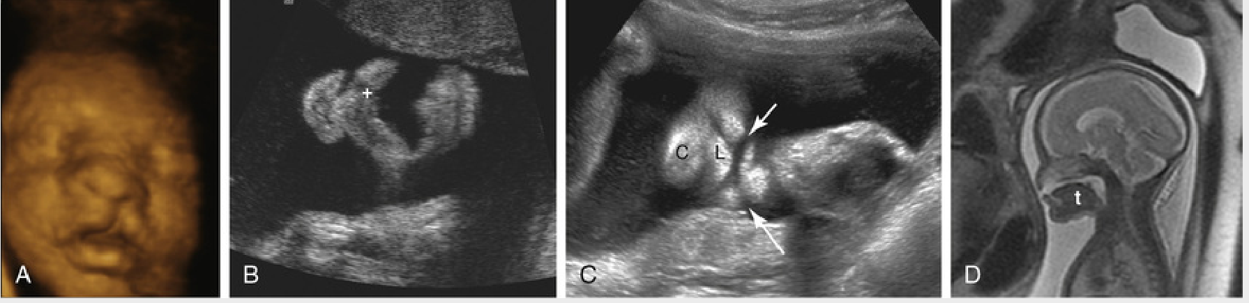
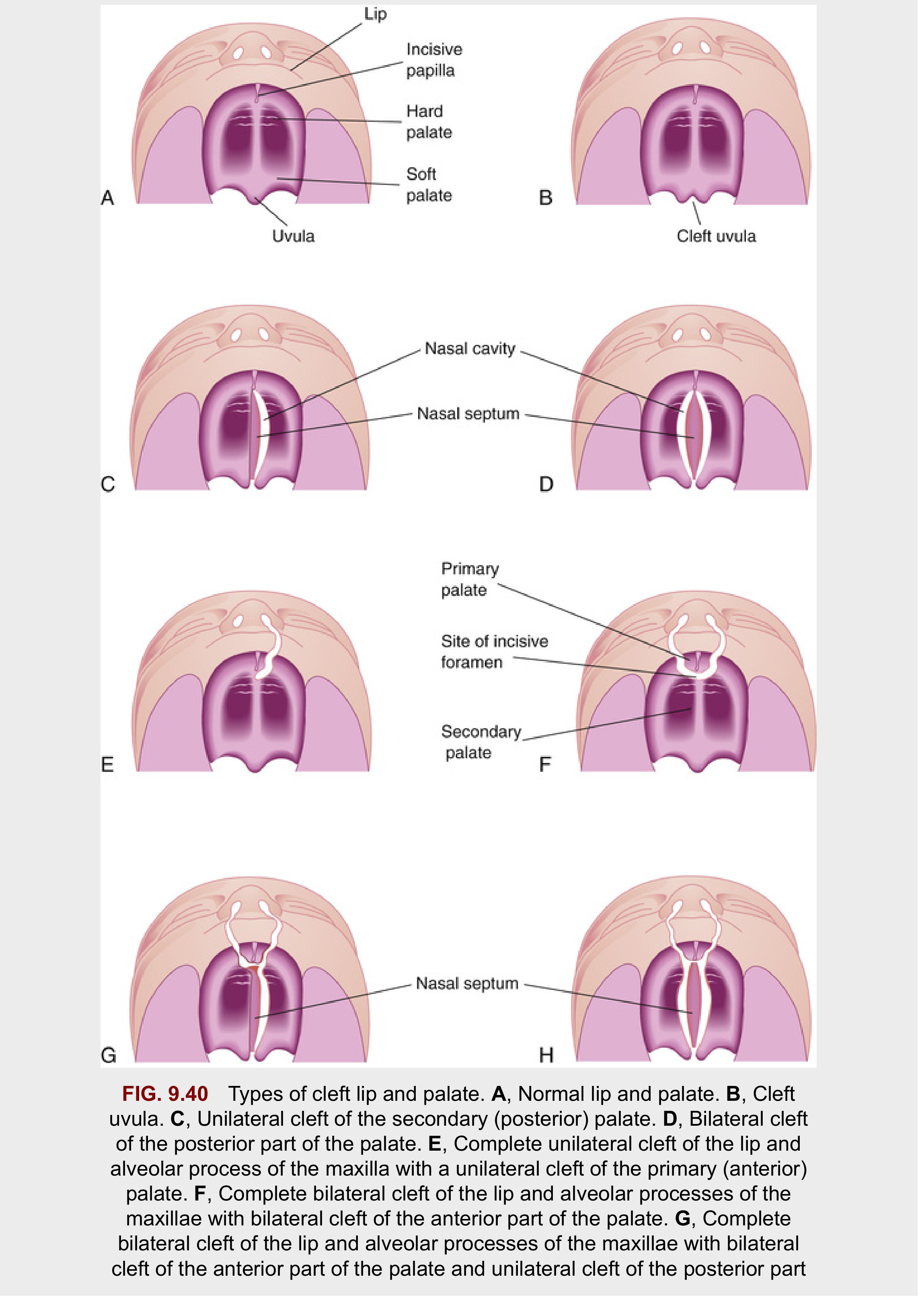
Clefts are classified using the incisive fossa as the landmark into two groups:
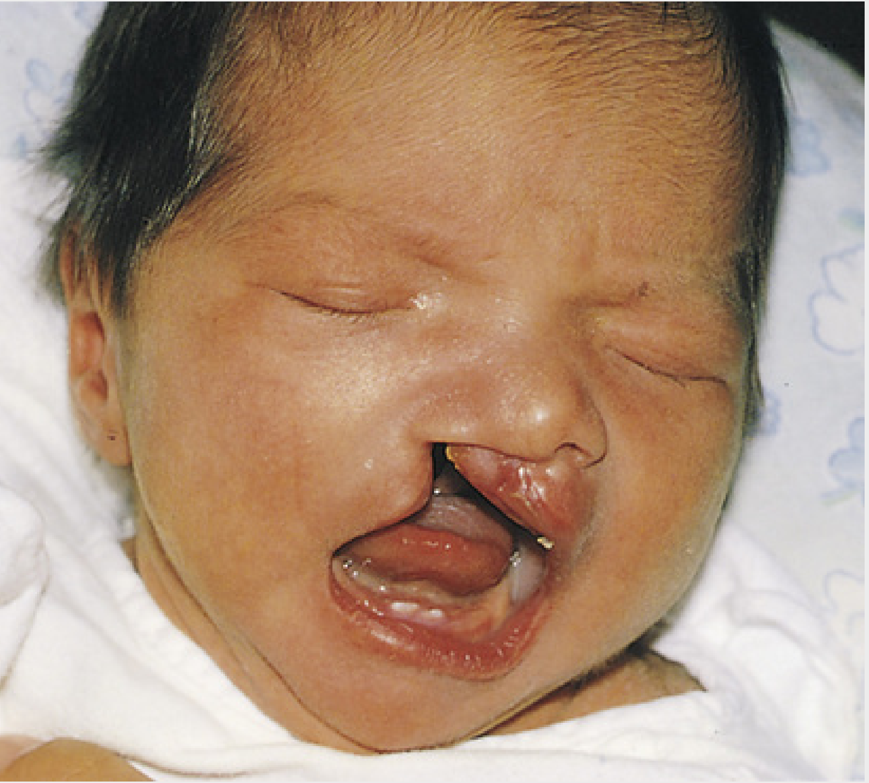
Anterior Cleft Defects (Cleft Lip ± cleft of alveolar maxilla)
- Result from failure of the maxillary prominence to fuse with the merged medial nasal prominences on one or both sides, due to deficient mesenchyme.
- Can be unilateral or bilateral, incomplete or complete (extending through lip, nostril floor, and alveolar process of maxilla to the incisive fossa).
- A persistent epithelial labial groove that fails to be filled/smoothed by mesenchyme leads to breakdown of the groove floor, splitting the lip. A residual bridge of tissue joining the two parts is called the Simonart band.
- Occurs in ~1/1000 births; 60-80% of affected infants are male.
Posterior Cleft Defects (Cleft of Secondary Palate)
- Result from failure of the palatal shelves (lateral palatine processes) to fuse with each other and/or with the nasal septum.
- Extend through the soft and hard palate to the incisive fossa, separating anterior and posterior palate.
- Contributing factors: width of the stomodeum, restricted shelf mobility, and delayed/defective epithelial breakdown at fusion sites.
Genetics
- Multifactorial inheritance is most common; the IRF6 gene is implicated in isolated (non-syndromic) clefts.
- Can occur as part of chromosomal syndromes (e.g., trisomy 13) or be induced by teratogens (e.g., anticonvulsants such as phenytoin).
- Genetic factors weigh more heavily in cleft lip (with/without cleft palate) than in isolated cleft palate.
- If neither parent is affected, recurrence risk in future siblings is about 4%.
B. Other Facial Defects
| Defect | Embryological basis |
|---|---|
| Microstomia (small mouth) | Excessive merging of maxillary and mandibular mesenchyme; may accompany mandibular hypoplasia |
| Single nostril | Only one nasal placode develops |
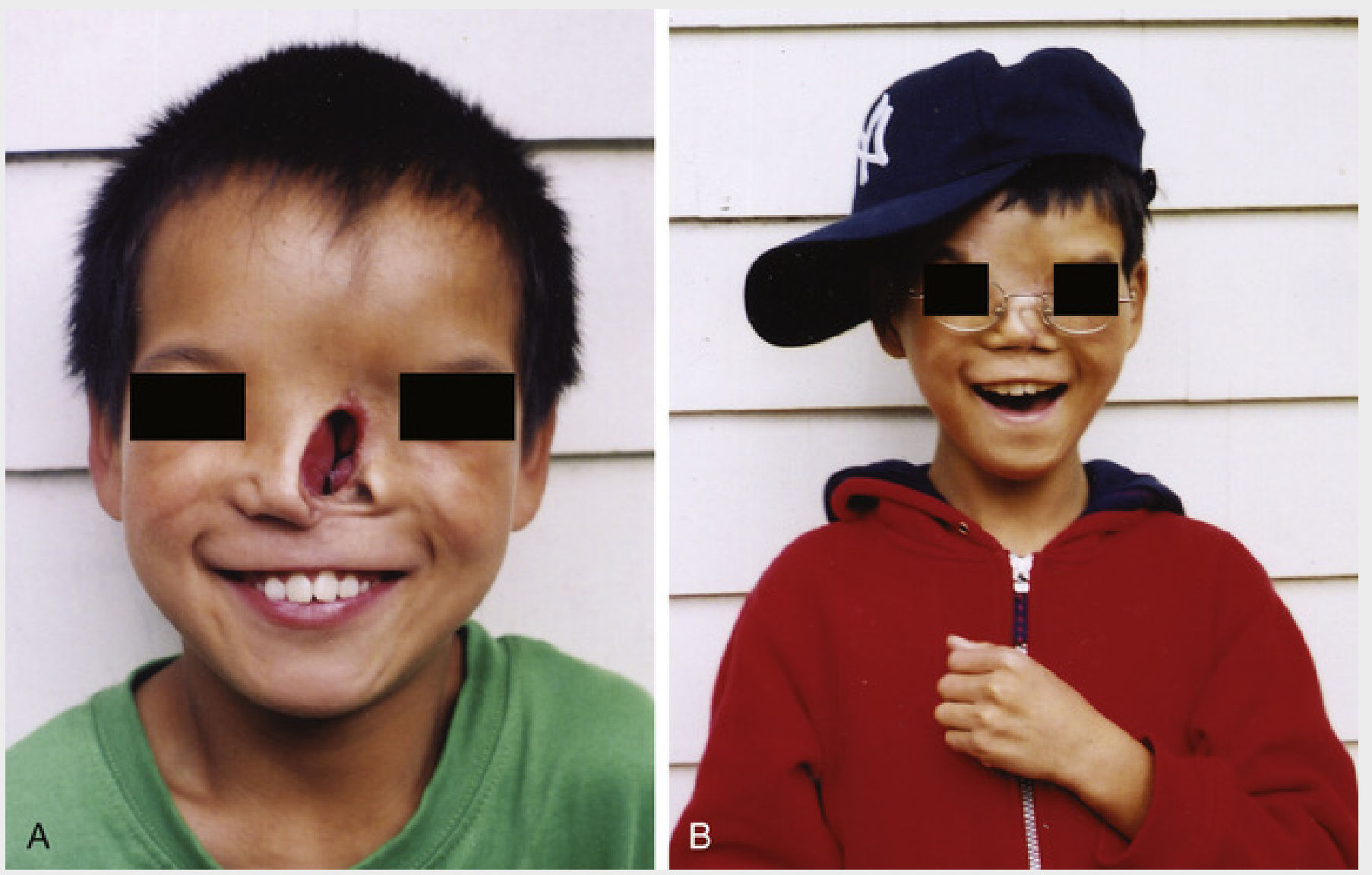
| Bifid nose | Incomplete merging of medial nasal prominences; widely separated nostrils, bifid nasal bridge (mild form = groove in nasal tip) |
| Oblique facial cleft | Failure of maxillary prominence mesenchyme to merge with lateral/medial nasal prominences; runs from upper lip to medial margin of orbit; leaves the nasolacrimal duct as an open groove |
| Transverse (lateral) facial cleft | Runs from mouth toward ear; bilateral clefts cause macrostomia (very large mouth), and severe cases extend almost to the ear |
C. First Pharyngeal Arch Syndrome
Insufficient migration of neural crest cells into the first pharyngeal arch in week 4 causes combined defects of the eyes, ears, mandible, and palate. Two major patterns:
Treacher Collins syndrome (mandibulofacial dysostosis)
- Autosomal dominant; caused by mutation in TCOF1 (encodes "treacle," involved in ribosomal RNA biogenesis needed for facial bone/cartilage development)
- Features: malar hypoplasia (zygomatic underdevelopment), down-slanting palpebral fissures, lower eyelid defects, deformed external ears, sometimes middle/inner ear defects
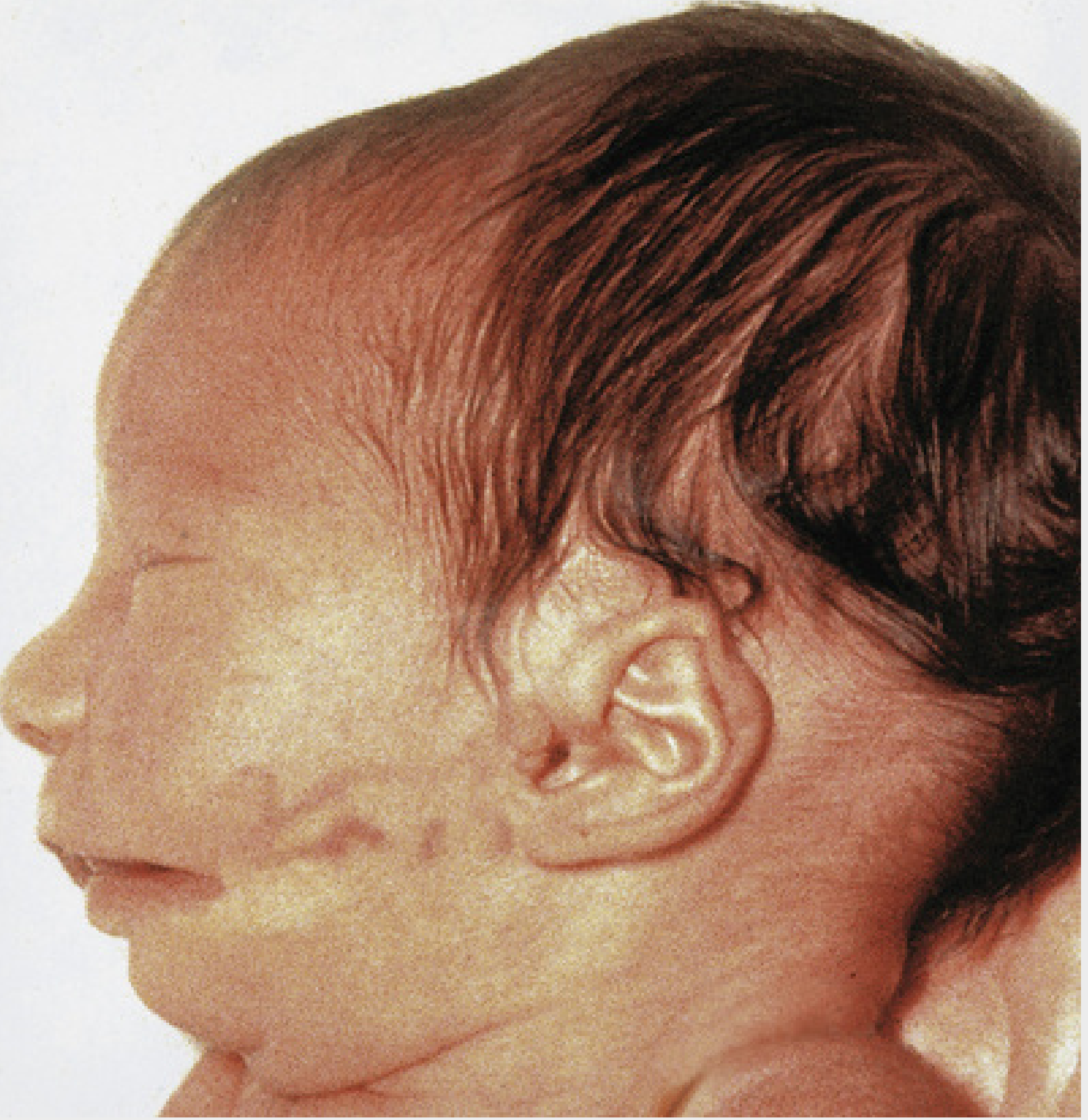
Pierre Robin sequence
- Usually occurs de novo (rarely autosomal dominant)
- Initiating defect: micrognathia (small mandible) → posterior tongue displacement → obstructs palatal shelf closure → bilateral cleft palate
- Associated eye and ear defects
D. DiGeorge Syndrome (22q11.2 deletion)
Though primarily a pharyngeal pouch disorder (absent thymus and parathyroids, cardiac outflow defects), it also produces facial features relevant to this topic: shortened philtrum of the upper lip, low-set notched ears, and nasal clefts, due to abnormal neural crest-derived pharyngeal apparatus development.
Quick Revision Table
| Structure | Forms From | Defect if fusion fails |
|---|---|---|
| Philtrum, primary palate | Medial nasal prominences (intermaxillary segment) | Median/anterior cleft, cleft of primary palate |
| Lateral upper lip | Maxillary prominence | Unilateral/bilateral cleft lip |
| Nasal alae | Lateral nasal prominence | Bifid nose, single nostril, oblique cleft |
| Secondary (hard + soft) palate | Palatal shelves of maxillary prominence | Posterior cleft palate |
| Lower lip/jaw | Mandibular prominences | Micrognathia, midline defects (rare) |
| Cheek, mouth angle | 1st arch (maxillary + mandibular) mesenchyme | Macrostomia/microstomia, transverse facial cleft |
Source: The Developing Human: Clinically Oriented Embryology (Moore, Persaud, Torchia), Chapter 9 - Pharyngeal Apparatus, Face, and Neck, pp. 484-550.
If you want, I can go deeper into any single subtopic (e.g., a step-by-step labeled diagram walkthrough of palate fusion, or a table comparing cleft lip vs cleft palate embryology for exam purposes).
This is a shared conversation. Sign in to Orris to start your own chat.