
Why after oxytocin baby movement is low
Reading File
Oxytocin is used to induce or augment labor by stimulating uterine contractions. Reduced fetal movement after oxytocin administration is mainly due to increased uterine contractions compressing placental blood flow and causing fetal distress. Let me look this up in the medical library for a detailed, evidence-based answer.
Searching sources...
Finding Sources
Finding Sources
Finding Sources
Good results found. Let me read the key sections in detail.
Reading File
Reading File
Finding Sources
Finding Sources
Finding Sources
Finding Sources
Finding Sources
Reading File
Reading File
I now have excellent textbook content to give a thorough answer. Let me fetch the relevant image of the oxytocin challenge test since it directly illustrates the mechanism.
Loading Image
Loading Image
I now have all the information needed for a thorough answer. Here is the complete explanation:
Why Baby Movement Decreases After Oxytocin
This is a clinically important question. When oxytocin is given to induce or augment labor, reduced fetal movement is primarily a sign of fetal stress from decreased oxygen delivery - and it needs to be taken seriously.
The Core Mechanism: Oxytocin Compresses Placental Blood Flow
Oxytocin works by stimulating uterine muscle contractions. With every contraction, the uterine wall squeezes, which transiently reduces placental blood flow to the baby. The placenta is embedded in the uterine wall, so when the uterus tightens, blood perfusion through the intervillous space (where oxygen transfer happens) drops.
"During the antepartum period, oxytocin induces uterine contractions that transiently reduce placental blood flow to the fetus." - [Katzung's Basic and Clinical Pharmacology, 16th ed.]
In a healthy pregnancy, the baby tolerates these brief pauses in oxygen delivery between contractions. But if the contractions become too frequent, too long, or too strong (called uterine hyperstimulation or tachysystole), the baby does not get enough recovery time, oxygen levels drop, and the baby responds by reducing its movements to conserve oxygen.
This is an adaptive protective response by the fetal brain - it reduces non-essential activity (movements) to redirect oxygen to vital organs like the heart and brain.
The Specific Problem: Oxytocin Overdose / Hyperstimulation
According to Morgan & Mikhail's Clinical Anesthesiology:
"Complications of oxytocin administration may include fetal distress due to hyperstimulation, uterine tetany..."
And from Katzung's:
"Excessive stimulation of uterine contractions before delivery can cause fetal distress, placental abruption, or uterine rupture."
Uterine tetany is the extreme form - a sustained, non-relaxing contraction that completely cuts off placental blood flow, causing acute fetal hypoxia.
How the Fetal Brain Responds to Low Oxygen
Fetal movement is controlled by the central nervous system. When oxygen is reduced, the fetal brain progressively shuts down "higher" functions first:
| CNS Activity | Sensitivity to Hypoxia |
|---|---|
| Fetal breathing movements | First to disappear |
| Fetal body movements | Second to decrease |
| Fetal tone (flexion/extension) | Third to be lost |
| Fetal heart rate reactivity | Last to be affected |
This is assessed clinically using the Biophysical Profile (BPP) scoring system, where fetal movement (at least 3 gross body movements in 30 minutes) is one of the 5 parameters. A score of 0 for fetal movement is a sign of possible fetal compromise.
"In chronic sustained fetal hypoxia, a protective redistribution of fetal cardiac output may occur, with blood being directed away from nonvital fetal organs (kidneys or lungs) toward vital fetal organs." - Pfenninger and Fowler's Procedures for Primary Care
The Oxytocin Challenge Test (OCT) - The Diagnostic Connection
This mechanism is actually used diagnostically in the Oxytocin Challenge Test (OCT) or Contraction Stress Test (CST). A small oxytocin infusion is given intentionally to provoke contractions, and the fetal heart rate response is monitored:
- Negative test (normal): No late decelerations - baby tolerates reduced blood flow well
- Positive test (abnormal): Late decelerations after contractions = the baby's oxygen reserve is insufficient
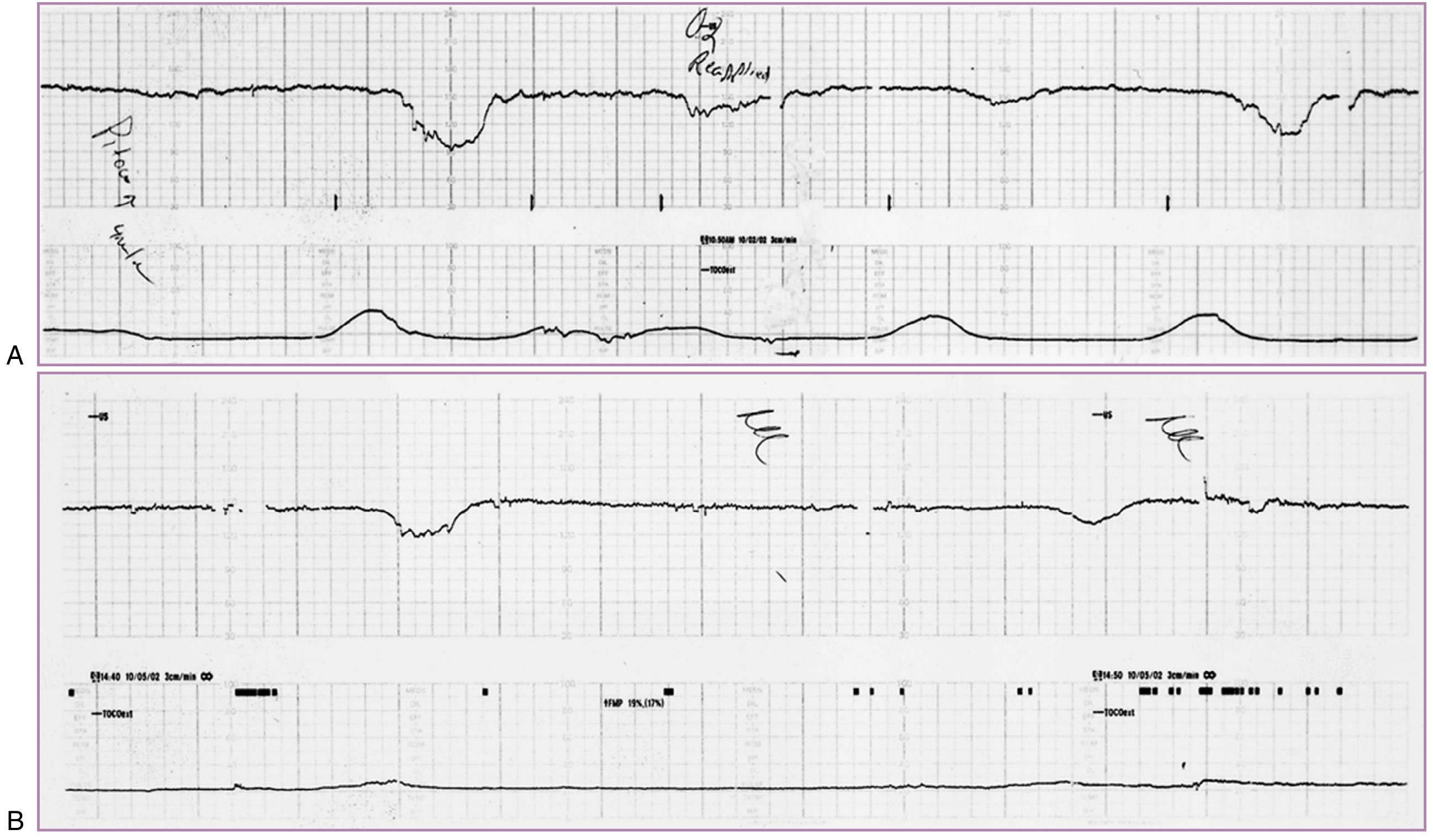
OCT tracing: Panel A shows repetitive late decelerations with each uterine contraction - a positive result indicating fetal compromise. Panel B shows late decelerations even with trivial contractions in a growth-restricted fetus.
Other Contributing Factors
Beyond hypoxia, there are additional reasons movement may feel reduced after oxytocin:
- Physical constriction - During a strong contraction, the baby is physically compressed inside the tightened uterus and has less room to move freely
- Fetal sleep state - Normal fetal sleep cycles (20-40 minutes of reduced activity) can coincide with oxytocin administration and be mistakenly attributed to the drug
- Sedative co-medications - If the mother received opioids or sedatives alongside oxytocin for pain relief, these cross the placenta and suppress fetal CNS activity and movements
When to Be Concerned (Clinical Red Flags)
Reduced fetal movement after oxytocin is a warning sign that warrants immediate attention if accompanied by:
- Late decelerations on the fetal heart rate monitor (drop in FHR after the peak of a contraction)
- Prolonged deceleration lasting >2 minutes
- Loss of fetal heart rate variability
- Uterine tachysystole (more than 5 contractions in 10 minutes)
In such cases, the oxytocin infusion should be slowed or stopped, the mother placed on her left side to improve uterine perfusion, oxygen given, and IV fluids increased - while the team prepares for potential emergency delivery.
In summary: Oxytocin reduces fetal movement mainly because its contractions intermittently cut off the placental oxygen supply. When this is excessive, the fetal brain adaptively reduces movement to conserve oxygen - a protective but serious sign that the baby is under stress.
This is a shared conversation. Sign in to Orris to start your own chat.