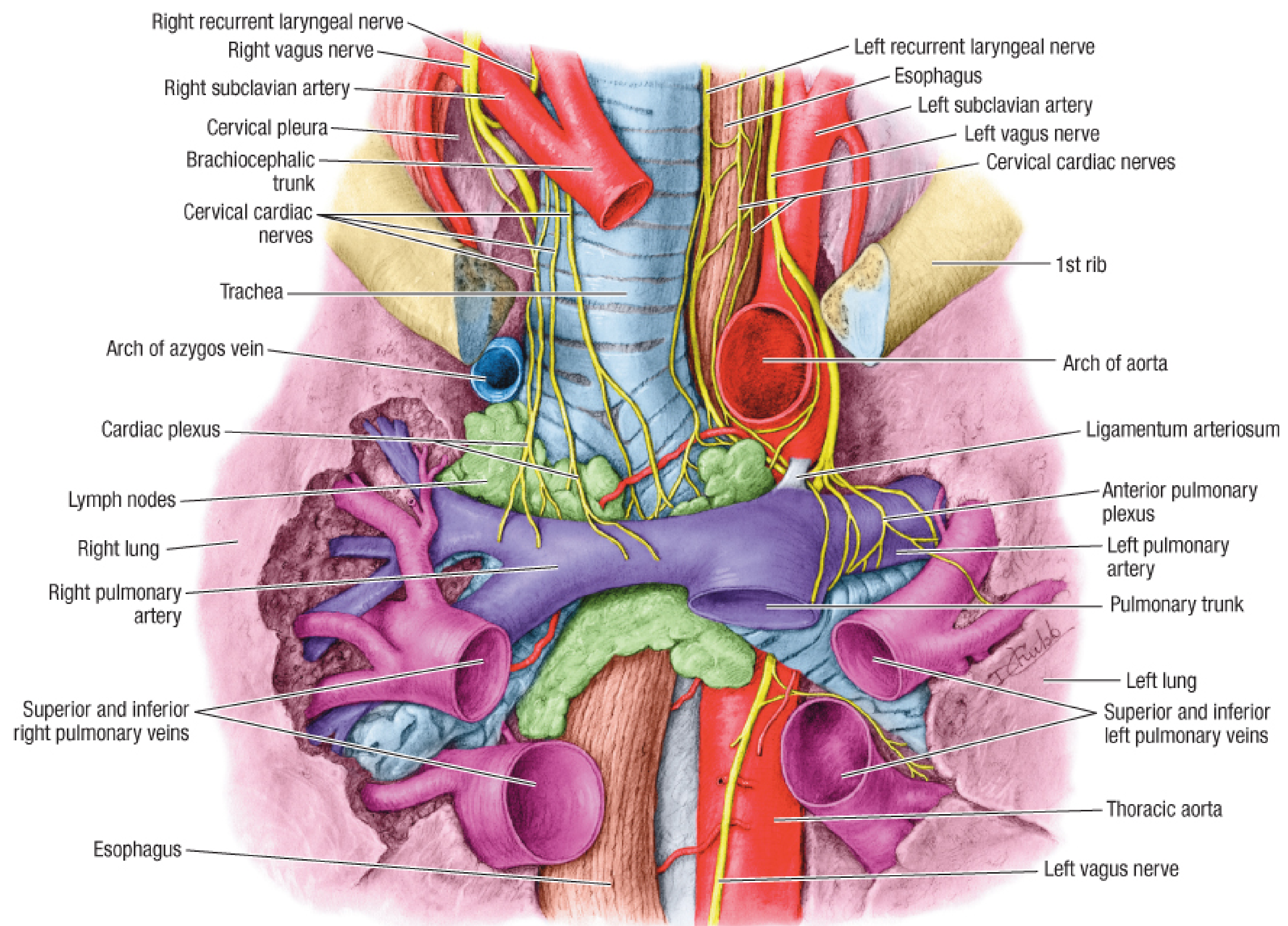
femoral sheath
Femoral Sheath
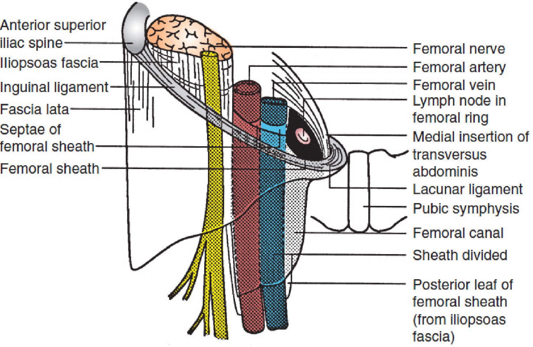
Definition and Formation
- Anteriorly: transversalis fascia (proper)
- Posteriorly/Laterally: iliopsoas fascia (itself a continuation of the transversalis/endoabdominal fascia)
- The iliopectineal arch joins the iliopubic tract to contribute to the proximal portion of the sheath
Compartments (Three)
| Compartment | Position | Contents |
|---|---|---|
| Lateral | Most lateral | Femoral artery |
| Intermediate | Middle | Femoral vein |
| Femoral canal | Most medial | Lymphatics + connective tissue; Cloquet's node at the ring |
The femoral nerve lies lateral to the sheath and is not enclosed within it (it lies in the lacuna musculorum, separated by the iliopectineal arch).
Femoral Canal
- The most medial compartment of the sheath
- Cone/cul-de-sac shaped, narrowing inferiorly as the medial septum fuses with the femoral vein adventitia
- Normally contains only wisps of connective tissue and small lymph nodes
- At its proximal (superior) opening = the femoral ring
Femoral Ring
- Anteriorly: inguinal ligament
- Posteriorly: iliopubic tract + pectineal (Cooper's) ligament
- Medially: curved edge of the transversus abdominis aponeurosis (NOT the lacunar ligament, which inserts more medially)
- Laterally: connective tissue septum between femoral ring and femoral vein
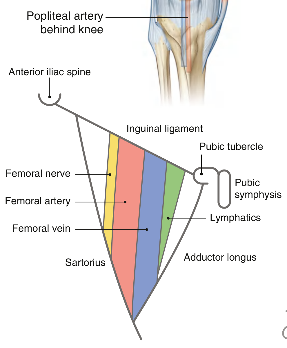
Femoral Triangle Context
Clinical Relevance
- Coronary angiography and angioplasty (arterial access)
- Right heart catheterization, pulmonary artery catheters (venous access)
- Access to renal, gonadal, cerebral, and thoracic vessels
only femoral shealth for 5 marks
Femoral Sheath (5 Marks)
Definition
Formation
- Anteriorly - transversalis fascia
- Posteriorly - iliopsoas fascia (iliac fascia)
- Above - continuous with abdominal fascia
- Below - fuses with adventitia of femoral vessels (~4 cm below inguinal ligament)
Compartments (3)
| Compartment | Contents |
|---|---|
| Lateral | Femoral artery |
| Middle | Femoral vein |
| Medial (femoral canal) | Lymphatics + Cloquet's node |
Note: Femoral nerve lies outside the sheath (lateral to it)
Femoral Canal
- Most medial, cone-shaped compartment
- Contains lymphatics and connective tissue
- Upper opening = femoral ring
- Anteriorly - inguinal ligament
- Posteriorly - pectineal (Cooper's) ligament
- Medially - lacunar ligament
- Laterally - femoral vein
Clinical Significance
- Femoral hernia - abdominal contents herniate through the femoral ring into the canal; more common in females
- Vascular access - femoral artery/vein used for cardiac catheterization and angiography
also add a digram
Femoral Sheath (5 Marks)
Definition
Formation
- Anteriorly - transversalis fascia
- Posteriorly - iliopsoas fascia (iliac fascia)
- Above - continuous with abdominal fascia
- Below - fuses with adventitia of femoral vessels (~4 cm below inguinal ligament)
Compartments (3)
| Compartment | Contents |
|---|---|
| Lateral | Femoral artery |
| Middle | Femoral vein |
| Medial (femoral canal) | Lymphatics + Cloquet's node |
Note: Femoral nerve lies outside the sheath (lateral to it)
Femoral Canal
- Most medial, cone-shaped compartment
- Contains lymphatics and connective tissue
- Upper opening = femoral ring
- Anteriorly - inguinal ligament
- Posteriorly - pectineal (Cooper's) ligament
- Medially - lacunar ligament
- Laterally - femoral vein
Diagram
Clinical Significance
- Femoral hernia - abdominal contents herniate through the femoral ring into the canal; more common in females
- Vascular access - femoral artery/vein used for cardiac catheterization and angiography
femoral canal
Femoral Canal
Definition
Dimensions
- Length: approximately 1.25 cm
- Wider above (at the femoral ring), narrows and closes below as it fuses with the adventitia of the femoral vein
Contents
- Areolar (loose connective) tissue
- Lymphatics
- Cloquet's node (Rosenmüller's node) - a large lymph node at the femoral ring
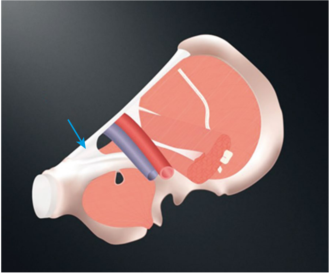
Femoral Ring (Upper Opening)
| Boundary | Structure |
|---|---|
| Anteriorly | Inguinal ligament (iliopubic tract) |
| Posteriorly | Pectineal (Cooper's) ligament |
| Medially | Lacunar (Gimbernat's) ligament |
| Laterally | Femoral vein (via its periadventitial septum) |
Important: The medial border is the lacunar ligament only in smaller canals; in reality the iliopubic tract forms the anterior/medial boundary in most individuals. The lacunar ligament is more medial still.
Diagram
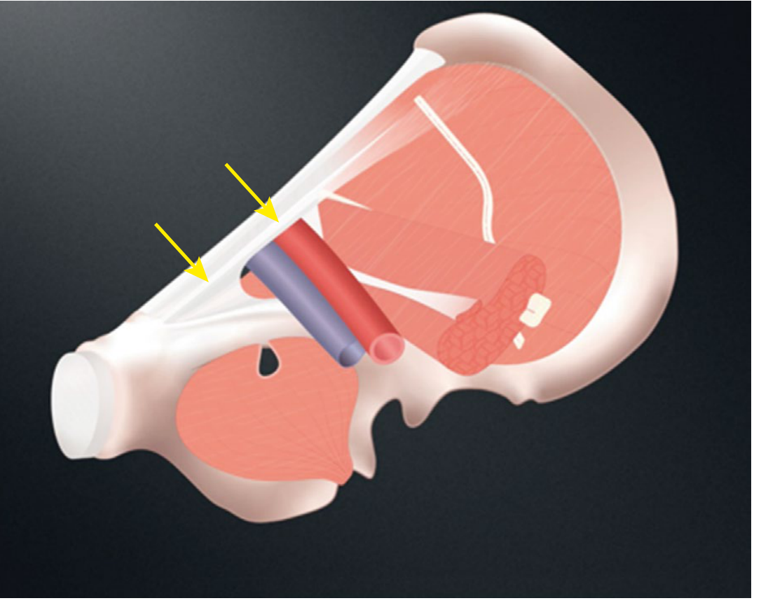
Function
- Allows the femoral vein to expand during increased venous return (e.g., during exercise)
- Acts as a potential dead space
Clinical Significance
Femoral Hernia
- The femoral ring is a potential weak point in the abdominal wall
- Abdominal contents (usually small bowel or omentum) can herniate through the ring into the canal
- More common in females due to wider pelvis and broader femoral ring
- There is no preformed peritoneal sac - the sac develops as the hernia pushes through
- The canal is rigid (bony + ligamentous walls) so strangulation is common
Dangerous Structures at Risk During Repair
- Femoral vein - can be compressed by sutures placed too laterally
- Anomalous obturator artery (present in ~30%) - may cross the femoral ring medially and be easily cut ("corona mortis" variant)
Repair Principle
- Close the femoral ring by suturing Cooper's ligament to the femoral sheath (McVay/Cooper's ligament repair)
- Alternatively, a mesh plug is used to obliterate the canal (the ring is relatively rigid and difficult to close with sutures from below)
femoral artery with diagram
Femoral Artery
Definition / Origin
Surface Marking
Course (Three Segments)
| Segment | Location |
|---|---|
| 1. Femoral triangle | Passes vertically downward within the femoral sheath |
| 2. Adductor (Hunter's) canal | Passes inferomedially beneath sartorius between vastus medialis and adductors |
| 3. Adductor hiatus | Passes through the opening in adductor magnus → becomes popliteal artery |
Relations in the Femoral Triangle
- Lateral: femoral nerve (outside the sheath)
- Medial: femoral vein (within the sheath)
- In the distal femoral triangle/adductor canal: the femoral vein shifts posterior then posterolateral to the artery
Branches
In the Femoral Triangle (Superficial branches - arise just below inguinal ligament):
- Superficial epigastric artery - to lower abdominal wall
- Superficial circumflex iliac artery - to skin over iliac crest
- Superficial external pudendal artery - to scrotum/labia
- Deep external pudendal artery - to perineum
Major Branch:
- Profunda femoris artery (Deep artery of thigh) - arises 4-5 cm below the inguinal ligament from the posterolateral aspect; the most important branch
- Medial circumflex femoral artery - main supply to head and neck of femur
- Lateral circumflex femoral artery - ascending, descending, and transverse branches
- 4 perforating arteries - supply posterior thigh (hamstrings)
Terminal Branch:
- Descending genicular artery - just before the adductor hiatus; contributes to knee anastomosis
Diagrams
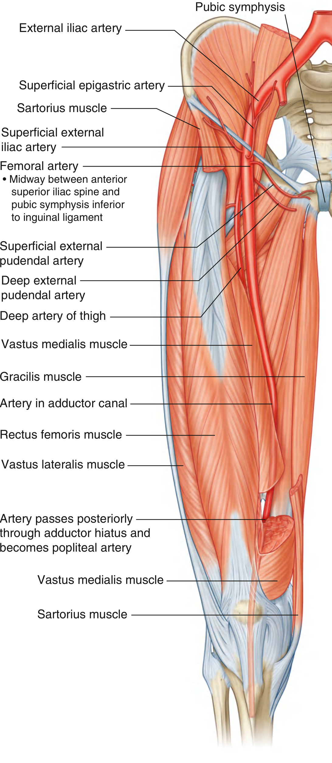
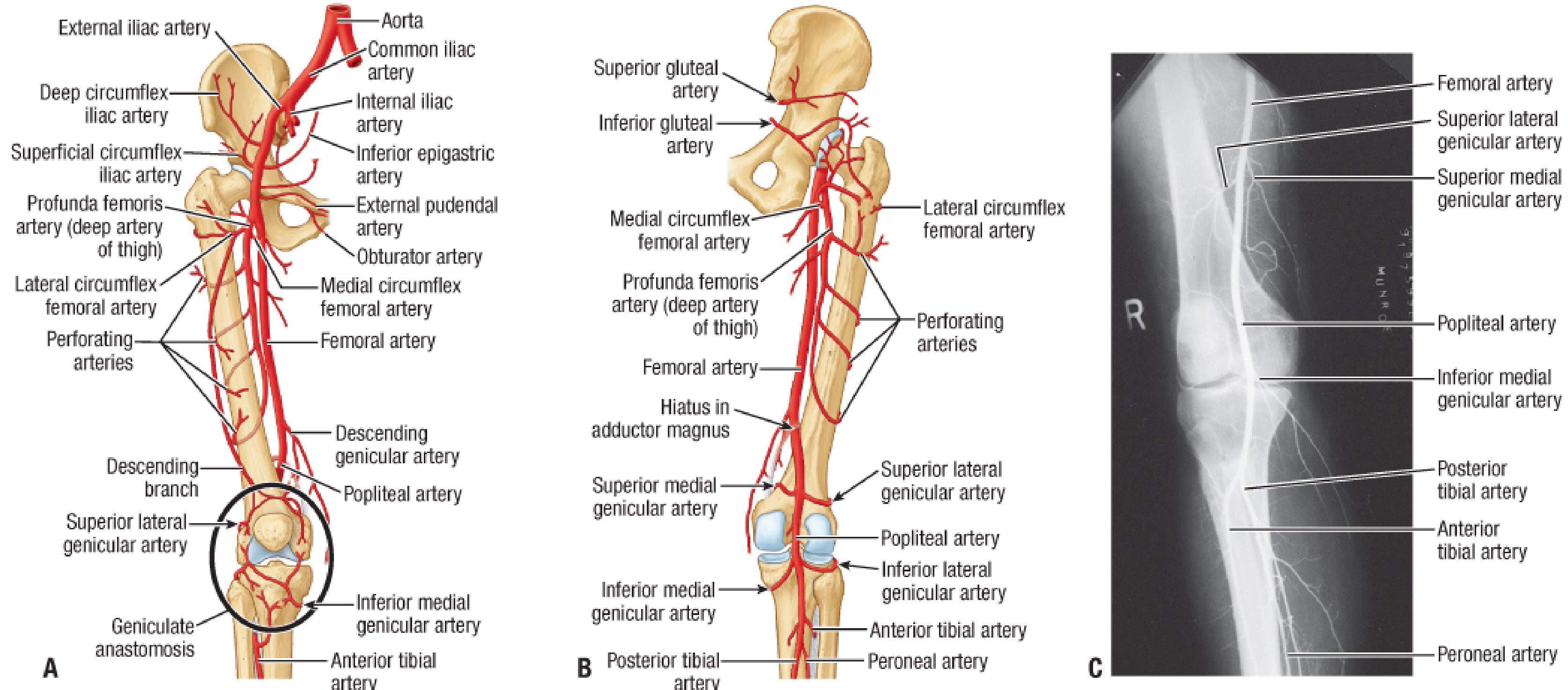
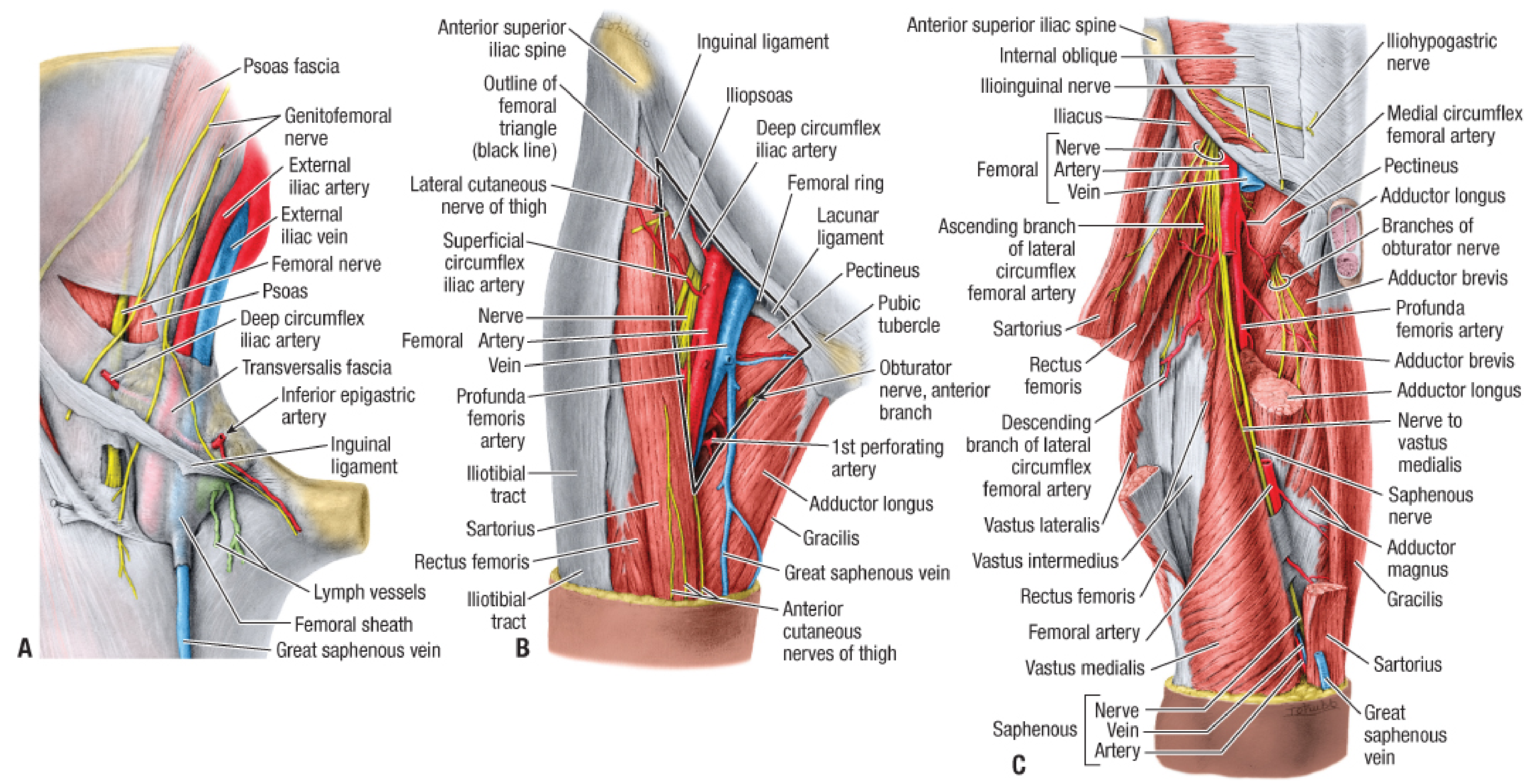
Anastomoses
Clinical Significance
- Vascular access - femoral artery puncture just below inguinal ligament for cardiac catheterization and angiography
- Femoral artery aneurysm - pulsatile groin mass
- Peripheral arterial disease - superficial femoral artery most commonly affected segment in the thigh
- At-risk nerves during surgery: saphenous nerve and nerve to vastus medialis lie lateral to the artery within the sheath - endangered by excessive lateral dissection
cubiotal fossa with clinical
Cubital Fossa
Definition
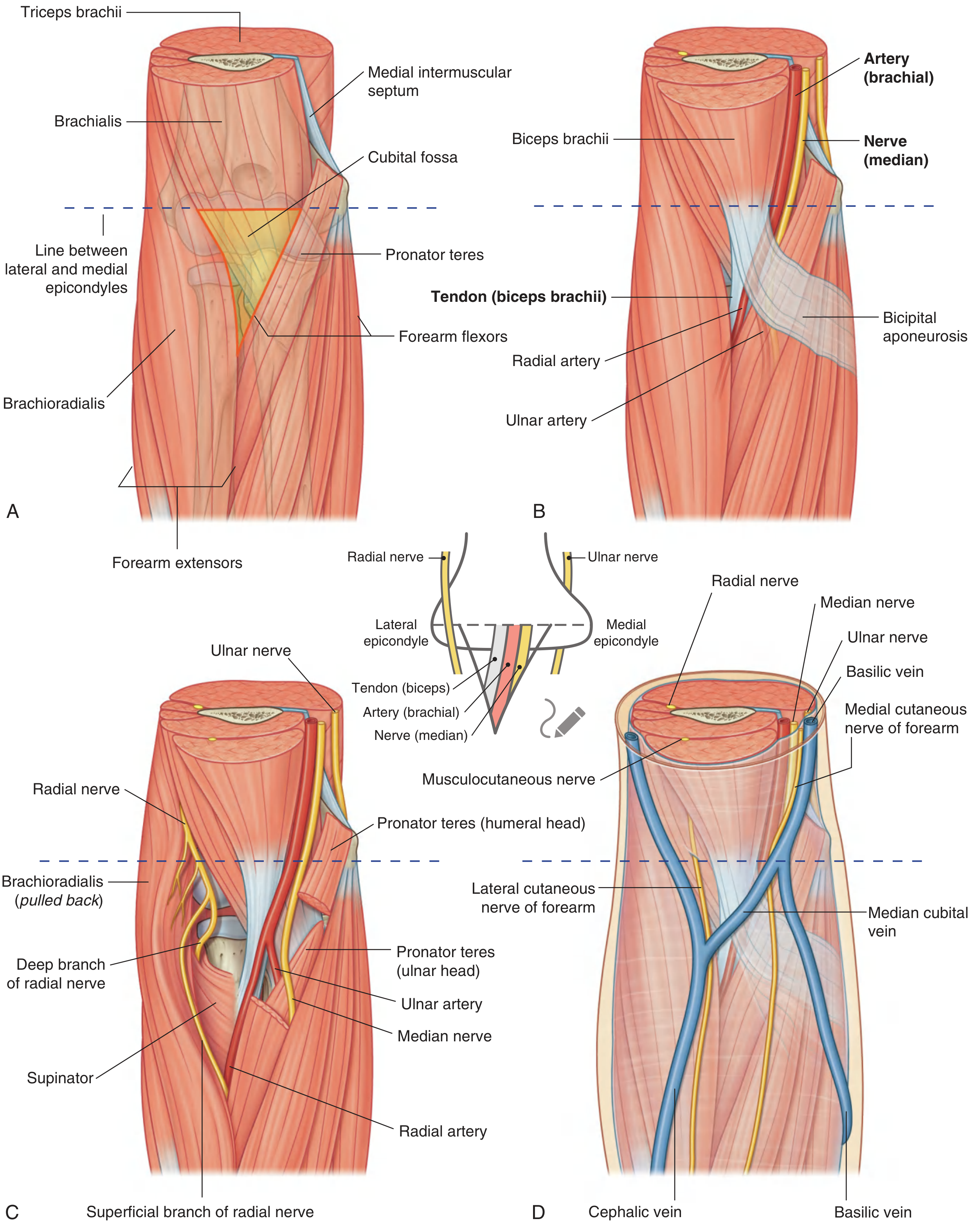
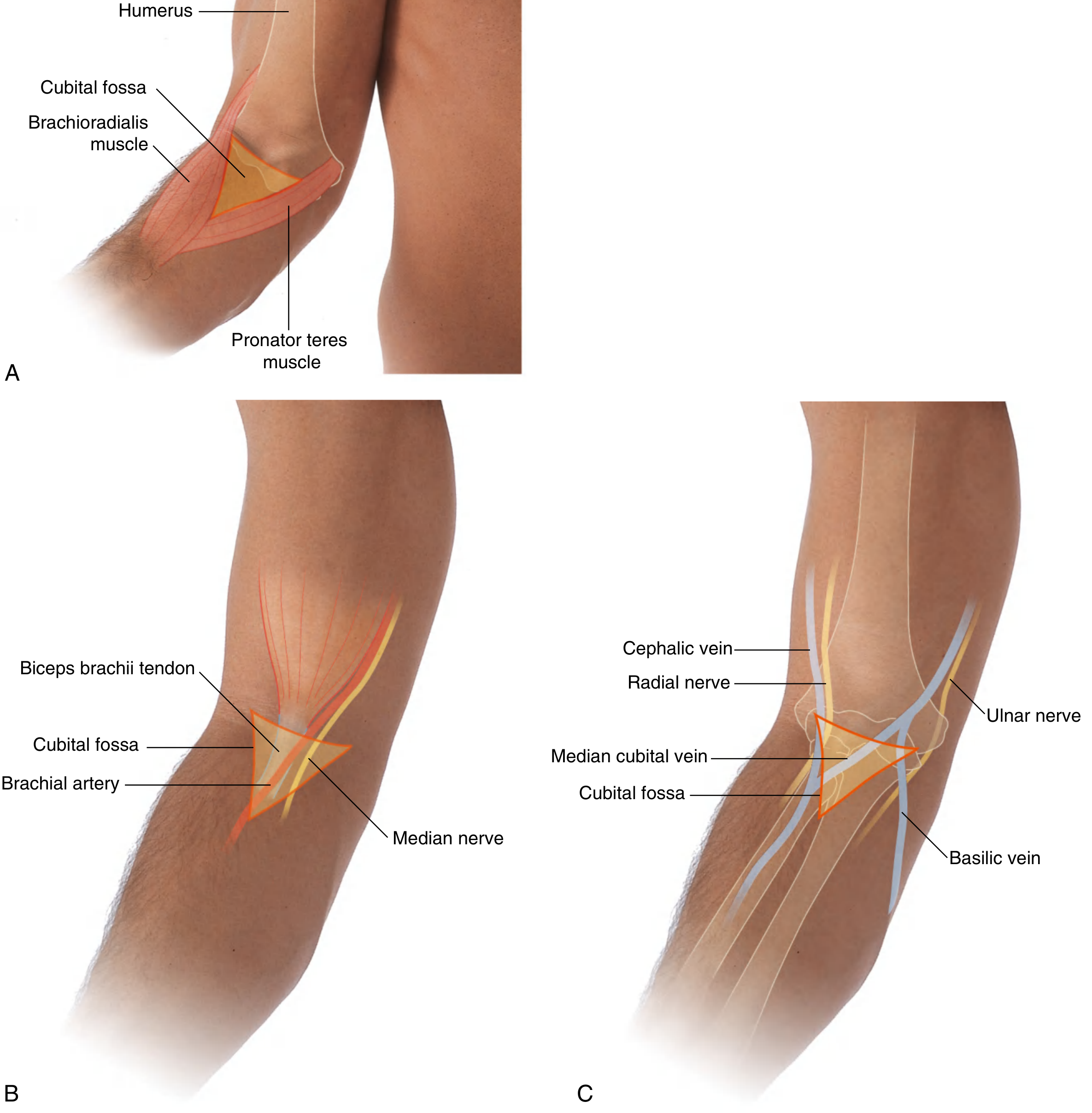
Boundaries
| Boundary | Structure |
|---|---|
| Base (superior) | Imaginary line between medial and lateral epicondyles of the humerus |
| Lateral border | Brachioradialis muscle (from lateral supraepicondylar ridge) |
| Medial border | Pronator teres muscle (from medial epicondyle) |
| Apex | Where brachioradialis and pronator teres meet inferiorly |
| Floor (bed) | Brachialis muscle (and supinator laterally) |
| Roof | Skin + superficial fascia (containing median cubital vein) + deep fascia reinforced by bicipital aponeurosis |
Contents (Lateral to Medial) - Mnemonic: TAN
| Structure | Notes |
|---|---|
| T - Tendon of biceps brachii | Palpable centrally; inserts into radial tuberosity |
| A - Brachial Artery | Bifurcates into radial and ulnar arteries at the apex |
| N - Median Nerve | Medial to brachial artery; exits by passing between two heads of pronator teres |
Radial nerve lies just deep to the lip of brachioradialis (lateral margin) and divides into superficial and deep branches here - but is NOT considered a major content of the fossa.
Ulnar nerve does NOT pass through the cubital fossa - it passes posterior to the medial epicondyle.
Diagrams
Bicipital Aponeurosis
- A flat connective tissue sheet passing from the medial side of the biceps tendon to the deep fascia of the forearm
- Covers and protects the brachial artery and median nerve in the distal fossa
- Its sharp medial margin is palpable
- Separates the superficial cubital veins (roof) from the deep brachial artery (floor) - clinically important during venepuncture
Clinical Significance
1. Venepuncture / IV Access
- Blood sampling
- IV cannulation
- Blood transfusions
2. Blood Pressure Measurement
3. Brachial Artery Injury
- In supracondylar fractures of the humerus (most common in children), the brachial artery can be compressed or lacerated, causing forearm ischemia
- Presents with the 5 Ps: Pain, Pallor, Pulselessness, Paraesthesia, Paralysis
- Can lead to Volkmann's ischaemic contracture if untreated
4. Median Nerve Injury
- Loss of pronation (pronator teres, pronator quadratus)
- Loss of wrist flexion (flexor carpi radialis)
- Loss of thumb opposition
- "Hand of benediction" deformity on attempted fist
5. Radial Nerve Injury (at lateral margin)
6. Intravenous Drug Abuse
- Thrombosed veins
- Abscess/cellulitis
- Pigmented "rail-road track" scars
- Risk of septicaemia, HIV, Hepatitis B/C
7. Lymph Nodes

give answers as well as diagram
Your image lists many histology topics. Which specific topic do you want the answer + diagram for?
HISTOLOGY IMPORTANT DIAGRAMS
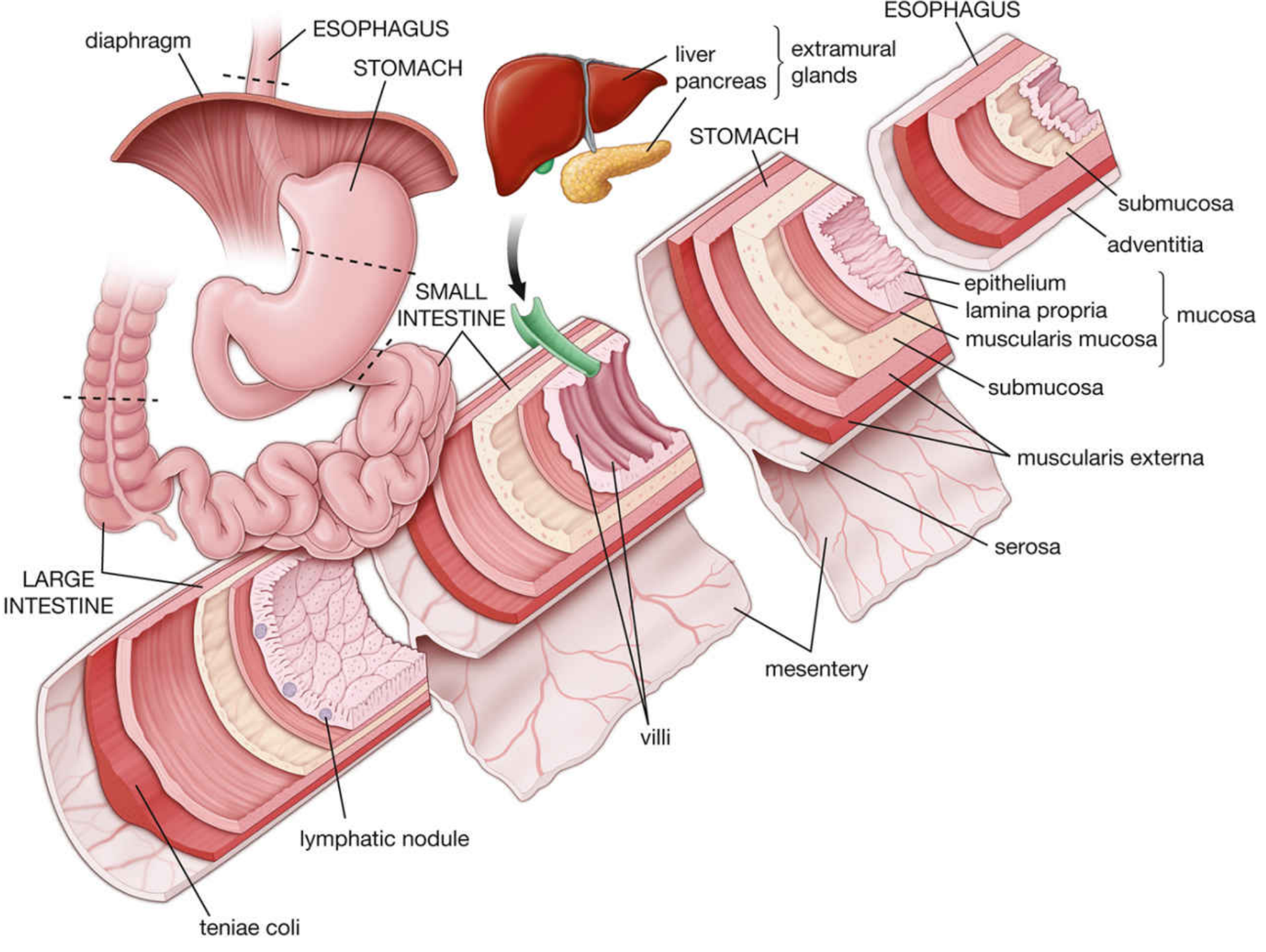
GIT Overview (applies to Oesophagus, Stomach, Small & Large Intestine)
HNF (Head, Neck, Face)
1. Cerebrum (Cerebral Cortex)
- Molecular layer (I) - sparse neurons, nerve fibres
- External granular layer (II) - small pyramidal + stellate cells
- External pyramidal layer (III) - medium pyramidal cells
- Internal granular layer (IV) - stellate cells; receives thalamic input
- Internal pyramidal (ganglionic) layer (V) - Betz cells (giant pyramidal) - motor cortex
- Multiform (polymorphic) layer (VI) - fusiform cells
- H&E: general architecture
- Nissl stain: neuronal cell bodies (rough ER = Nissl substance)
- Golgi stain: entire neuron morphology
2. DRG (Dorsal Root Ganglion)
- Large pseudounipolar neurons with prominent nuclei and nucleoli
- Neurons surrounded by satellite cells (flattened cells forming a capsule)
- Myelinated and unmyelinated nerve fibres between neurons
- Connective tissue capsule (epineurium)
- No synapses (sensory relay only)
3. Thyroid Gland
- Follicles - spherical units lined by simple cuboidal/low columnar epithelium
- Colloid - pink homogeneous material filling follicles (thyroglobulin)
- Follicular cells - height varies with activity (tall = active, flat = inactive)
- Parafollicular (C) cells - pale cells between follicles; produce calcitonin
- Thin connective tissue capsule with septa
4. Salivary Gland
| Type | Serous | Mucous | Mixed |
|---|---|---|---|
| Acini | Round, dark nuclei, zymogen granules | Pale cytoplasm, flat basal nuclei | Mucous acini with serous demilunes |
| Example | Parotid | Sublingual | Submandibular |
| Secretion | Watery, enzyme-rich | Thick, mucus-rich | Both |
- Striated ducts - columnar cells with basal infoldings (modify saliva)
- Intercalated ducts - small, cuboidal cells connecting acini to striated ducts
- Myoepithelial cells - surrounding acini, aid in expulsion
5. Tongue
- Dorsal surface: stratified squamous epithelium with papillae
- Filiform - most numerous; no taste buds; keratinized; pointed
- Fungiform - mushroom-shaped; few taste buds; scattered
- Circumvallate (vallate) - large; surrounded by a trench; most taste buds; Von Ebner glands in base
- Foliate - on lateral margins; leaf-like folds; taste buds
- Taste buds: barrel-shaped; contain taste cells, supporting cells, basal cells; open via taste pore
- Core: skeletal muscle in 3 planes (longitudinal, transverse, vertical)
- Ventral surface: thin, non-keratinized stratified squamous epithelium
6. Trachea
- Mucosa - pseudostratified ciliated columnar epithelium (respiratory epithelium) with goblet cells; lamina propria with elastic fibres
- Submucosa - loose CT; mixed seromucous glands
- C-shaped hyaline cartilage rings (16-20); open posteriorly; trachealis muscle (smooth) bridges gap
- Adventitia - loose CT
7. Oesophagus
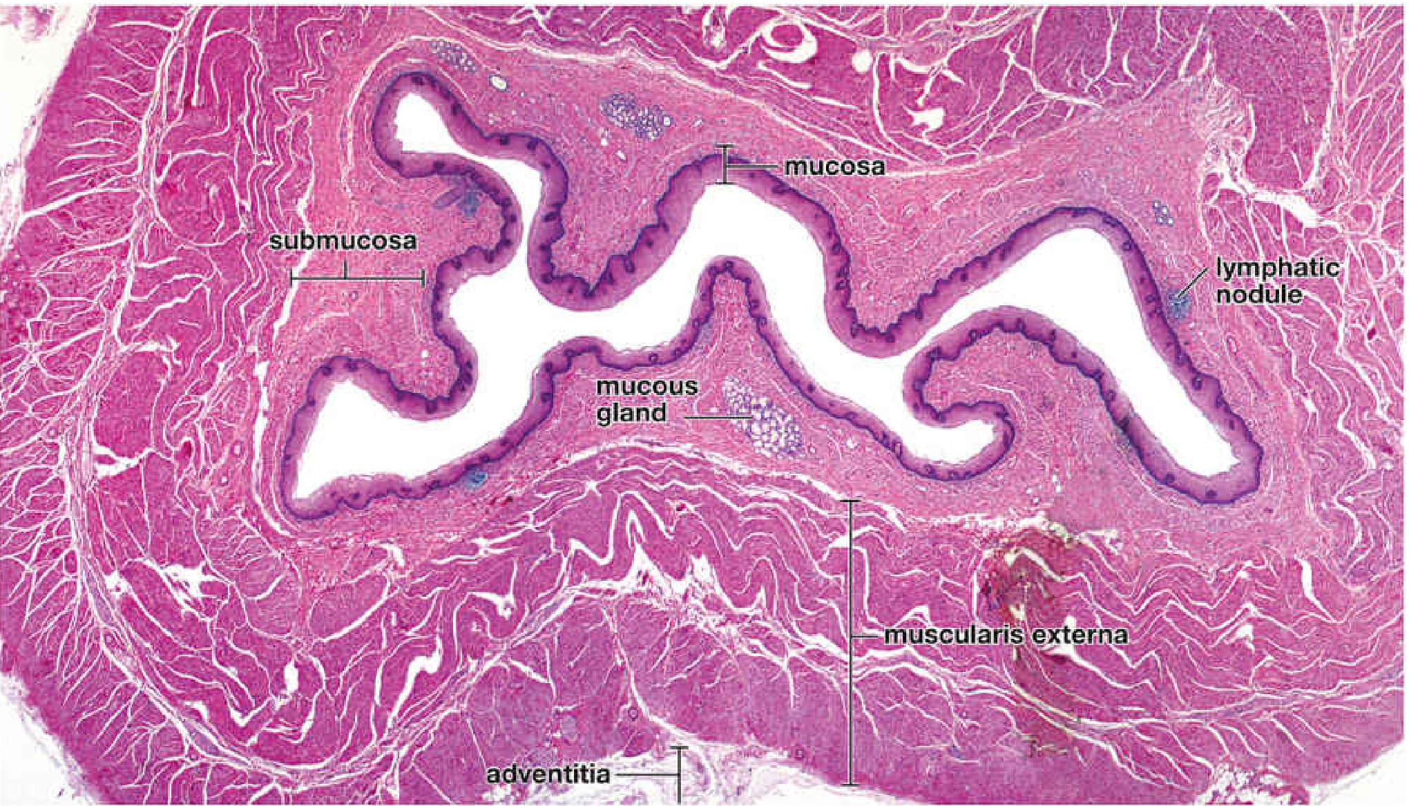
- Mucosa - non-keratinized stratified squamous epithelium; muscularis mucosae (longitudinal smooth muscle)
- Submucosa - mucous glands (esophageal glands proper); Meissner's plexus
- Muscularis externa - upper 1/3: skeletal; middle 1/3: mixed; lower 1/3: smooth; Auerbach's plexus between layers
- Adventitia (no serosa except short abdominal part)
THORAX
8. Lungs
- Bronchi - hyaline cartilage plates (not rings); pseudostratified ciliated epithelium; smooth muscle
- Bronchioles - no cartilage; Clara (Club) cells; simple columnar/cuboidal ciliated; smooth muscle
- Terminal bronchioles - last purely conducting airway
- Respiratory bronchioles - first with alveoli in walls
- Alveoli - thin walls; Type I pneumocytes (gas exchange, flat); Type II pneumocytes (surfactant, cuboidal); alveolar macrophages ("dust cells")
- Interalveolar septum - Type I + II cells, capillaries, elastic fibres
9. Cardiac Muscle
- Branching cylindrical fibres (NOT parallel like skeletal)
- Centrally placed single nucleus (1-2 per cell)
- Intercalated discs - transverse dark lines at cell junctions; contain fascia adherens, desmosomes, gap junctions
- Cross-striations present (like skeletal)
- Rich in mitochondria (abundant energy demand)
- No satellite cells; limited regeneration
| Feature | Skeletal | Cardiac | Smooth |
|---|---|---|---|
| Nucleus | Peripheral, multiple | Central, 1-2 | Central, 1 |
| Striations | Yes | Yes | No |
| Branching | No | Yes | No |
| Intercalated discs | No | Yes | No |
10. Artery and Veins
- Tunica intima: endothelium + internal elastic lamina (prominent)
- Tunica media: thick; multiple layers of smooth muscle
- Tunica adventitia: collagen + external elastic lamina
- Tunica intima: thin
- Tunica media: thin smooth muscle
- Tunica adventitia: thickest layer (collagen bundles)
- Valves present
- Lumen: larger, irregular, often collapsed
11. Lymph Node
- Capsule - dense CT; trabeculae extend inward
- Cortex - lymphoid follicles (B cell zone)
- Primary follicles: dense, dark-staining
- Secondary follicles: germinal centre (light, pale) + mantle zone
- Paracortex - T cell zone; between cortex and medulla
- Medulla - medullary cords (plasma cells, macrophages) + medullary sinuses
- Afferent lymphatics → subcapsular sinus → cortical sinuses → medullary sinuses → efferent lymphatic (at hilum)
- Blood vessels enter/leave at hilum
ABDOMEN
12. Stomach
- Mucosa - simple columnar epithelium; gastric pits; gastric glands
- Fundic glands (body/fundus): Chief cells (pepsinogen, blue/basophilic), Parietal cells (HCl + intrinsic factor, pink/eosinophilic, fried-egg nucleus), Mucous neck cells, Enteroendocrine cells
- Cardiac glands: mucous
- Pyloric glands: mucous + G cells (gastrin)
- Submucosa - no glands (except in pylorus)
- Muscularis externa - 3 layers (oblique inner, circular middle, longitudinal outer)
- Serosa
13. Duodenum
- Villi - tall, finger-like (tallest in duodenum); covered by simple columnar epithelium with brush border
- Microvilli (brush border) on enterocytes
- Goblet cells scattered
- Crypts of Lieberkühn - intestinal glands at base of villi
- Brunner's glands in submucosa - pathognomonic of duodenum; mucous glands; secrete alkaline mucus to neutralize acid chyme
- Paneth cells in crypts (acidophilic granules, lysozyme)
14. Ileum
- Villi - shorter, leaf-like/finger-like
- Peyer's patches - large aggregated lymphoid nodules in lamina propria and submucosa - pathognomonic of ileum
- Goblet cells more numerous than duodenum
- No Brunner's glands
- Enterocytes + goblet cells + Paneth cells + M cells (over Peyer's patches)
15. Appendix
- Small, round lumen (often collapsed/irregular)
- Mucosa - simple columnar epithelium; crypts of Lieberkühn (few, short)
- Abundant lymphoid follicles in lamina propria and submucosa (may obliterate the submucosa)
- No villi
- Muscularis externa - complete (unlike large intestine with teniae coli)
- Mesoappendix (serosa)
16. Liver
- Classic hepatic lobule (hexagonal):
- Central vein (central hepatic venule) at centre
- Portal triads at corners (portal vein, hepatic artery, bile duct)
- Hepatocytes in radiating cords (plates)
- Sinusoids between plates (contain blood from portal vein + hepatic artery)
- Kupffer cells (macrophages) lining sinusoids
- Space of Disse (perisinusoidal space) - between hepatocytes and sinusoids; contains Ito cells (stellate, store vitamin A)
- Portal lobule - triangle with bile duct at centre
- Acinus (of Rappaport) - zones 1/2/3 around portal tract
17. Kidney
- Cortex - renal corpuscles (glomerulus + Bowman's capsule); proximal + distal convoluted tubules
- Medulla - loops of Henle; collecting ducts; renal pyramids
- Renal corpuscle: glomerulus (fenestrated capillaries) + Bowman's capsule (parietal = simple squamous; visceral = podocytes)
- PCT - cuboidal cells, brush border (microvilli), pale pink, indistinct lumen
- DCT - cuboidal cells, no brush border, distinct lumen
- Loop of Henle - thin segment: simple squamous; thick segment: cuboidal
- Collecting duct - clear cells (principal) + dark cells (intercalated); distinct cell borders
18. Ureter
- Lumen - star-shaped (stellate) when contracted
- Mucosa - transitional epithelium (urothelium) - 4-5 layers; umbrella cells on surface
- Lamina propria - dense CT
- Muscularis - inner longitudinal + outer circular (proximal 2/3); additional outer longitudinal in distal 1/3
- Adventitia - loose CT, fat
19. Spleen
- Capsule - dense CT with trabeculae; myoepithelial cells in animals
- White pulp - lymphoid tissue around central artery (PALS = periarteriolar lymphoid sheath; T cells); lymphoid follicles (B cells)
- Red pulp - splenic sinusoids (blood-filled) + splenic cords (Billroth's cords) - macrophages
- Marginal zone - between white and red pulp
PELVIS
20. Ovary
- Germinal epithelium - simple cuboidal covering (NOT germinal in function)
- Tunica albuginea - dense CT beneath epithelium
- Cortex - follicles in various stages:
- Primordial: oocyte + single flat granulosa cells
- Primary: oocyte + single cuboidal granulosa layer
- Secondary: multiple granulosa layers + theca interna/externa
- Graafian (antral): large antrum (follicular fluid); cumulus oophorus; zona pellucida
- Corpus luteum - post-ovulation; granulosa lutein cells (large, pale) + theca lutein cells
- Corpus albicans - white scar tissue after corpus luteum regresses
- Medulla - loose CT, vessels
21. Testis
- Tunica albuginea - thick fibrous capsule
- Seminiferous tubules - lined by spermatogenic cells + Sertoli cells
- Spermatogonia → primary spermatocytes → secondary spermatocytes → spermatids → spermatozoa
- Sertoli cells - tall, pale, irregular nuclei with prominent nucleolus; blood-testis barrier
- Interstitium - Leydig cells (interstitial cells) - large, pink, round; produce testosterone; may have Reinke's crystals
- Rete testis → efferent ductules → epididymis
22. Prostate (Theory)
- Glandular tissue - tubuloalveolar glands in 3 concentric zones:
- Transition zone (periurethral)
- Central zone
- Peripheral zone (most cancers arise here)
- Epithelium - pseudostratified/simple columnar; secretory cells + basal cells
- Corpora amylacea - concentric laminated bodies in lumina (prostatic concretions); increase with age
- Stroma - fibromuscular (smooth muscle + fibrous CT)
- Prostatic urethra passes through
scalp diagram for 5 mark question
Scalp (5 Marks)
Definition & Extent
Layers - Mnemonic: SCALP
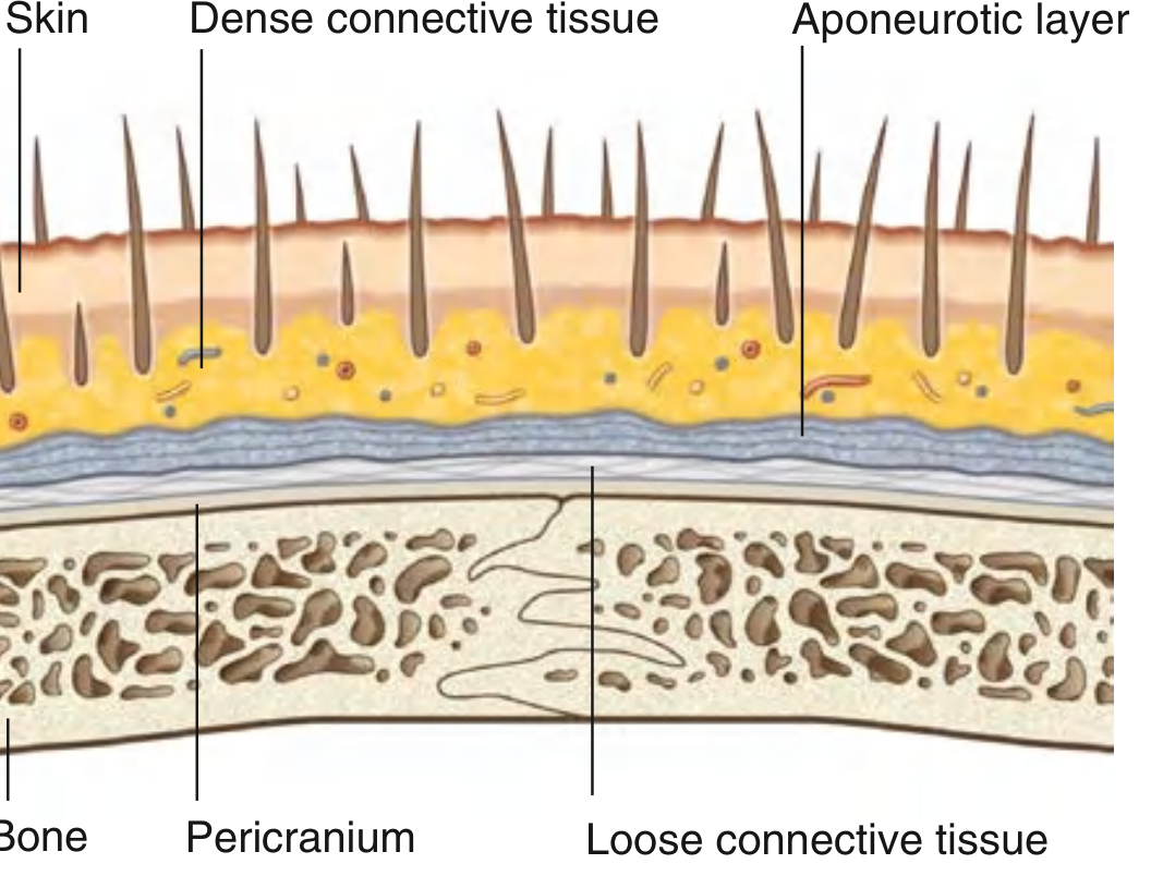
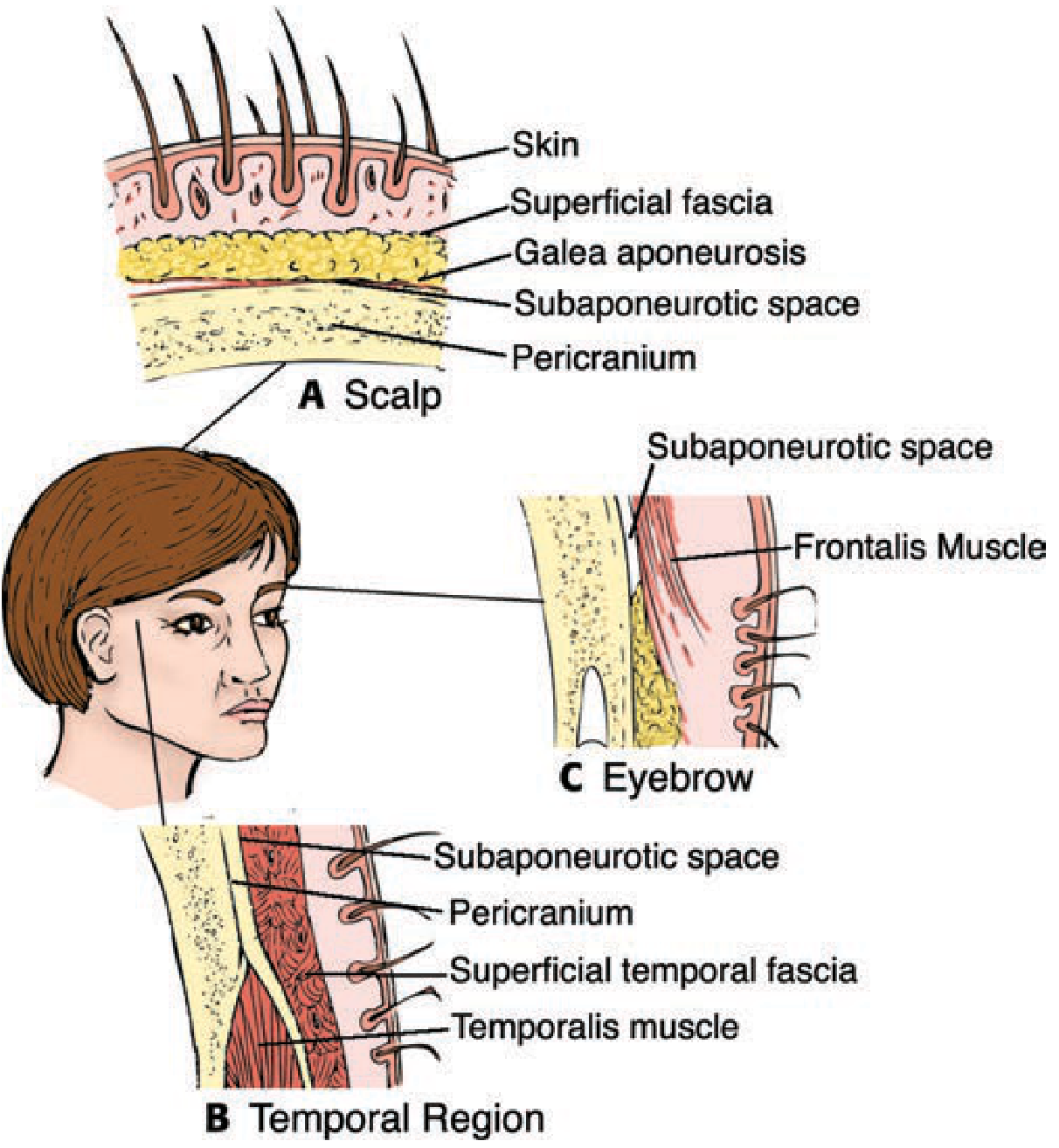
The 5 Layers in Detail
| Layer | Key Features |
|---|---|
| S - Skin | Thick; contains hair follicles, sebaceous glands, sweat glands |
| C - Connective tissue (dense) | Firmly binds skin to galea; contains arteries, veins, nerves; vessels held open by dense CT → profuse bleeding when cut |
| A - Aponeurotic layer (Galea aponeurotica) | Flat tendinous sheet of occipitofrontalis muscle; frontal belly anteriorly, occipital belly posteriorly; moves the scalp |
| L - Loose connective tissue | "Danger zone"; allows scalp movement; infections/haematomas spread freely here; emissary veins pass through |
| P - Pericranium | Periosteum of outer skull; firmly attached to bone but removable except at sutures |
Scalp proper = first 3 layers (S + C + A) - tightly bound together and move as one unit; torn together in scalping injuries
Blood Supply
| Region | Artery | Source |
|---|---|---|
| Anterior | Supratrochlear, Supraorbital | Ophthalmic → ICA |
| Lateral | Superficial temporal | External carotid |
| Posterior | Occipital, Posterior auricular | External carotid |
- All arteries anastomose freely across the scalp
- Vessels are held open by dense CT → scalp wounds bleed profusely
Nerve Supply
| Region | Nerve |
|---|---|
| Anterior (forehead) | Supratrochlear + Supraorbital (V1) |
| Temporal | Auriculotemporal (V3) + Zygomaticotemporal (V2) |
| Posterior | Greater occipital (C2) + Lesser occipital (C2) + Greater auricular (C2,3) |
Clinical Significance
- Profuse bleeding in scalp wounds - dense CT holds vessels open; treat by pressure or full-thickness suturing
- "Dangerous area" - Layer L (loose CT) allows spread of infection or blood widely under the galea; pus/blood can track from orbit to orbit or forehead to occiput
- Emissary veins in loose CT - connect scalp veins to intracranial venous sinuses → infection can spread intracranially (meningitis, cavernous sinus thrombosis)
- Subgaleal haematoma - blood in loose CT layer; can cross suture lines (unlike cephalhaematoma which is limited by sutures)
- Cephalhaematoma - blood between pericranium and bone; cannot cross sutures (periosteum attached at sutures)
- Scalp flaps in surgery - based on peripheral vessels; easy to raise because of loose areolar plane
Dangerous Areas of Face diagram
Dangerous Area of the Face
Definition
Boundaries of the Danger Triangle
Nose bridge
/ \
/ \
Corner Corner
of mouth --- of mouth
- Apex: Bridge of the nose (nasion)
- Base: A line joining the corners of the mouth
- Sides: Along the nasolabial folds
- The area includes the upper lip, nose, nasolabial folds, and central cheek
The region above the mouth is the critical danger zone.
Why It Is Dangerous - The Venous Pathway
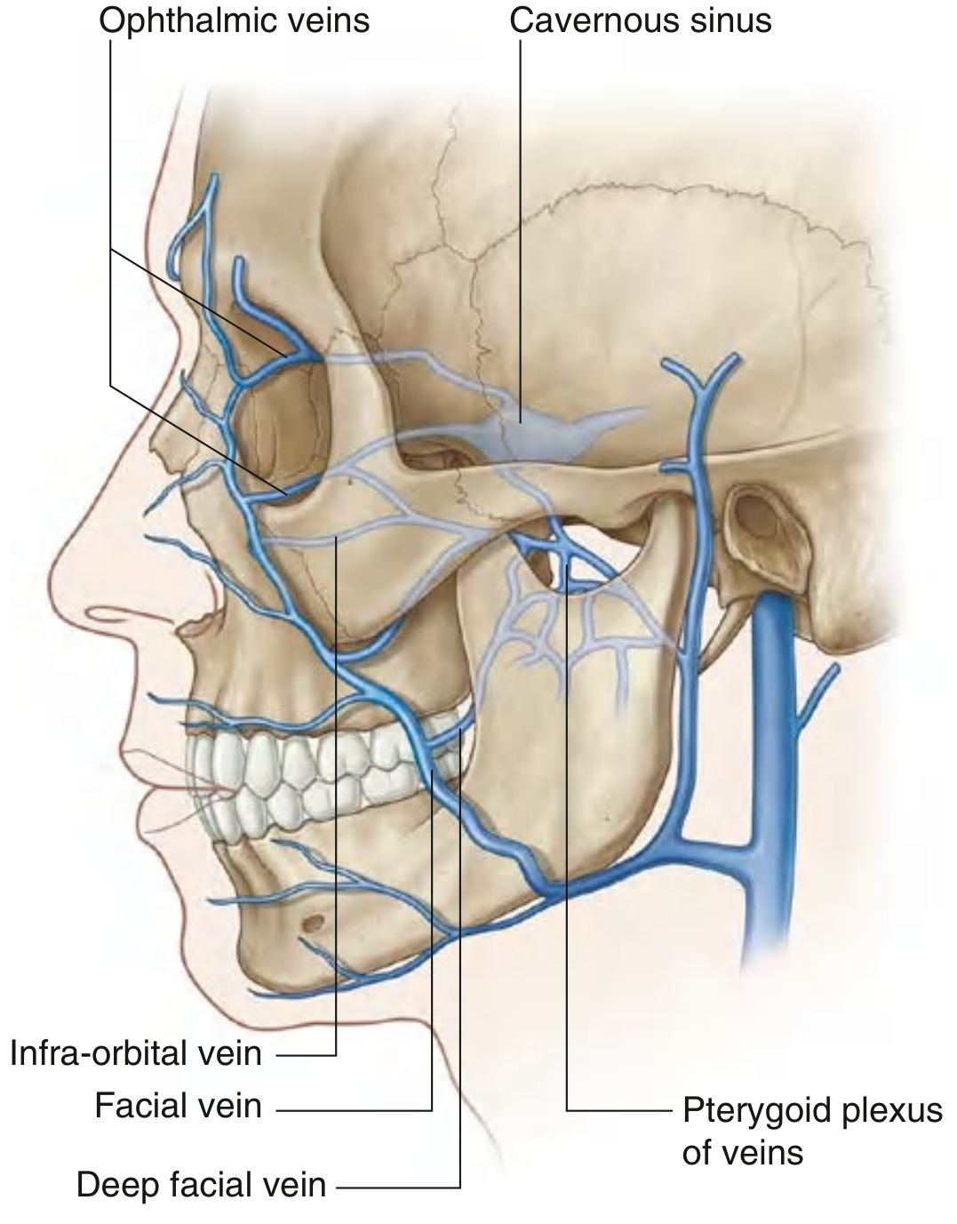
Route of Spread
Facial vein → Angular vein (at medial corner of eye) → Superior ophthalmic vein → Cavernous sinus
Facial vein → Deep facial vein → Pterygoid plexus → Inferior ophthalmic vein / emissary veins → Cavernous sinus
Facial vein → Infraorbital vein (via infraorbital foramen) → Pterygoid plexus → Cavernous sinus
Summary Table
| Feature | Detail |
|---|---|
| Area | Upper lip, nose, nasolabial folds, central cheek |
| Key vessel | Facial vein (valveless) |
| Communication | Angular vein → superior ophthalmic vein |
| Intracranial destination | Cavernous sinus |
| Complication | Cavernous sinus thrombosis |
Clinical Consequences of Infection Spread
Cavernous Sinus Thrombosis
| Structure | Effect |
|---|---|
| CN III (oculomotor) | Ptosis, mydriasis, loss of eye movements |
| CN IV (trochlear) | Loss of downward/inward gaze |
| CN VI (abducens) | Lateral gaze palsy (most common - lies inside sinus) |
| CN V1 + V2 | Facial pain/numbness (ophthalmic + maxillary divisions) |
| Internal carotid artery | Horner's syndrome, arteritis |
- Severe headache, high fever, rigors
- Proptosis (forward protrusion of eye)
- Periorbital oedema
- Chemosis (conjunctival oedema)
- Painful ophthalmoplegia
- Signs may be bilateral (venous sinuses communicate across midline)
Practical Warning
"Never squeeze a pimple, boil, or furuncle in the dangerous area of the face"
- Nasal furuncle (boil inside nose)
- Infected pimple/acne on upper lip or nose
- Dental abscess (upper teeth)
- Nasal cellulitis
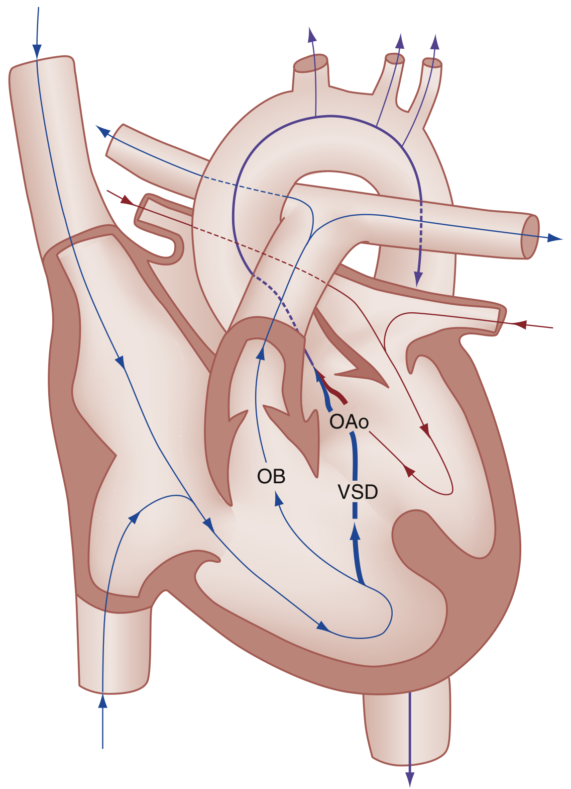
Tetralogy of Fallot (TOF)
labeled diagram of tetralogy of Fallot showing ventricular septal defect overriding aorta pulmonary stenosis and right ventricular hypertrophy
"tetralogy of Fallot"
American Heart Association tetralogy of Fallot congenital heart defect
https://www.heart.org/en/health-topics/congenital-heart-defe…
Tetralogy of Fallot (TOF)
Four Features - PROVe
- Pulmonary stenosis / Right ventricular outflow tract obstruction
- Right ventricular hypertrophy
- Overriding of aorta
- Ventricular septal defect (VSD)
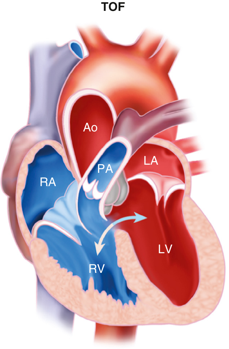
Pathophysiology
- Cyanosis
- Decreased pulmonary blood flow
- Hypoxaemia
- Right ventricular hypertrophy
Clinical Features
- Cyanosis, often worsening during crying, feeding, or exertion
- Dyspnoea and poor feeding in infants
- Cyanotic spells (Tet spells): sudden severe cyanosis, rapid breathing, irritability or syncope
- Older children may squat after exercise. Squatting raises systemic vascular resistance, reducing right-to-left shunting.
- Ejection systolic murmur at the left upper sternal border due to pulmonary stenosis
- Clubbing and polycythaemia in chronic cases
Investigations
- Chest X-ray: “Boot-shaped heart” (coeur en sabot), decreased pulmonary vascular markings
- ECG: right ventricular hypertrophy and right-axis deviation
- Echocardiography: confirms VSD, overriding aorta, RV outflow obstruction, and RV hypertrophy
Management
- During a tet spell: knee-chest position, oxygen, calm the child, IV fluids, and urgent medical treatment.
- Definitive treatment is surgical repair, usually in infancy: VSD closure plus relief of right ventricular outflow obstruction.
Arch of aorta
labeled anatomy diagram aortic arch branches brachiocephalic trunk left common carotid left subclavian and relations
"aortic arch" anatomy
aortic arch anatomy branches relations StatPearls
Arch of Aorta
Definition and Situation
- Begins: behind the lower half of manubrium at the level of the sternal angle
- Ends: on the left side of T4/T5 vertebra, where it continues as the descending thoracic aorta
- Its highest point reaches the middle of the manubrium sterni.
Diagram
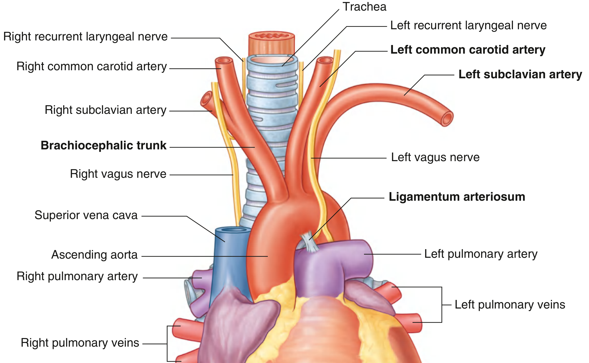
Branches
-
Brachiocephalic trunk
- Largest branch
- Divides behind the right sternoclavicular joint into:
- Right common carotid artery
- Right subclavian artery
-
Left common carotid artery
- Supplies left side of head and neck
-
Left subclavian artery
- Main arterial supply to left upper limb
Brachiocephalic trunk - Common carotid - Subclavian artery.
Relations
| Aspect | Relations |
|---|---|
| Anterior and left | Left brachiocephalic vein, thymus, left vagus nerve, left phrenic nerve, left pleura and lung |
| Posterior and right | Trachea, oesophagus, thoracic duct, deep cardiac plexus |
| Inferior | Left pulmonary artery, left main bronchus, ligamentum arteriosum, left recurrent laryngeal nerve |
| Superior | Its three branches |
Important Relation
Clinical Importance
- Coarctation of aorta commonly occurs just distal to the origin of the left subclavian artery, near the ligamentum arteriosum.
- Aortic aneurysm may compress the left recurrent laryngeal nerve, causing hoarseness of voice due to left vocal cord paralysis.
- Patent ductus arteriosus is persistence of the fetal ductus arteriosus, which normally becomes the ligamentum arteriosum after birth.
- Sudden deceleration trauma can tear the aorta near the aortic isthmus, close to the ligamentum arteriosum.
Arches of aorta
labeled embryology diagram pharyngeal aortic arches derivatives first second third fourth sixth arch
"aortic arch" embryology
aortic arch embryology derivatives pharyngeal arch arteries
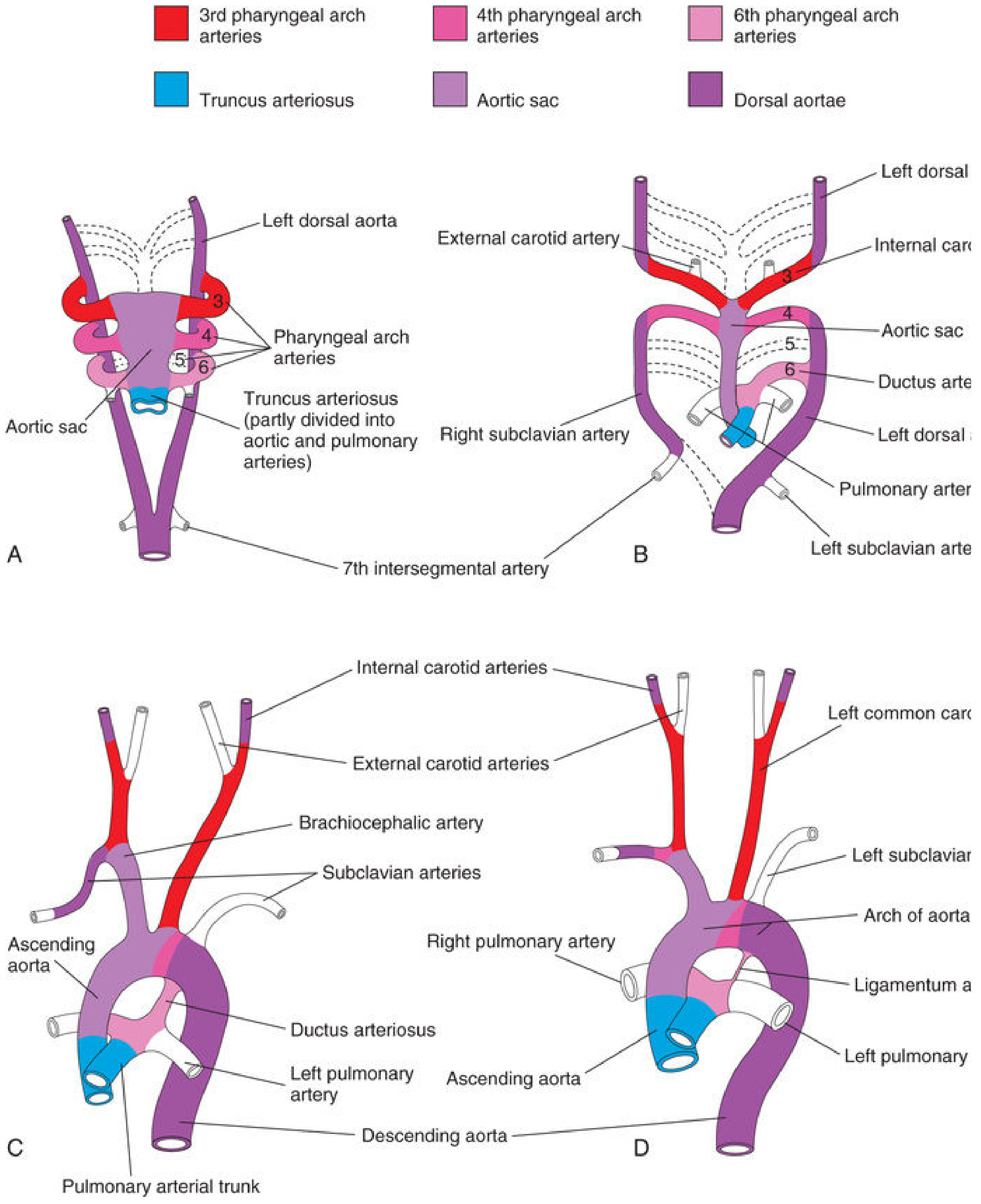
Aortic Arches (Pharyngeal Arch Arteries)
There are six numbered arches, but the 5th arch is absent or regresses, so only I, II, III, IV, and VI are important.
Diagram
Derivatives of Aortic Arches
| Aortic arch | Main derivative |
|---|---|
| 1st arch | Part of maxillary artery |
| 2nd arch | Stapedial artery and hyoid artery, mostly disappears |
| 3rd arch | Common carotid artery and proximal part of internal carotid artery |
| 4th arch - Right | Proximal part of right subclavian artery |
| 4th arch - Left | Part of arch of aorta |
| 5th arch | Rudimentary or absent, disappears completely |
| 6th arch - Right | Proximal part of right pulmonary artery; distal part disappears |
| 6th arch - Left | Proximal part of left pulmonary artery and distal part forms ductus arteriosus |
Easy Mnemonic
- 1 - Maxillary artery
- 2 - Stapedial artery
- 3 - Carotid arteries
- 4 - Systemic arch: aorta on left, right subclavian on right
- 6 - Pulmonary arteries and ductus arteriosus
Important Additional Points
- Aortic sac right horn forms the brachiocephalic trunk.
- Aortic sac left horn forms the proximal part of the arch of aorta.
- Left seventh intersegmental artery forms the left subclavian artery.
- The right recurrent laryngeal nerve hooks around the right subclavian artery.
- The left recurrent laryngeal nerve hooks around the arch of aorta near the ligamentum arteriosum.
Clinical Correlation
-
Patent ductus arteriosus (PDA)
Failure of the left sixth arch distal part to close after birth. -
Coarctation of aorta
Narrowing of aorta, usually near the ductus arteriosus or ligamentum arteriosum. -
Right-sided aortic arch / double aortic arch
Abnormal persistence or regression of parts of the fourth arches or dorsal aortae. It can form a vascular ring and compress the trachea or oesophagus. -
Aberrant right subclavian artery
May pass behind the oesophagus, causing dysphagia lusoria.
Quick answer
Thoracic duct Thoracic part of oesophagus Thoracic part of trachea Structures supplied by phrenic nerve
"phrenic nerve" AND anatomy
thoracic duct thoracic oesophagus thoracic trachea phrenic nerve anatomy
1. Thoracic Duct
Definition
Extent and Course
- Begins as continuation of cisterna chyli at about T12
- Enters thorax through the aortic hiatus of diaphragm
- Ascends in the posterior mediastinum
- At T5, crosses from right to left behind the oesophagus
- Ascends in the superior mediastinum and root of neck
- Ends at the left venous angle: junction of left internal jugular and left subclavian veins
Relations in Posterior Mediastinum
- Anterior: Oesophagus
- Right: Azygos vein
- Left: Descending thoracic aorta
- Posterior: Vertebral bodies
Area Drained
- Right side of head and neck
- Right upper limb
- Right side of thorax
Clinical Importance
- Injury during oesophageal or thoracic surgery can cause chylothorax, accumulation of milky chyle in pleural cavity.
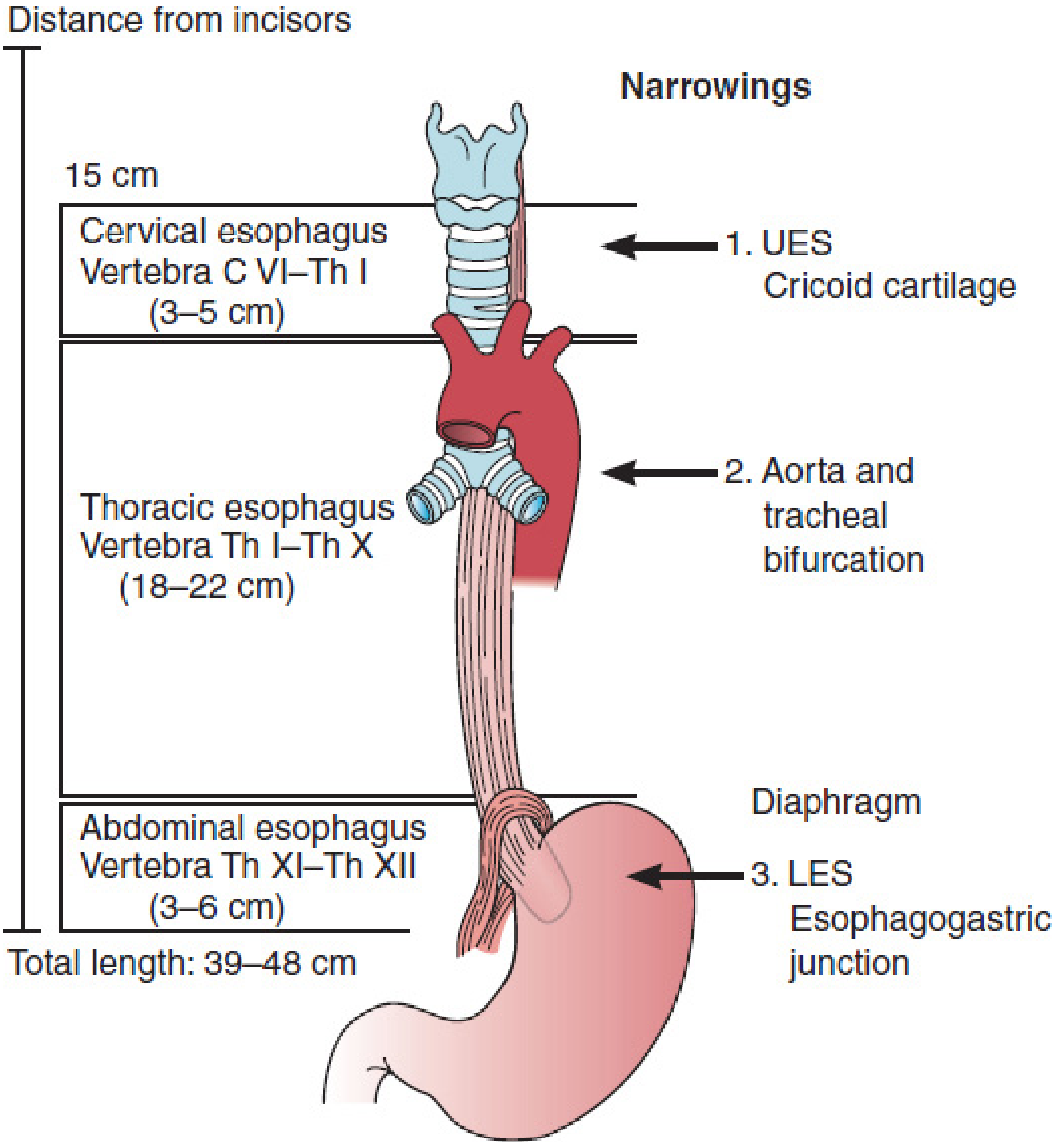
2. Thoracic Part of Oesophagus
Extent
- Begins at thoracic inlet, at T1
- Ends at oesophageal hiatus of diaphragm at T10
- Length: about 18-20 cm
Course
- Initially lies posterior to trachea.
- In upper thorax, lies to the right of arch of aorta.
- Passes behind bifurcation of trachea and left main bronchus.
- Moves to the left and passes through diaphragm at T10.
Important Relations
| Anterior | Posterior |
|---|---|
| Trachea | Vertebral bodies |
| Left main bronchus | Thoracic duct |
| Pericardium and left atrium | Azygos and hemiazygos veins |
| Left vagus nerve | Descending thoracic aorta in lower part |
| Right | Left |
|---|---|
| Azygos vein | Arch of aorta |
| Right pleura | Descending thoracic aorta |
| Right vagus nerve | Left pleura |
| Left vagus nerve |
Constrictions of Oesophagus
- At pharyngooesophageal junction: C6
- Where crossed by arch of aorta: T4
- Where crossed by left main bronchus: T5-T6
- At oesophageal hiatus: T10
Clinical Importance
- Foreign bodies can lodge at these constrictions.
- Oesophageal varices may occur due to portal hypertension at lower end.
- Thoracic duct lies close to oesophagus and may be injured during oesophageal surgery.
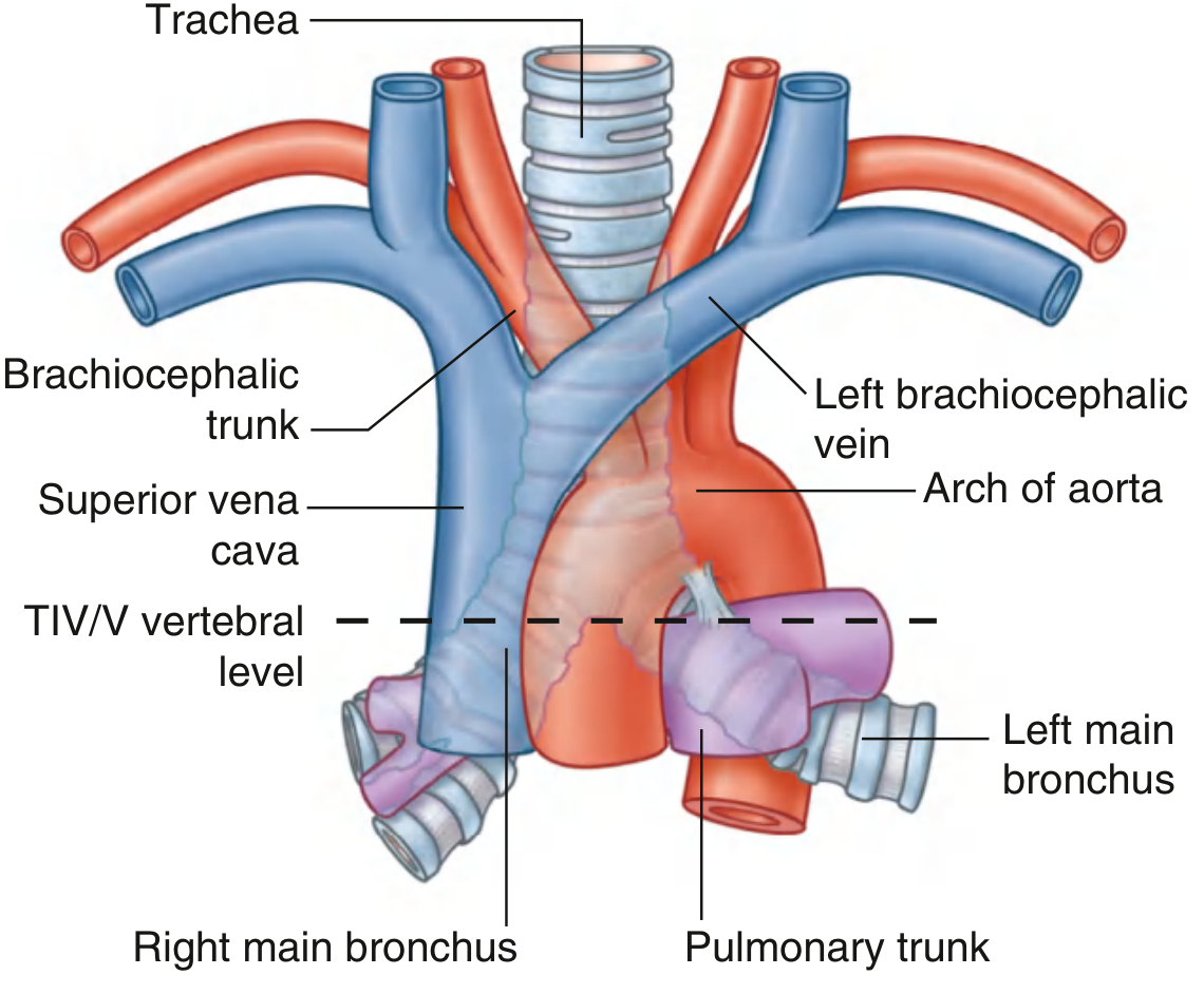
3. Thoracic Part of Trachea
Extent
- Begins at thoracic inlet
- Ends at the carina, where it divides into right and left main bronchi
- Bifurcation occurs at the level of sternal angle, T4/T5
Course
- Enters superior mediastinum just to the right of the median plane
- Descends anterior to oesophagus
- Ends at carina at T4/T5
Relations
| Anterior | Posterior |
|---|---|
| Manubrium sterni | Oesophagus |
| Thymus | Recurrent laryngeal nerves near tracheo-oesophageal grooves |
| Left brachiocephalic vein | Vertebral column indirectly through oesophagus |
| Arch of aorta |
| Right | Left |
|---|---|
| Brachiocephalic trunk | Arch of aorta |
| Right vagus nerve | Left vagus nerve |
| Azygos vein arch | Left recurrent laryngeal nerve |
| Right pleura | Left pleura |
Blood Supply
- Bronchial arteries
- Inferior thyroid arteries
Nerve Supply
- Parasympathetic: vagus nerve and recurrent laryngeal nerves
- Sympathetic: sympathetic trunk
Clinical Importance
- Carina is highly sensitive. Irritation produces a strong cough reflex.
- Enlargement of arch of aorta or mediastinal lymph nodes may compress the trachea.
- Aspiration of foreign body is more common in the right main bronchus because it is wider, shorter, and more vertical.
4. Structures Supplied by Phrenic Nerve
Root Value
Mnemonic: “C3, 4, 5 keep the diaphragm alive.”
Motor Supply
- Diaphragm: the phrenic nerve is the only motor nerve supply of the diaphragm.
Sensory Supply
- Fibrous pericardium
- Parietal layer of serous pericardium
- Mediastinal pleura
- Central part of diaphragmatic pleura
- Central part of diaphragmatic peritoneum
- Central part of diaphragm
Peripheral diaphragm receives sensory fibres from lower intercostal nerves.
Course in Thorax
- Between mediastinal pleura and fibrous pericardium
- Anterior to root of lung
| Right phrenic nerve | Left phrenic nerve |
|---|---|
| Passes along right side of SVC | Crosses arch of aorta |
| Runs over pericardium covering right atrium | Runs over pericardium covering left ventricle |
| Enters diaphragm near caval opening with IVC | Pierces diaphragm separately |
Clinical Importance
- Injury causes ipsilateral paralysis of diaphragm.
- Irritation causes referred pain at the shoulder tip, supplied by C3-C5 dermatomes.
- Pericarditis, subphrenic abscess, or diaphragmatic pleurisy may cause shoulder pain.