
Mucocele of appendix
Mucocele of appendix
mucocele of appendix pathology gross specimen
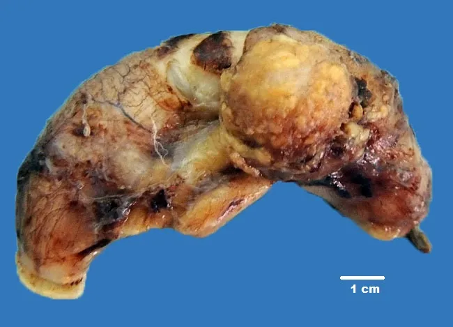
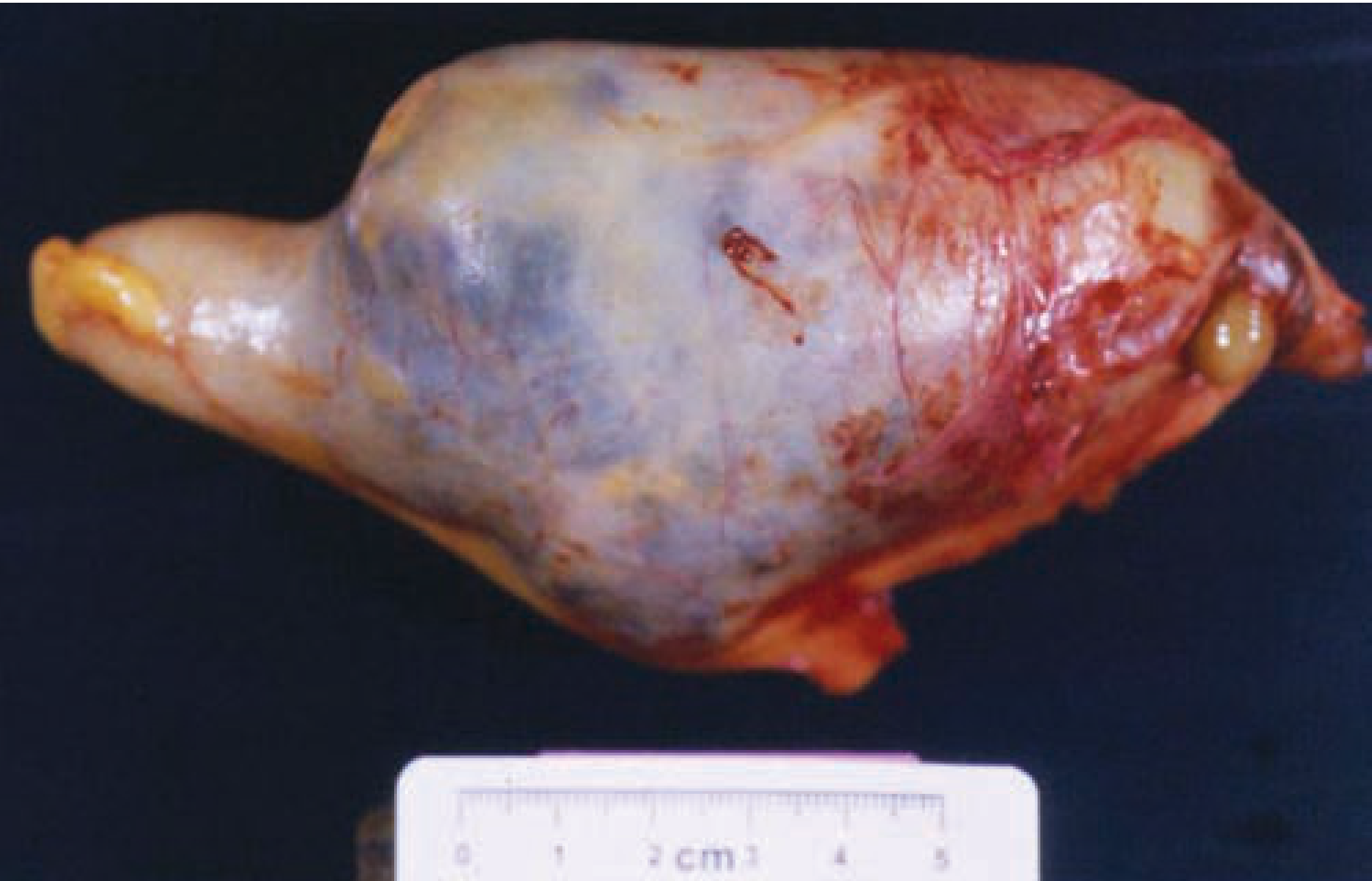
Gross pathology of an appendectomy specimen reveals an enlarged, dilated appendix with multiple cystic loci containing viscous mucinous material. The specimen measures approximately 8 cm in length with a maximum external diameter near 5 cm; the mucin-filled chambers create a lobulated, sponge-like appearance along the distal portion of the organ. The cut surfaces show translucent to pale-yellow cystic spaces, rimmed by a thin fibrous wall. Histology (described in the accompanying histology image) demonstrates simple mucinous columnar epithelium lining the cystic lumina with goblet cells and abundant mucin, consistent with a mucinous cystadenoma of the appendix. There is no obvious invasion into the serosa reported in this case, supporting a benign mucinous appendiceal neoplasm. Clinically, elderly patients may present with right lower quadrant pain and an ultrasound showing an enlarged appendix; mucinous appendiceal neoplasms can present as appendiceal mucocele and may carry risk of rupture with peritoneal seeding, such as pseudomyxoma peritonei if spilled during surgery. The diagnostic significance lies in distinguishing benign mucinous cystadenoma from mucinous cystadenocarcinoma; surgical resection is curative for localized lesions. Differential diagnoses include mucinous cystadenocarcinoma, mucinous hyperplasia, and nonneoplastic mucocele. This image and description provide essential context for gross-pathology-radiology correlation, surgical planning, and educational prompts on appendiceal mucinous neoplasms.
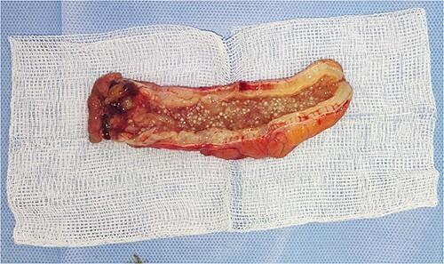
Clinical photograph showing a gross pathological specimen of a surgically resected appendix, which has been longitudinally opened to reveal its contents. The specimen demonstrates the classic presentation of myxoglobulosis, a rare variant of appendiceal mucocele. The oblong structure features a thickened, hyperemic serosal surface and a distended lumen. Key diagnostic features include the presence of numerous small, translucent-to-white, rounded gelatinous globules measuring a few millimeters in diameter, often described as having a 'fish egg' or 'sago grain' appearance. These globules are composed of organized mucin and are distributed throughout the luminal space. One end of the specimen shows areas of dark congestion or hemorrhage. The specimen is displayed on a sterile white gauze pad. This image illustrates the gross morphological findings critical for distinguishing myxoglobulosis from other mucinous neoplasms of the appendix within the context of gastroenterology and surgical pathology.
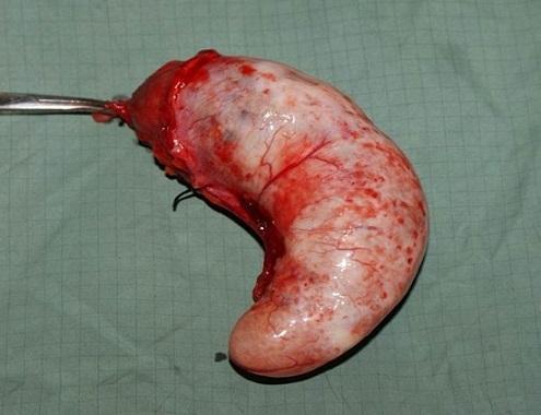
This clinical photograph shows a gross surgical specimen of an appendiceal mucocele, presented in a lateral view against a green surgical drape. The specimen is an oblong, distended, comma-shaped cystic mass approximately 15 cm in length. The surface is predominantly pearly white and glistening, indicative of a stretched serosal layer, with diffuse areas of erythema and prominent superficial vascularization. There are focal subserosal hemorrhages or petechiae visible across the curved body of the appendix. At the proximal end (the base), the tissue is intensely congested and hemorrhagic, showing the site of surgical excision where a metallic surgical instrument and a dark suture are visible. The specimen demonstrates the characteristic macroscopic appearance of a mucinous cystadenoma of the appendix, illustrating the significant luminal dilation caused by intraluminal mucin accumulation. This image is relevant for surgical pathology and gastroenterology education, specifically regarding the diagnosis and surgical management of appendiceal neoplasms.
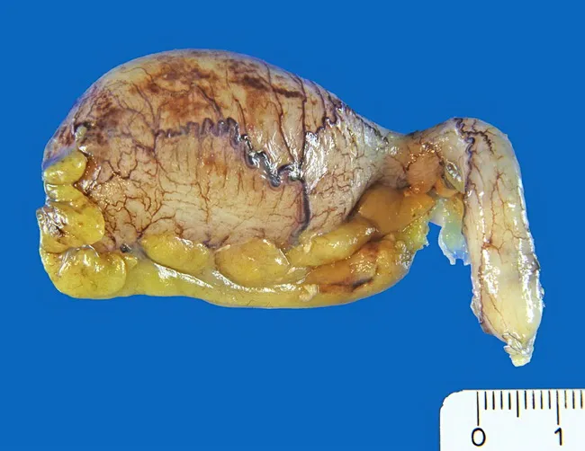
Gross pathology photograph of a surgically excised vermiform appendix. The specimen demonstrates bulbous dilation of the distal (tip) portion with a lobulated, pale-tan surface and fine vascular markings; a portion shows cystic, mucin-filled spaces projecting from the tip. Scale markings (centimeter ruler) are visible for size reference. The interior of the dilated segment contains mucinous material, and the cystic lining is lined by mucin-producing epithelium with cytologic atypia, without obvious stromal invasion on this gross view. The overall pattern is compatible with a benign mucinous cystadenoma (low-grade appendiceal mucinous neoplasm); however, the presence of atypia warrants careful histologic assessment to exclude mucinous cystadenocarcinoma. Radiographic or gross features may include appendiceal mucocele-like dilation. Notably, the lesion has a risk of rupture with potential peritoneal seeding if not completely excised. This image highlights key gross features: bulbous tip expansion, mucin-filled lacunae, delicate mural vasculature, and mucinous epithelium lining. Clinically relevant use cases include educational reference for gastrointestinal pathology, differential diagnosis between benign mucinous cystadenoma and malignant mucinous neoplasms, and correlation with histopathology to guide surgical management and prognosis. This representation is valuable for medical students, residents, and pathologists studying appendiceal mucinous neoplasms and pseudomyxoma peritonei risk assessment in various clinical education settings.
mucocele appendix
Mucocele of the Appendix
Definition
- Maingot's Abdominal Operations, p. 659
- Sabiston Textbook of Surgery, p. 2048
Classification / Spectrum of Underlying Causes
| Type | Nature | Size | Key Features |
|---|---|---|---|
| Retention cyst (simple cyst) | Non-neoplastic | Usually <2 cm | Non-neoplastic occlusion of lumen; incidental finding |
| Mucosal hyperplasia | Non-neoplastic | Small | Proliferative but non-neoplastic mucosa |
| Mucinous cystadenoma (LAMN/HAMN) | Benign neoplasm | Up to 8 cm+ | Majority of "mucoceles"; slow-growing; formerly called mucinous cystadenoma, now Low-grade Appendiceal Mucinous Neoplasm (LAMN) |
| Mucinous cystadenocarcinoma | Malignant | Variable | Histological evidence of malignancy in ~1/3 of cases; risk of pseudomyxoma peritonei |
- Robbins & Kumar Basic Pathology, p. 1020
- Sabiston Textbook of Surgery, p. 2048
- Grainger & Allison's Diagnostic Radiology, p. 273
Epidemiology
- Appendiceal neoplasms (including mucoceles) affect less than 1% of appendectomies.
- Up to 50% of patients with an appendiceal neoplasm present with acute appendicitis as the initial symptom.
- Risk factors for harboring an underlying neoplasm: age >40, immunocompromised status, periapendiceal abscess or phlegmon.
- Associated with synchronous colonic/rectal carcinoma - colonoscopy is recommended after surgery.
- Maingot's Abdominal Operations, p. 657; Grainger & Allison's, p. 273
Pathology
Gross
- Distended, smooth, sausage/balloon-shaped appendix filled with viscous mucin
- Wall calcification is characteristic (visible on plain X-ray or CT)
- Size ranges from <2 cm (simple retention cyst) to >14 cm (mucinous cystadenoma/carcinoma)
Histology
- LAMN (Low-Grade Appendiceal Mucinous Neoplasm): flat or undulating neoplastic mucinous epithelium; pushing (non-infiltrative) pattern of invasion; abundant extracellular mucin; low cytologic atypia
- HAMN (High-Grade): marked cytologic atypia but still pushing border
- Mucinous adenocarcinoma: infiltrative invasion with >50% extracellular mucin; can be well, moderately, or poorly differentiated
Clinical Features
- Often incidental - found on imaging or during surgery/colonoscopy for another reason
- Can mimic acute appendicitis - right lower quadrant pain, nausea, vomiting
- Palpable right lower quadrant mass
- Urologic symptoms (mass effect on ureter/bladder)
- Intussusception
- In cystadenocarcinoma: abdominal pain, weight loss, increasing abdominal girth (suggests pseudomyxoma peritonei)
- Sabiston Textbook of Surgery, p. 2048; Maingot's, p. 659
Imaging
CT (investigation of choice)
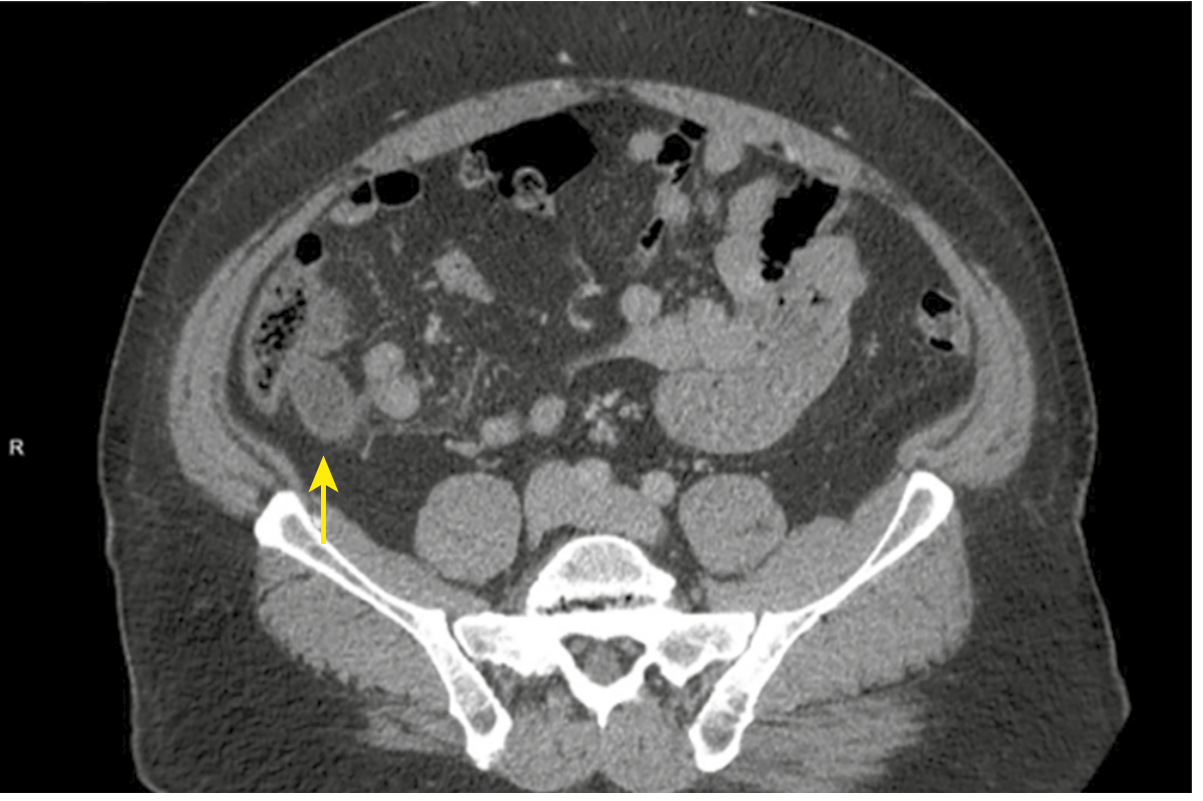
- Well-defined, thin-walled, low-attenuation cystic mass in the right iliac fossa
- Mural calcification - highly characteristic (curvilinear or eggshell pattern)
- Luminal diameter >2 cm, lack of periappendiceal fat stranding, and wall irregularity raise suspicion for neoplasm
- Can identify peritoneal mucin deposits if pseudomyxoma has developed
Ultrasound
- Anechoic to hypoechoic tubular/elongated cystic structure in the right lower quadrant
CT appearance of mucocele (Sabiston, Fig. 94.9):
Gross pathology - mucinous cystadenoma specimen:
Complications
Pseudomyxoma Peritonei (PMP)
- Results from rupture of the mucocele with peritoneal seeding of mucin-secreting cells
- Abdomen fills with tenacious, semi-solid mucin ("jelly belly")
- In women, peritoneal implants may mimic mucinous ovarian tumors
- Diffuse PMP is highly predictive of malignancy - in one series, 95% of patients with pseudomyxoma had an associated mucinous cystadenocarcinoma
- May be held in check for years by repeated debulking but is ultimately fatal in most advanced cases
- Robbins & Kumar Basic Pathology, p. 1020; Maingot's, p. 2897
Management
Surgical Principles (Critical)
- Avoid rupture at all costs - spillage of mucin-secreting cells can seed the peritoneum
- Laparoscopic appendectomy is NOT currently recommended due to risk of malignancy and mucin spillage
- Open surgery with intact removal is preferred
By Pathological Subtype
| Lesion | Recommended Surgery |
|---|---|
| Simple retention cyst (<2 cm) | Appendectomy |
| LAMN / Mucinous cystadenoma (benign, base uninvolved) | Appendectomy; remove any residual mucin |
| LAMN with rupture (localized PMP) | Appendectomy + mucin removal (usually curative if benign) |
| Mucinous cystadenocarcinoma | Right hemicolectomy + debulking; omentectomy at some centers |
| Diffuse PMP (malignant) | Right hemicolectomy + repeated debulking; hyperthermic intraperitoneal chemotherapy (HIPEC) at specialized centers |
- 5-year survival for mucinous cystadenocarcinoma: 75% after right hemicolectomy vs. <50% after appendectomy alone
- All mucinous appendiceal masses ≥2 cm should be surgically removed
- Maingot's Abdominal Operations, p. 2878-2897
Post-operative
- Colonoscopy recommended - due to association with synchronous colorectal carcinoma
- Tumor markers: CEA, CA-125, CA19-9 for staging in adenocarcinoma
- Multidisciplinary tumor board discussion at a high-volume center
- Cross-sectional imaging (CT/MRI abdomen and pelvis)
Clinical Pathology Images
Key Points Summary
- Mucocele = descriptive term only; always requires histological subtyping (LAMN, HAMN, mucinous adenocarcinoma)
- Wall calcification on imaging is characteristic
- Malignancy present in approximately 1/3 of cases of appendiceal mucocele
- Rupture leads to pseudomyxoma peritonei - a serious, potentially fatal complication
- Avoid laparoscopic surgery and rupture intraoperatively
- Right hemicolectomy for malignant lesions; appendectomy is sufficient for confirmed benign LAMN
- Always perform post-operative colonoscopy to screen for synchronous colonic tumors
Lucid interval
extradural epidural hematoma CT scan lucid interval
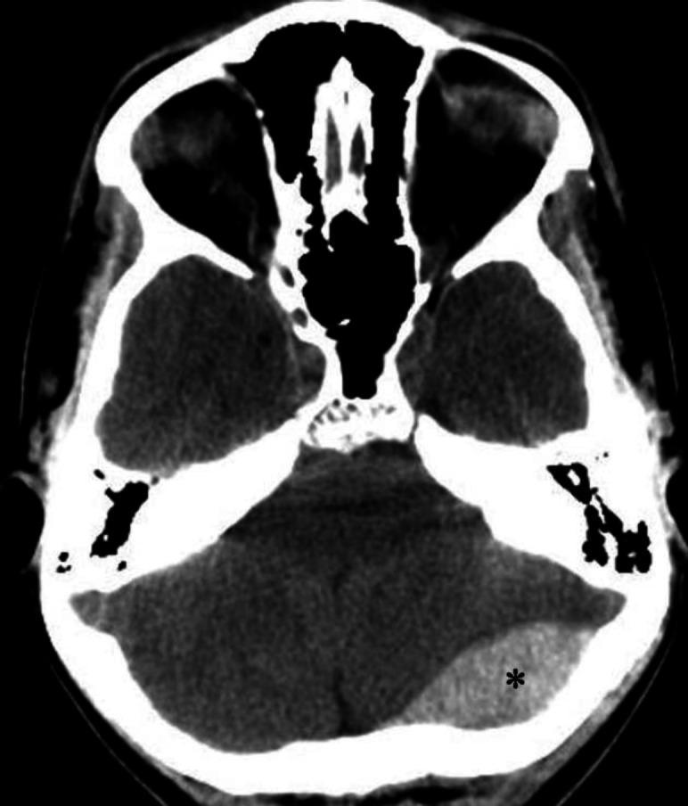
An axial non-contrast computerized tomography (CT) scan of the brain demonstrating an acute epidural (extradural) hematoma. The primary finding is a well-defined, biconvex (lens-shaped), hyperdense fluid collection located in the left occipital region, marked with an asterisk. This hyperdensity is characteristic of an acute hemorrhage within the epidural space, which typically does not cross cranial sutures. Secondary mass effect is visible, characterized by a mild left-to-right midline shift and compression of adjacent brain parenchyma. Anatomical landmarks include the skull base, posterior fossa structures, and the cerebellum. The image illustrates a classic neurosurgical emergency often associated with traumatic skull fractures and arterial bleeding, typically from the middle meningeal artery. This diagnostic image is intended for educational use in emergency medicine, radiology, and neurology to distinguish epidural hematomas from crescent-shaped subdural hematomas.
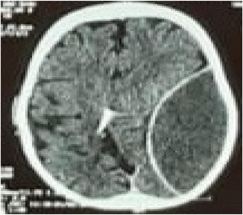
An axial non-contrast CT scan of the brain demonstrating a large, acute epidural (extradural) hematoma in the left hemisphere. The hematoma presents as a well-defined, biconvex (lenticular), hyperdense collection located between the skull and the dura mater. There is a significant mass effect evidenced by the compression of the left cerebral parenchyma, effacement of the adjacent cortical sulci, and midline shift toward the right. In the contralateral hemisphere, a radiopaque ventricular catheter is visible, extending into the posterior aspect of the right lateral ventricle near the occipital horn. This diagnostic image is representative of neurosurgical trauma emergencies, illustrating the classic appearance of arterial bleeding (typically from the middle meningeal artery) and secondary intracranial hypertension markers. The image serves as an educational resource for identifying acute traumatic intracranial hemorrhage and the presence of neurosurgical shunting devices.
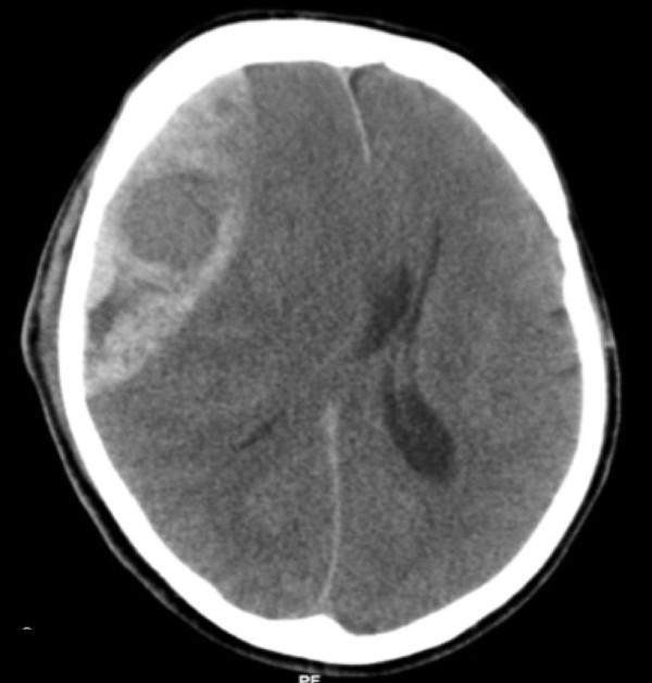
This diagnostic image is an axial non-contrast Computed Tomography (CT) scan of the brain, demonstrating a large, acute epidural (extradural) hematoma. The lesion is located on the right side of the skull and exhibits a classic biconvex or lens-shaped (lentiform) morphology, characterized by a hyperdense appearance relative to the brain parenchyma. The hematoma is clearly demarcated and constrained by the cranial sutures, positioned between the inner table of the skull and the dura mater. The image illustrates a significant mass effect, resulting in the effacement of adjacent sulci, partial compression of the right lateral ventricle, and a notable leftward midline shift of the falx cerebri. These findings are highly characteristic of an acute arterial bleed, typically involving the middle meningeal artery. This scan serves as a critical educational tool for identifying neurosurgical emergencies, understanding intracranial pressure dynamics, and recognizing the radiological hallmarks that differentiate epidural hematomas from crescent-shaped subdural hematomas.
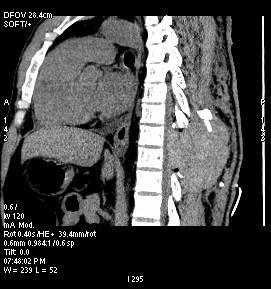
This diagnostic image is a sagittal CT scan of the dorsal spine, demonstrating a large spinal epidural hematoma. The imaging shows a localized, hyperdense collection (increased density) within the posterior extradural space at the thoracic level. This mass effect causes significant anterior displacement and compression of the spinal cord against the posterior aspect of the vertebral bodies. Anatomical landmarks include the thoracic vertebral column and the adjacent paraspinal tissues. Notably, the hematoma has displaced the overlying spinous process, pushing it posteriorly/superiorly from its normal alignment. This clinical finding is characteristic of an acute postoperative or traumatic complication, specifically a spinal epidural hematoma (SEH) resulting in spinal cord compression. The scan provides critical evidence for emergency surgical intervention, such as re-exploration and evacuation, to prevent permanent neurological deficit.
Lucid Interval
Definition
- S Das - Manual on Clinical Surgery, p. 274
- The Essentials of Forensic Medicine and Toxicology 36th ed., p. 4895
Mechanism (Pathophysiology)
- Head injury - usually a blow to the temporal/pterional region
- Concussion - initial brief unconsciousness from diffuse brain disruption
- Recovery - patient wakes up; dura is slowly being stripped off the inner skull by the accumulating blood clot
- Compensation phase (the lucid interval) - intracranial pressure is initially kept normal by displacement of CSF into the spinal canal. The patient appears normal or near-normal
- Compensation failure - as blood continues to accumulate, ICP rises; the medial aspect of the temporal lobe herniates through the tentorial hiatus, compressing the midbrain and reticular activating system
- Secondary unconsciousness - progressive coma, ipsilateral pupil dilation, contralateral hemiplegia, respiratory failure, and death if untreated
- S Das - Manual on Clinical Surgery, p. 274
Classic Cause: Extradural (Epidural) Hematoma
Stages of Consciousness in EDH:
| Stage | Clinical State | Mechanism |
|---|---|---|
| Stage 1 | Initial unconsciousness | Concussion (diffuse axonal disruption) |
| Stage 2 | Lucid interval | ICP compensation by CSF displacement |
| Stage 3 | Progressive confusion ("appears drunk") | Rising ICP |
| Stage 4 | Ipsilateral pupil dilation (blown pupil) | CN III compression by uncal herniation |
| Stage 5 | Contralateral hemiplegia | Cerebral peduncle compression |
| Stage 6 | Deep coma, bilateral pupil fixation, decerebrate rigidity | Brainstem compression |
| Stage 7 | Death | Respiratory failure from brainstem compression |
- The Essentials of Forensic Medicine and Toxicology, p. 4891-4908; S Das, p. 273-274
Key Statistics
- Lucid interval is seen in only 30-40% of extradural hemorrhage cases (Essentials of Forensic Medicine)
- In another reference, nearly 50% of patients with EDH present with the classic lucid interval (Rosen's Emergency Medicine)
- It is absent when the initial brain injury is severe enough that the unconsciousness from concussion and from hematoma compression overlap
- A lucid interval is not pathognomonic for EDH - it can also occur in other expanding mass lesions (e.g., subdural hematoma in 12-36% of cases)
- Rosen's Emergency Medicine, p. 375; Essentials of Forensic Medicine, p. 4897
"Talk and Die" Syndrome
- Expansion of a subdural hematoma
- Expansion of an epidural hematoma
- Worsening brain edema around a contusion
- Delayed appearance of an epidural clot
- Adams and Victor's Principles of Neurology 12th Edition, p. 901
CT Scan Appearance of Extradural Hematoma

Differentiation: EDH vs SDH Lucid Interval
| Feature | Extradural Hemorrhage | Subdural Hemorrhage |
|---|---|---|
| Relative incidence | Less common (1:6) | 6× more common |
| Lucid interval | Classic, well-defined | Much shorter or absent |
| Onset of compression | Later | Earlier (blood accumulates faster) |
| Source of bleeding | Middle meningeal artery (arterial) | Bridging veins (venous) |
| Skull fracture on X-ray | Common (temporal) | Not typical |
| Midline shift on echo/CT | Present (unilateral) | Present; 50% bilateral |
| Compensation time | Longer (CSF displacement) | Shorter |
- S Das - Manual on Clinical Surgery, p. 274
Lucid Interval in Insanity (Forensic/Medico-legal Context)
| Feature | Head Injury | Insanity |
|---|---|---|
| History | Of head trauma | Of mental disease |
| Preceded by | Phase of concussion | Phase of insanity symptoms |
| Followed by | Cerebral irritation and brain compression | Recurrence of insanity |
| Frequency | Only once | More than once (recurrent) |
- P C Dikshit Textbook of Forensic Medicine and Toxicology, p. 9725-9736
- Essentials of Forensic Medicine 36th ed., Table 23.1
Summary of Key Points
- Lucid interval = period of apparent normality between two episodes of unconsciousness after head injury
- Classic cause: extradural hemorrhage from rupture of the middle meningeal artery
- Mechanism: initial concussion → ICP compensated by CSF displacement → compensation fails → uncal herniation → coma
- Present in only 30-50% of EDH cases; absence does NOT exclude serious injury
- Not pathognomonic for EDH - also occurs in subdural hematoma and other expanding lesions
- CT shows lentiform hyperdense collection - neurosurgical emergency requiring immediate evacuation
- Coined "talk and die" syndrome - never ignore a patient who has seemingly recovered from a head injury