Compare histological structures of artery and vein according to ib singh
artery vein histology
IB Singh human histology artery vein differences
histology microscopic comparison medium muscular artery and companion vein tunica media lumen
https://ndl.ethernet.edu.et/bitstream/123456789/12975/1/329.…
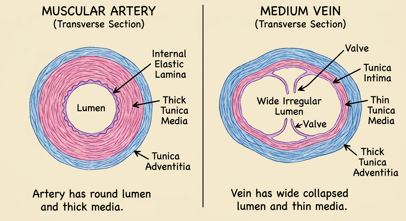
Histological comparison of an artery and a vein
| Feature | Artery | Vein |
|---|---|---|
| Wall thickness | Thick wall relative to lumen | Thin wall relative to lumen |
| Lumen | Comparatively narrow, usually round due to a firm wall | Wide, irregular, and often collapsed in a section |
| Tunica intima | Endothelium, basal lamina, subendothelial connective tissue; internal elastic lamina is distinct, especially in muscular arteries | Endothelium and thin subendothelial tissue; internal elastic lamina is absent or inconspicuous |
| Tunica media | Thickest and most prominent layer; many circular smooth-muscle layers with elastic fibers. External elastic lamina may be visible | Thin; has few smooth-muscle cells and relatively more collagen. Elastic tissue and muscle are much less than in an artery |
| Tunica adventitia | Usually thinner than the media in a medium-sized artery | Relatively thick and commonly the most prominent coat; contains collagen, elastic fibers, and in larger veins longitudinal smooth muscle |
| Elastic laminae | Internal elastic lamina is clear; external elastic lamina may also be seen | Usually poorly defined or absent |
| Separation of layers | Three tunics are clearly demarcated | Three tunics are less sharply demarcated |
| Valves | Absent | May be present, particularly in medium veins of the limbs; they are folds of tunica intima |
| Shape in histological section | Retains circular shape because of thick muscular and elastic wall | Often appears collapsed, oval, or irregular because the wall is thin |
| Main function reflected by structure | Withstands and regulates high-pressure blood flow from the heart | Returns low-pressure blood to the heart and acts as a capacitance reservoir |
Practical slide identification
- Artery: small, round lumen with a thick wall. The tunica media is conspicuous and the internal elastic lamina may appear as a wavy line.
- Vein: larger, irregular lumen with a thinner wall. The media is thin, while the adventitia is relatively thicker. A valve, if present, confirms a vein.
Create a concise artery-versus-vein histology diagram
artery vein histology
artery vein histology comparison tunica media lumen
A concise labeled educational histology diagram comparing a transverse section of a muscular artery on the left with a medium vein on the right. Clearly show and label lumen, tunica intima, internal elastic lamina, thick tunica media, and tunica adventitia for artery; show wide irregular lumen, tunica intima, thin tunica media, thick tunica adventitia, and a valve for vein. Include short comparison captions: artery has round lumen and thick media; vein has wide collapsed lumen and thin media. Suitable for first-year medical anatomy students.
Histology of cerebrum
cerebral cortex histology
cerebral cortex histology six layers molecular external granular pyramidal internal granular ganglionic multiform
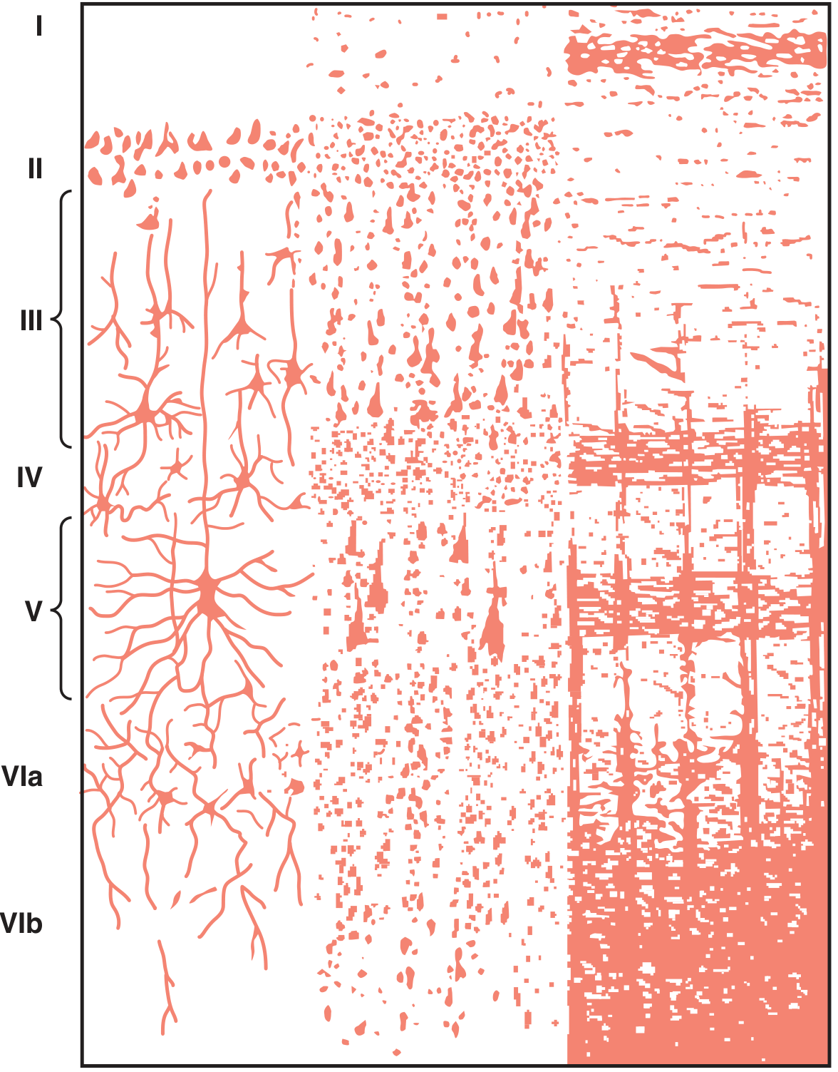
human cerebral cortex histology six layers labeled micrograph
Histology of the Cerebrum
- Gray matter: contains neuronal cell bodies, neuropil, glial cells, capillaries, dendrites, and unmyelinated nerve fibers.
- White matter: lies deep to the cortex and consists mainly of myelinated nerve fibers with neuroglial cells. It contains very few neuronal cell bodies.
- The surface of the cortex is covered by pia mater. It is thrown into gyri and sulci.
Neocortex
| Layer | Name | Main histological features |
|---|---|---|
| I | Molecular or plexiform layer | Few cells. Mainly nerve fibers, dendritic branches of deeper pyramidal cells, neuroglia, and horizontal cells of Cajal. |
| II | External granular layer | Small stellate or granule cells and small pyramidal cells. |
| III | External pyramidal layer | Medium-sized pyramidal cells. Their axons form association fibers and commissural fibers, including fibers crossing through the corpus callosum. |
| IV | Internal granular layer | Numerous stellate cells. It is the major recipient of thalamocortical sensory input and is especially well developed in sensory cortex. |
| V | Internal pyramidal or ganglionic layer | Large pyramidal cells. In the primary motor cortex, it contains giant Betz cells. Axons project to subcortical centers, brainstem, and spinal cord. |
| VI | Multiform, polymorphic, or fusiform layer | Fusiform and other variably shaped cells. Fibers project mainly to the thalamus. It merges with the underlying white matter. |
Important cells in cerebral cortex
- Pyramidal cells: chief efferent neurons of the cortex; triangular cell body, apical dendrite directed toward the pial surface, and axon directed toward white matter.
- Stellate or granular cells: small interneurons, prominent in granular layers.
- Fusiform cells: spindle-shaped neurons, mainly in layer VI.
- Neuroglial cells: astrocytes, oligodendrocytes, microglia, and other supportive glial elements.
Regional variations
- Motor cortex: called agranular cortex because layer IV is poorly developed. Layer V is very thick and contains Betz cells.
- Sensory cortex: called granular cortex because layer IV is well developed due to abundant thalamic sensory input.
- Archicortex: has fewer than six layers, classically three layers; the hippocampus is an example.
- Paleocortex: also has fewer layers and is associated with olfaction.
Slide identification points
- Outer pia mater.
- Outer dark-staining gray matter and inner pale-staining white matter.
- Layered arrangement of neurons in the cortex.
- Large pyramidal neurons, particularly in deeper layers.
- White matter composed predominantly of myelinated fibers and glial nuclei.
Use ib singh text book of histology
Inderbir Singh Textbook of Human Histology cerebrum histology cerebral cortex layers
cerebral cortex histology
Histology of Cerebrum - according to Inderbir Singh
- An outer layer of gray matter, the cerebral cortex
- Inner white matter
Layers of the cerebral cortex
| Lamina | Name | Important cells/features |
|---|---|---|
| I | Plexiform or molecular layer | Few nerve cells; mainly nerve fibers, neuroglia, and horizontal cells of Cajal. |
| II | Outer granular layer | Mainly small stellate cells and small pyramidal cells. |
| III | Outer pyramidal layer | Pyramidal cells, increasing in size in deeper parts. |
| IV | Inner granular layer | Numerous stellate (granule) cells; well developed in sensory areas. |
| V | Inner pyramidal layer | Large pyramidal cells. Giant pyramidal cells, or Betz cells, are present in the motor cortex. |
| VI | Polymorphic or multiform layer | Fusiform cells and cells of varied shapes; blends with underlying white matter. |
Cells seen in the cerebral cortex
- Pyramidal cells
- Stellate cells
- Fusiform cells
- Cells of Martinotti
- Basket cells
- Chandelier cells
- Horizontal cells of Cajal
- Double bouquet cells
Identification points in a slide of cerebrum
- Outer gray matter and inner white matter are clearly distinguishable.
- Gray matter shows a six-layered cerebral cortex.
- Pyramidal cells are seen in most layers, especially layers III and V.
- The deep cortex merges gradually with white matter containing predominantly nerve fibers and glial cells.
- The surface is covered by pia mater.
Important regional differences
- Motor cortex: Layer V is very prominent and shows giant Betz cells. Layer IV is poorly developed, hence it is called agranular cortex.
- Sensory cortex: Layer IV is prominent due to numerous granule cells, hence called granular cortex.
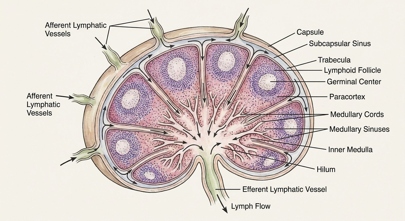
Histology of lymph node
histology lymph node labeled capsule subcapsular sinus cortex germinal center medulla
lymph node histology
Inderbir Singh textbook human histology lymph node cortex paracortex medulla sinuses
https://book.bsmi.uz/web/kitoblar/152372172.pdf
Histology of Lymph Node - according to Inderbir Singh
Main parts
-
Capsule
- Thin connective-tissue capsule, chiefly collagen fibers.
- May contain a few elastic fibers and smooth-muscle fibers.
- The capsule sends inward septa called trabeculae.
-
Subcapsular sinus
- A clear space immediately beneath the capsule.
- Receives lymph from afferent lymphatic vessels.
-
Cortex
- Outer, dark-staining region due to densely packed lymphocytes.
- Contains rounded lymphatic follicles (nodules).
- A secondary follicle shows a pale-staining germinal center, surrounded by a darker mantle of closely packed lymphocytes.
- The deep cortex, between follicles and medulla, is the paracortex, rich in T lymphocytes. It contains high endothelial venules.
-
Medulla
- Lighter-staining central region.
- Has branching, anastomosing medullary cords, which contain lymphocytes, plasma cells, macrophages, and reticular cells.
- The spaces between cords are medullary sinuses.
-
Hilum
- Depressed part of the node through which blood vessels enter and leave.
- Efferent lymphatic vessel(s) leave through the hilum.
Course of lymph
Slide identification points
- A thin capsule and inward trabeculae
- A clear subcapsular sinus
- Outer dark cortex with lymphoid follicles
- Pale germinal centers in secondary follicles
- Inner pale medulla with medullary cords and sinuses
- Numerous dark-staining lymphocyte nuclei throughout
| Region | Main feature |
|---|---|
| Capsule | Fibrous outer covering |
| Subcapsular sinus | Clear space below capsule |
| Cortex | Follicles, mainly B-cell area |
| Paracortex | T-cell-rich area; high endothelial venules |
| Medulla | Medullary cords and sinuses |
| Hilum | Exit of efferent lymphatic vessel; blood vessels |
Create a labeled diagram of lymph node histology
Labeled educational diagram of a lymph node in longitudinal section for medical histology. Show a bean-shaped node with a thin fibrous capsule, trabeculae, subcapsular sinus, outer cortex with several lymphoid follicles and pale germinal centers, paracortex, inner medulla with branching medullary cords and medullary sinuses, hilum, afferent lymphatic vessels entering the convex surface, and one efferent lymphatic vessel leaving the hilum. Add clear arrows showing lymph flow: afferent vessels to subcapsular sinus to cortical/trabecular sinuses to medullary sinuses to efferent vessel. Use concise readable labels and accurate anatomy, suitable for MBBS histology study.