
Underlying organs under abdomen quadrants
Reading File
This is a visual anatomy topic. Let me search the medical library for abdominal quadrant content and images.
Finding Sources
Finding Sources
Gray's Anatomy for Students has a dedicated section on abdominal surface topography. Let me read it and also look for images.
Reading File
Finding Sources
Excellent! I found a dedicated section in Gray's Anatomy for Students: "Using abdominal quadrants to locate major viscera." Let me read that and fetch the quadrant diagram image.
Reading File
Loading Image
Now let me also fetch the detailed viscera diagram and the nine-region diagram:
Loading Image
Loading Image
I now have everything needed for a thorough, image-rich answer. Here is the complete breakdown:
Organs Under the Abdominal Quadrants
The abdomen is divided into 4 quadrants by two planes that intersect at the umbilicus:
- Vertical (median) plane - runs down the midline
- Horizontal (transumbilical) plane - passes through the umbilicus and the L3/L4 intervertebral disc
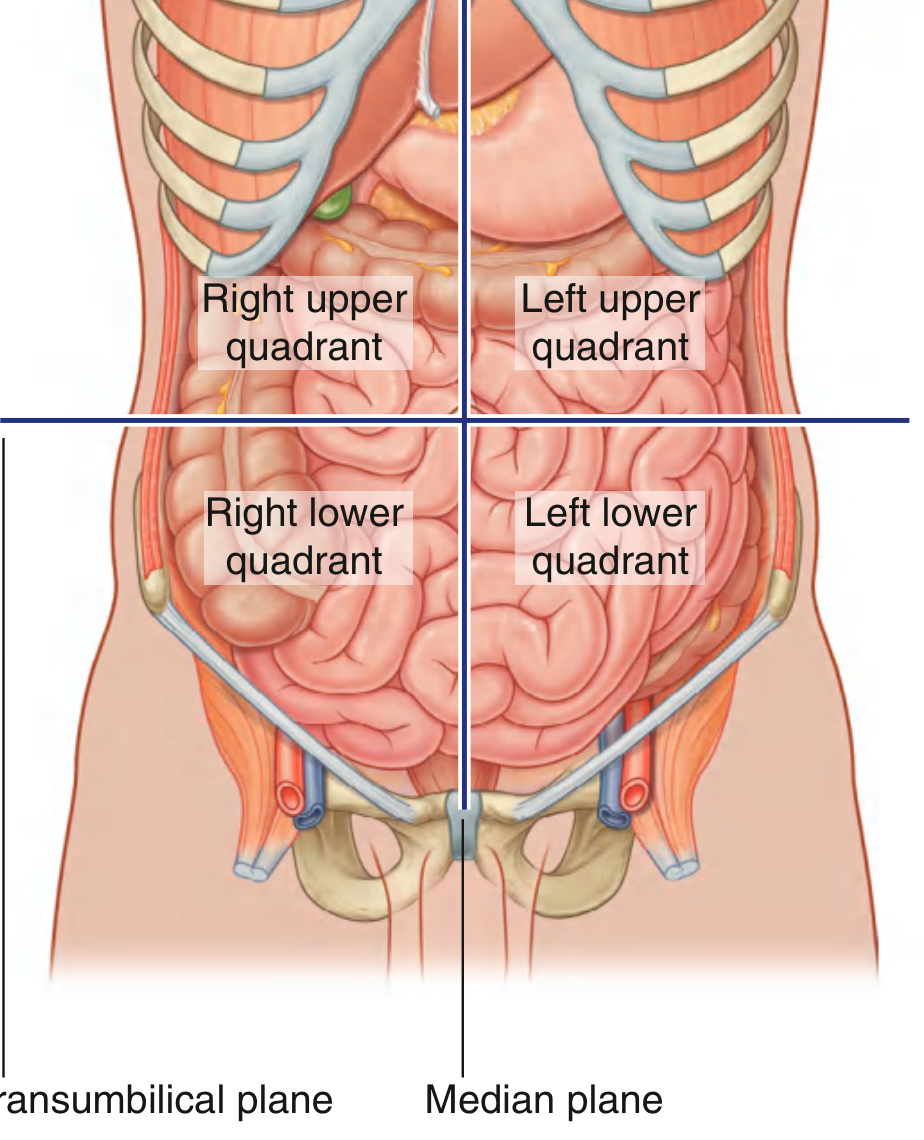
Organs by Quadrant
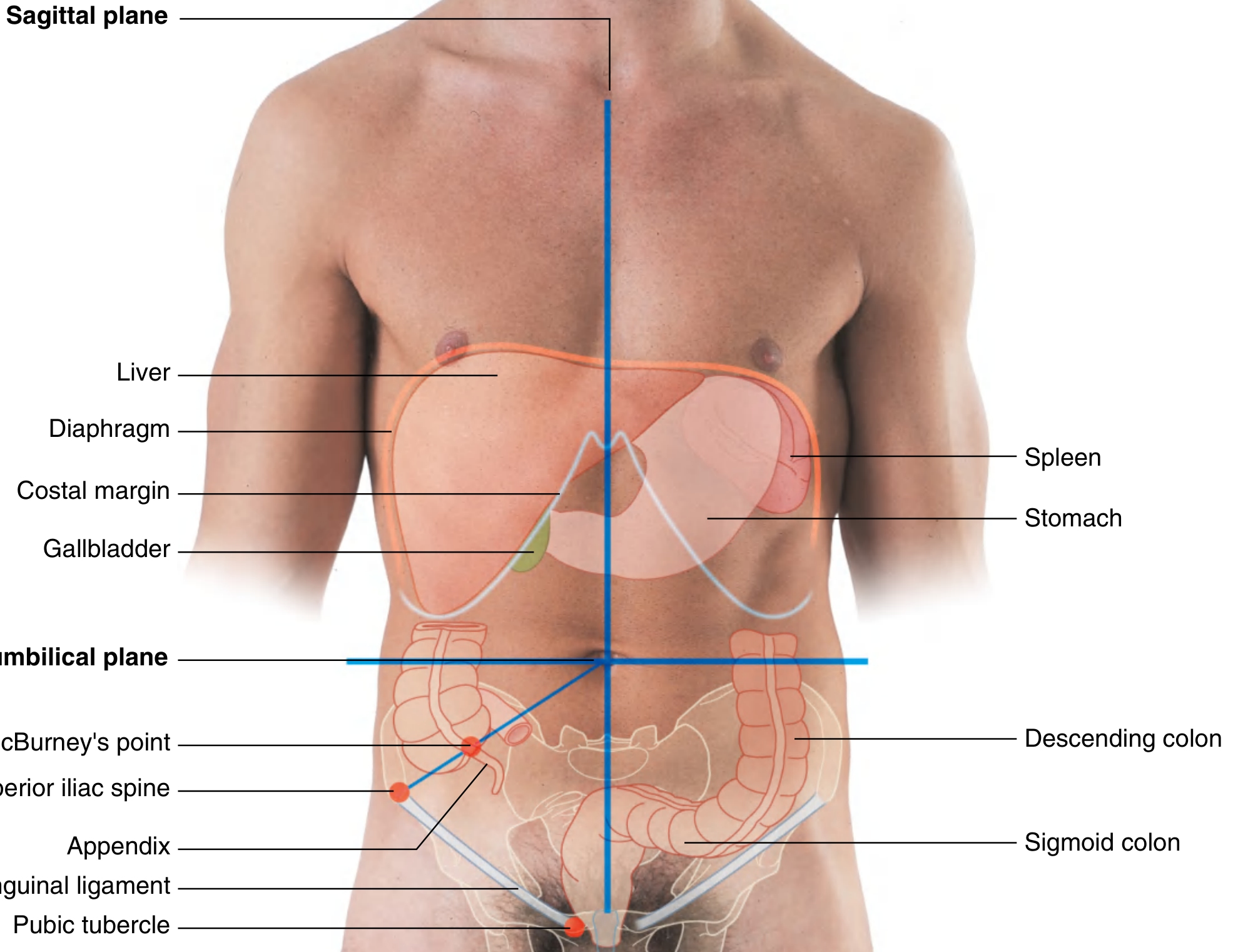
Right Upper Quadrant (RUQ)
| Organ | Notes |
|---|---|
| Liver (right lobe, most of it) | Sits under the right dome of the diaphragm; palpable below the costal margin on deep inspiration |
| Gallbladder | Tucked under the inferior surface of the liver |
| Right kidney (upper pole) | Retroperitoneal |
| Duodenum (parts 1-3) | Wraps around the head of the pancreas |
| Head of pancreas | Lies in the curve of the duodenum |
| Right adrenal gland | Retroperitoneal, on top of the right kidney |
| Hepatic flexure of colon | Right colic flexure where ascending colon turns |
| Part of the ascending colon | |
| Part of the transverse colon |
Left Upper Quadrant (LUQ)
| Organ | Notes |
|---|---|
| Stomach | Most of it sits here |
| Spleen | Under the left dome of the diaphragm, protected by ribs 9-11 |
| Left kidney (upper pole) | Retroperitoneal |
| Left adrenal gland | Retroperitoneal |
| Body and tail of pancreas | Retroperitoneal, extending toward the spleen |
| Splenic flexure of colon | Left colic flexure |
| Part of the transverse colon | |
| Part of the descending colon | |
| Liver (left lobe - small portion) |
Right Lower Quadrant (RLQ)
| Organ | Notes |
|---|---|
| Cecum | Beginning of the large intestine |
| Appendix | McBurney's point = 1/3 of the way from the right ASIS to the umbilicus |
| Terminal ileum | The last part of the small intestine |
| Right ureter (mid-section) | Retroperitoneal |
| Right ovary & fallopian tube | In females |
| Right spermatic cord | In males |
| Part of ascending colon |
Left Lower Quadrant (LLQ)
| Organ | Notes |
|---|---|
| Sigmoid colon | S-shaped loop connecting descending colon to rectum |
| Descending colon (lower part) | |
| Left ureter (mid-section) | Retroperitoneal |
| Left ovary & fallopian tube | In females |
| Left spermatic cord | In males |
Midline structures (span multiple quadrants)
- Aorta and inferior vena cava - span upper and lower quadrants along the midline
- Uterus and bladder - when enlarged, project into the lower quadrants
- Small intestine (jejunum & ileum) - fills all four quadrants, predominantly lower
Bonus: Nine-Region Pattern
Clinicians sometimes use a more detailed 9-region system for localizing pain. The abdomen is divided by two horizontal planes (subcostal and intertubercular) and two midclavicular vertical planes:
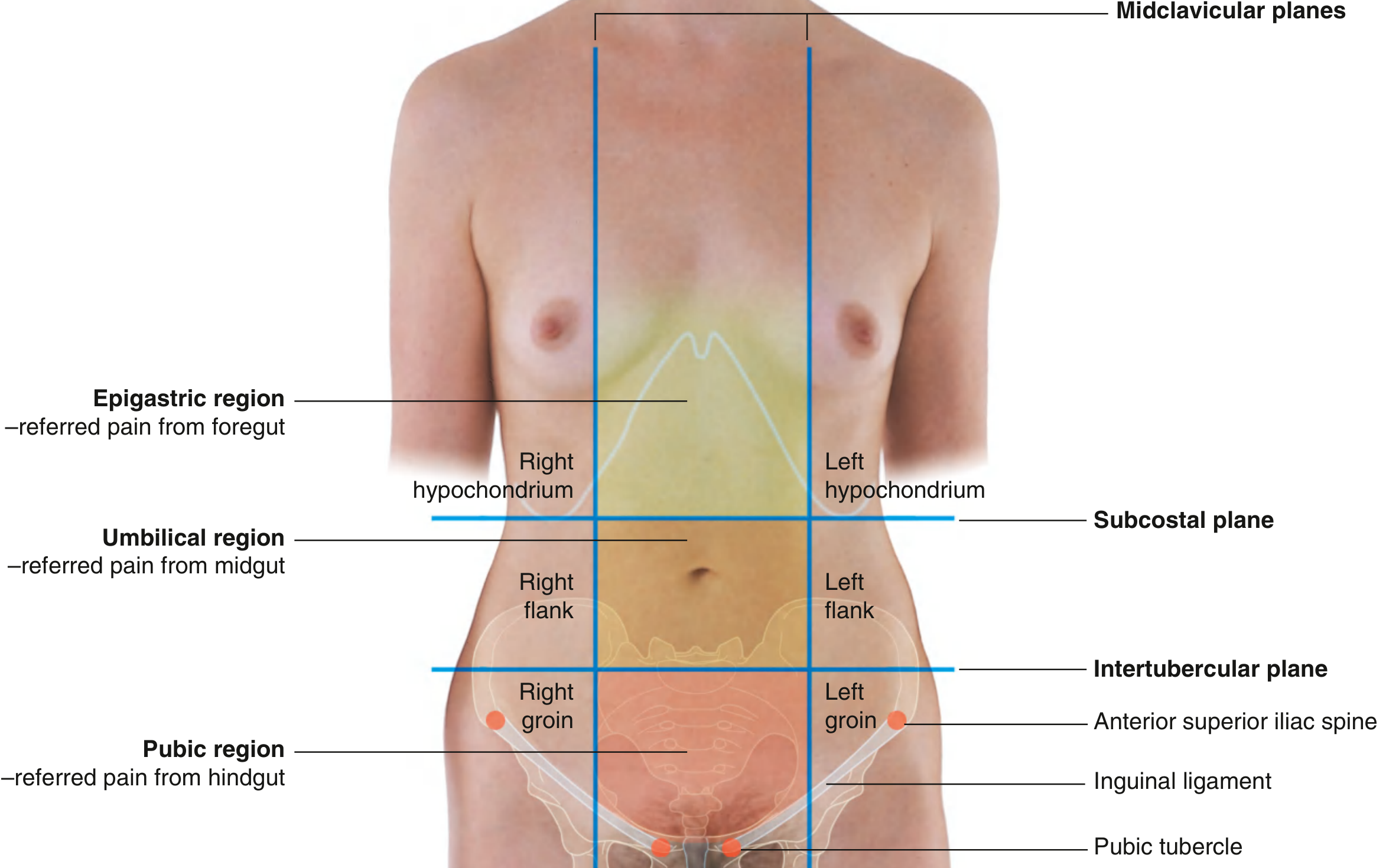
| Region | Pain Origin |
|---|---|
| Epigastric | Foregut (stomach, duodenum, liver, pancreas) |
| Umbilical | Midgut (small intestine, appendix) |
| Pubic (hypogastric) | Hindgut (sigmoid, rectum, bladder, uterus) |
| Right/Left Hypochondrium | Under the ribs (liver right; spleen left) |
| Right/Left Flank | Kidneys, ureters, ascending/descending colon |
| Right/Left Groin (inguinal) | Appendix/cecum right; sigmoid left; gonads bilaterally |
Clinical relevance: Knowing which organs occupy each quadrant is the foundation of localizing abdominal pain. For example, RUQ pain points toward gallbladder/liver pathology, RLQ pain toward appendicitis, and LLQ toward diverticulitis.
Source: Gray's Anatomy for Students, Fig. 4.185 & 4.186
This is a shared conversation. Sign in to Orris to start your own chat.