
Spinal cord lessons
Asking for Preferences
What aspect of spinal cord lesions are you interested in? · What is your background/purpose?
Great - clinical spinal cord syndromes for a medical student. Let me pull authoritative textbook content on this.
Searching sources...
Reading File
Finding Sources
Finding Sources
Finding Sources
Reading File
Reading File
Reading File
Reading File
Finding Sources
Now let me grab the diagram image and the complete cord syndrome figure that was referenced:
Loading Image
Reading File
I have all the content I need. Now let me compile a thorough, well-organized medical student guide on clinical spinal cord syndromes.
Clinical Spinal Cord Syndromes
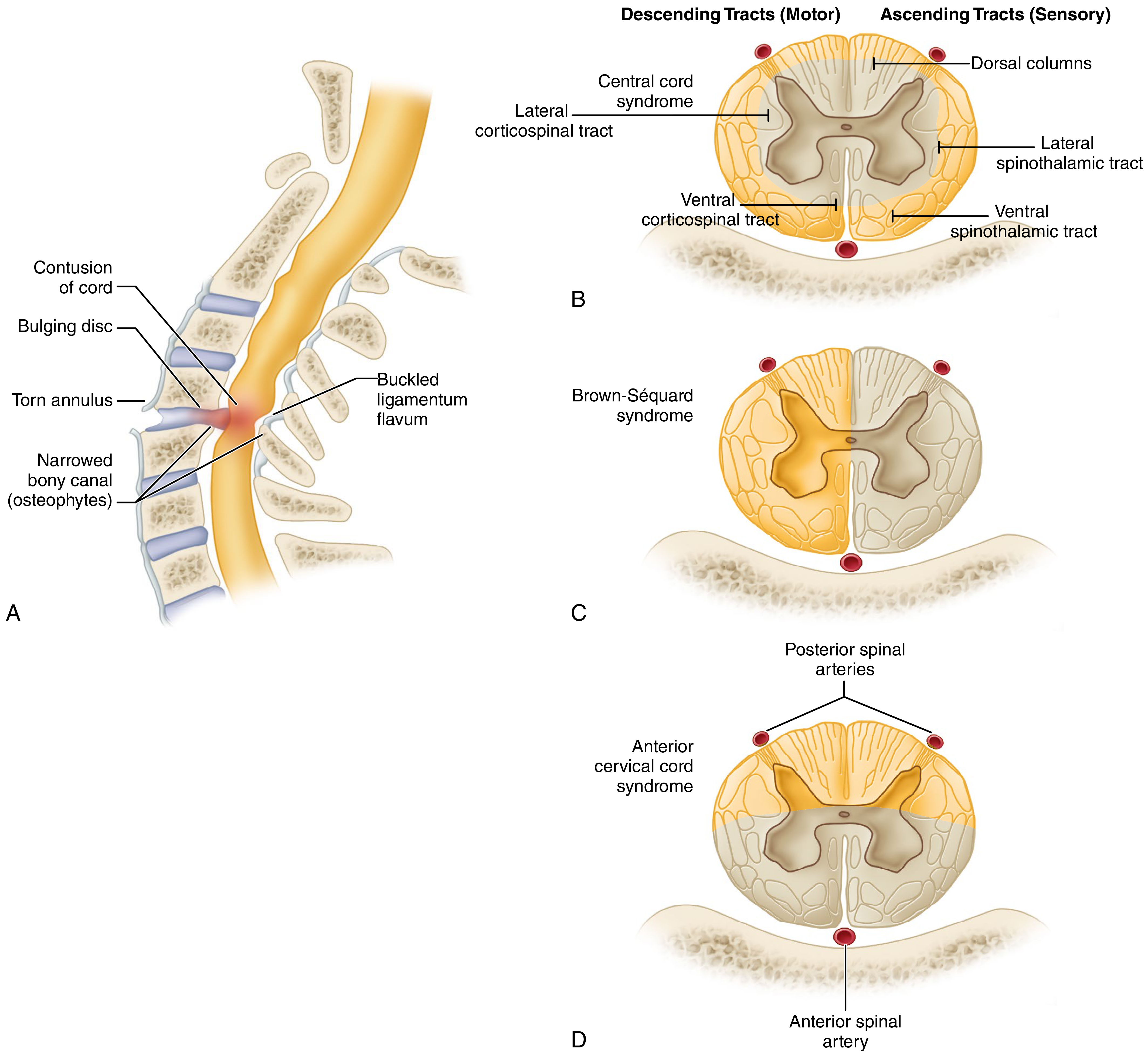
Cross-sectional anatomy of the major spinal cord syndromes. (Campbell's Operative Orthopaedics, 15th ed.)
Quick Anatomy Recap
Before the syndromes make sense, you need the three key tracts:
| Tract | Location | Decussation | Function |
|---|---|---|---|
| Corticospinal (CST) | Lateral white matter | At medulla | Voluntary motor (ipsilateral below) |
| Dorsal columns | Posterior white matter | At medulla | Vibration, proprioception, fine touch (ipsilateral below) |
| Spinothalamic | Lateral/ventral white matter | At entry level (1-2 segments up) | Pain and temperature (contralateral below) |
This explains why most syndromes produce dissociated sensory loss - different sensory modalities are lost on opposite sides.
1. Central Cord Syndrome
Most common incomplete SCI (~9% of all traumatic cord injuries).
Mechanism: Hyperextension in a cervical spine with preexisting spondylosis. The cord gets pinched anteriorly by disc/osteophyte complex and posteriorly by a buckled ligamentum flavum.
Key clinical features:
- Upper extremity weakness > lower extremity weakness (the classic hallmark)
- Greater distal dysfunction than proximal in the arms (hand and finger weakness is prominent)
- Variable sensory loss below the injury
- Urinary retention / bladder dysfunction
Why arms > legs? The corticospinal tracts are somatotopically arranged - cervical fibers run most medially, lumbar fibers most laterally. A central injury disproportionately hits the arm-supplying fibers.
Prognosis: Generally good. >50% regain bladder control and become ambulatory. Recovery typically begins in the lower extremities and progresses upward. Patients older than 50 with preexisting myelopathy tend to do worse.
Management: Controversial - operative vs. nonoperative. Surgery within 24 hours may offer slightly better neurologic outcomes, but older patients carry higher operative risk.
2. Brown-Séquard Syndrome (Cord Hemisection)
Accounts for 1%-4% of traumatic SCIs. Results from functional or anatomic hemisection of one lateral half of the cord.
Mechanism: Most commonly penetrating trauma (stab wounds), unilateral pedicle/laminar fractures, or rotational subluxation. Most often in the cervical spine.
Classic deficits (below the lesion):
| Deficit | Side |
|---|---|
| Motor loss (UMN, spastic) | Ipsilateral - CST does not cross until medulla |
| Loss of vibration, proprioception, fine touch | Ipsilateral - dorsal columns don't cross until medulla |
| Loss of pain and temperature | Contralateral (1-2 dermatomes below) - spinothalamic tract already crossed |
At the level of the lesion: ipsilateral LMN signs (flaccid weakness, loss of all sensation) due to gray matter destruction.
Brown-Séquard Plus: The more common variant - asymmetric hemiplegia with hypoalgesia more prominent on the less paretic side. Classic BSS actually has a worse prognosis than the Plus variant.
Prognosis: Best of all cord syndromes. Gait typically recovers within 6 months. Most patients regain bowel and bladder continence.
3. Anterior Cord Syndrome
Occurs in ~2.7% of traumatic SCIs. Involves the ventral two-thirds of the cord; posterior columns are spared.
Mechanism:
- Anterior spinal artery occlusion (classic cause)
- Hyperflexion injuries with retropulsed bone or disc compressing the anterior cord
Clinical features:
- Complete motor loss below the lesion (CST disrupted)
- Loss of pain and temperature below the lesion (spinothalamic tract disrupted)
- Preserved vibration, proprioception, and fine touch (posterior columns intact - this is the key distinguishing feature)
- Patients may describe hyperesthesia or hypoalgesia
Prognosis: Worst of the incomplete syndromes. Significant neurologic recovery is unlikely.
4. Posterior Cord Syndrome
Rare (<1% incidence).
Mechanism: Hyperextension injury; also seen with tumors and in the context of pernicious anemia (subacute combined degeneration - a non-traumatic cause).
Clinical features:
- Loss of vibration and proprioception (dorsal columns damaged)
- Preserved pain, temperature, and motor function
- Patients may have a "sensory ataxia" - wide-based unsteady gait, positive Romberg
Note: Because the International Standards for Neurological Classification of SCI does not formally recognize this as a separate syndrome, it is less commonly tested but clinically important to know.
5. Complete (Transverse) Cord Syndrome
Total loss of all neurological function (motor AND all sensory) below the level of injury.
- Spinal shock occurs acutely: flaccid paralysis, areflexia, loss of all sensation, urinary retention
- After spinal shock resolves (days to weeks): UMN signs emerge (spasticity, hyperreflexia, Babinski)
6. Conus Medullaris Syndrome
Injury to the sacral cord (conus) and lumbar nerve roots within the spinal canal. Level: typically T11-L2.
Features (mixed UMN + LMN):
- Areflexic bladder - urinary retention with overflow incontinence
- Areflexic bowel with fecal incontinence
- Lower extremity weakness - initially flaccid (may later develop some tone)
- Absent bulbocavernosus reflex and perianal wink (indicates irreversible sacral damage)
- Sexual dysfunction
Important: If nerve root sparing occurs at L1-L4, some lower extremity motor function may be preserved.
7. Cauda Equina Syndrome
Injury to the lumbosacral nerve roots below the conus (below L1-L2). These are peripheral nerves, not cord tissue.
Features (purely LMN):
- Areflexic bladder and bowel
- Saddle anesthesia (perineum, inner thighs, perianal area)
- Lower limb weakness (variable, asymmetric)
- Absent reflexes throughout lower limbs
- All lower extremity reflexes lost (bulbocavernosus, anal wink)
Cauda equina vs. conus: The distinction can be blurry clinically, but cauda equina tends to be asymmetric (roots affected variably), while conus tends to be more symmetric. Cauda equina is a surgical emergency - outcomes improve with rapid decompression.
8. Cervicomedullary Syndrome
Injury at the upper cervical cord extending to the medulla (C1-C2 level / atlantoaxial complex).
Features:
- Respiratory compromise (may need ventilation)
- Hypotension
- Tetraparesis - arms > legs (mimics central cord)
- Onion-skin / Déjerine sensory pattern on the face - perioral area affected with lower medullary lesions, facial periphery with more caudal injuries
- This happens because trigeminal fibers enter and descend to different levels depending on their facial origin
Summary Comparison Table
| Syndrome | Motor | Vibration/Proprioception | Pain/Temp | Key Cause | Prognosis |
|---|---|---|---|---|---|
| Central Cord | Arms > Legs (bilateral) | Variable | Variable | Cervical hyperextension + spondylosis | Good |
| Brown-Séquard | Ipsilateral loss | Ipsilateral loss | Contralateral loss | Penetrating trauma | Best |
| Anterior Cord | Complete bilateral loss | Preserved | Bilateral loss | Anterior spinal artery / hyperflexion | Worst |
| Posterior Cord | Preserved | Bilateral loss | Preserved | Hyperextension / pernicious anemia | Good |
| Conus Medullaris | Flaccid LE weakness | Variable | Variable | T11-L2 fracture | Irreversible sacral |
| Cauda Equina | Asymmetric LMN weakness | Variable | Saddle anesthesia | Lumbar disc, fracture | Surgical emergency |
High-Yield Points for Exams
- Central cord = most common incomplete SCI; arms > legs; hyperextension in elderly with spondylosis
- Brown-Séquard = best prognosis; ipsilateral motor + dorsal column loss, contralateral pain/temp loss
- Anterior cord = worst prognosis; motor + pain/temp lost; vibration/proprioception preserved (classic MCQ trap)
- Posterior cord - isolated vibration/proprioception loss; think pernicious anemia if non-traumatic
- Anterior spinal artery supplies ventral 2/3 of cord - occlusion = anterior cord syndrome
- Cauda equina is an emergency - saddle anesthesia + urinary retention = urgent MRI + surgical decompression
- Conus: mixed UMN/LMN signs; cauda equina: pure LMN (peripheral nerve)
Sources: Bradley and Daroff's Neurology in Clinical Practice; Campbell's Operative Orthopaedics, 15th ed. (2026); Rosen's Emergency Medicine; Tintinalli's Emergency Medicine.
Blood brain barrier
The blood-brain barrier (BBB) is a highly selective semipermeable border formed by specialized brain endothelial cells, tight junctions, astrocyte endfeet, and pericytes. It keeps the neural environment stable by restricting passage of most molecules from blood into the CNS, while allowing oxygen, CO2, glucose, and lipid-soluble drugs to cross freely.
Searching sources...
Reading File
Finding Sources
Finding Sources
Reading File
Reading File
Reading File
Reading File
Reading File
Reading File
Loading Image
Finding Sources
I have rich, authoritative content from multiple textbooks. Let me now compile the full answer.
The Blood-Brain Barrier (BBB)
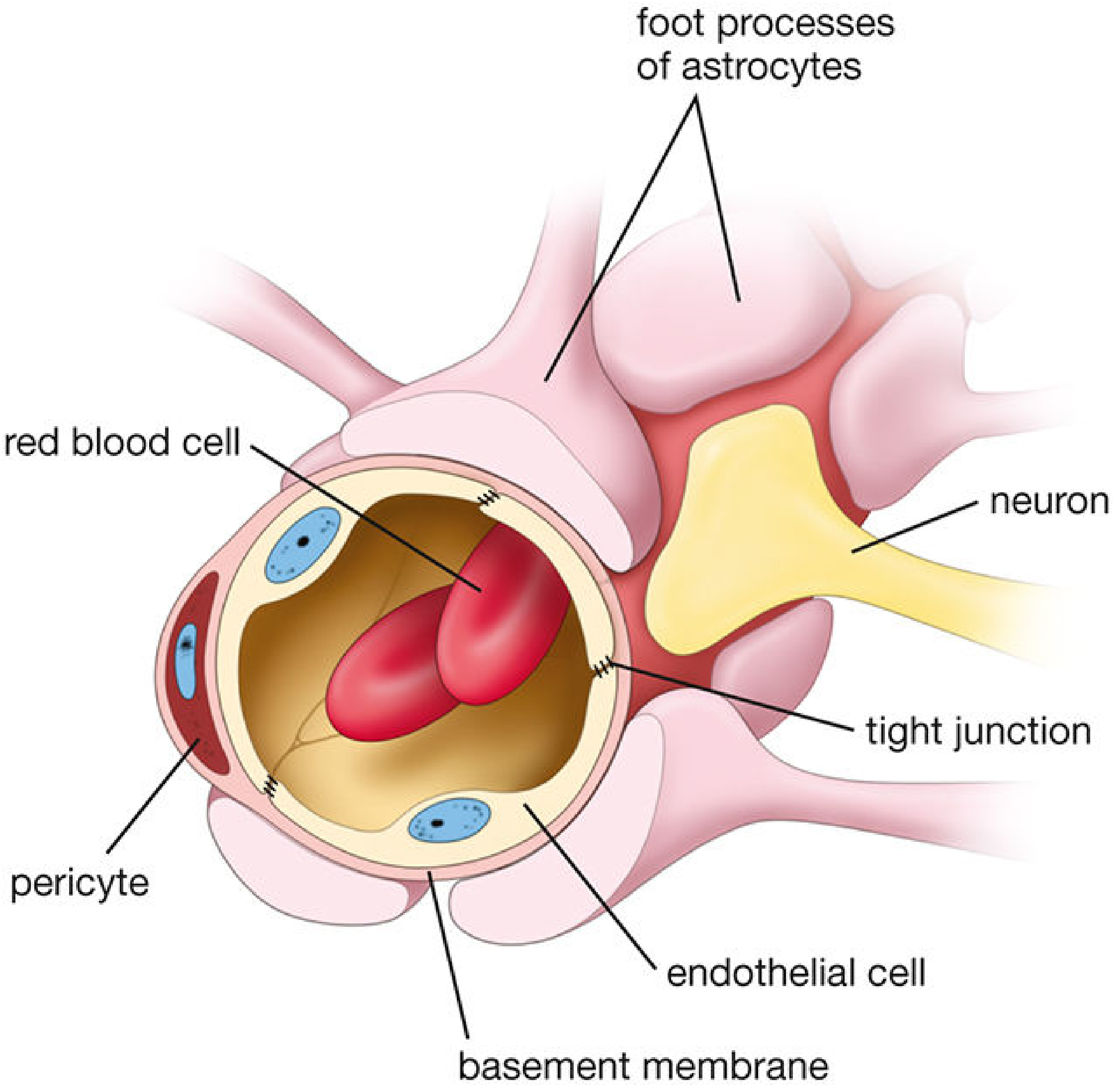
Schematic of the BBB: endothelial cells joined by tight junctions, wrapped by pericytes, basement membrane, and astrocyte endfeet. (Histology: A Text and Atlas, 9th ed.)
1. What Is the BBB?
The BBB is a highly selective, protective interface between the circulating blood and the CNS extracellular fluid. The concept arose from 19th-century observations that vital dyes injected into the bloodstream stained nearly every organ - except the brain. The BBB is the reason why.
Its three core functions are:
- Protect the brain from blood-borne toxins and pathogens
- Supply the brain with essential nutrients
- Regulate the ionic and molecular homeostasis the nervous system needs to function
2. Anatomy - The Four Layers
The BBB is not a single structure but a composite barrier with four components:
(A) Brain Capillary Endothelial Cells - the principal component
Unlike capillaries elsewhere in the body (which have fenestrations / clefts allowing free passage), brain capillary endothelial cells form a continuous, non-fenestrated lining sealed by tight junctions. Pinocytosis (vesicle-mediated transcytosis) across these cells is also severely restricted. Any substance crossing the BBB must pass through the endothelial cell, not between cells.
(B) Tight Junctions (Zonula Occludens)
The hallmark of the BBB. Proteins like claudins, occludins, and ZO-1 physically weld adjacent endothelial cells together, eliminating paracellular gaps. These junctions more closely resemble epithelial tight junctions than typical endothelial ones - reflecting the exceptional impermeability required. Molecules >500 Da generally cannot cross.
(C) Basement Membrane
A non-cellular extracellular matrix (collagen IV, laminin, fibronectin) that surrounds the endothelial tube and provides structural scaffolding. Pericytes are embedded in this membrane - they regulate blood flow and support tight junction maintenance.
(D) Astrocyte Endfeet (Perivascular Footplates)
Astrocytes extend "end-foot" processes that wrap tightly around the capillaries. They are critical for inducing and maintaining the barrier - they release soluble factors that upregulate tight junction proteins and increase barrier properties. However, the endfeet themselves are not the primary physical seal; that role belongs to the endothelial tight junctions. The BBB develops embryologically through the interaction between capillary endothelial cells and glial astrocytes.
This combined unit (endothelial cells + pericytes + basement membrane + astrocyte endfeet + nearby neurons) is called the neurovascular unit. - Goldman-Cecil Medicine
3. What Can Cross the BBB?
| Category | Examples | Mechanism |
|---|---|---|
| Freely crosses | O₂, CO₂ | Simple diffusion |
| Lipid-soluble (small) | Ethanol, steroid hormones, most general anesthetics | Transcellular diffusion |
| Glucose | Glucose | GLUT1 (facilitated transport, non-energy dependent) |
| Amino acids | Large neutral amino acids, L-DOPA | SLC transporters (amino acid transporter) |
| Active transport | Ions, vitamins, nucleotides | ABC transporters, SLC proteins |
| Cannot cross | Dopamine, most antibiotics, most chemotherapy, charged/polar molecules, large proteins | No transporter; blocked by tight junctions |
Clinical Example - L-DOPA vs. Dopamine
Dopamine itself cannot cross the BBB (polar, charged). L-DOPA, its precursor, uses a large neutral amino acid transporter to enter the brain, where it is converted to dopamine. This is why we give L-DOPA to Parkinson's patients, not dopamine directly. - Katzung's Basic & Clinical Pharmacology, 16th ed.
Drug Polarity Matters
Second-generation antihistamines (e.g., cetirizine, loratadine) are engineered to be more polar than first-generation ones (e.g., diphenhydramine), so they cannot cross the BBB and therefore cause less sedation. - Katzung's, 16th ed.
4. Transporters in Detail
Brain endothelial cells are highly polarized with two sets of transporters:
- Influx transporters (on the luminal/blood side): bring in glucose (GLUT1), amino acids (SLC7A1), ions, vitamins, and proteins
- Efflux transporters (on the abluminal/brain side): ATP-binding cassette (ABC) transporters (e.g., P-glycoprotein/ABCB1) actively pump drugs and toxins back into the blood against concentration gradients, using ATP
P-glycoprotein is a major reason many CNS drugs fail - it actively ejects them back into the bloodstream before they can act. Drug resistance in epilepsy and CNS infections is partly mediated by upregulation of these efflux pumps.
5. Aquaporin-4 and Water Balance
AQP4 (aquaporin-4) water channels are densely expressed in astrocyte endfeet at the BBB. They regulate water movement across the barrier. In pathological conditions like cerebral edema, AQP4 plays a key role in redistributing water to restore osmotic balance. This is an active area of research in stroke and traumatic brain injury. - Histology: A Text and Atlas
6. Where the BBB Does NOT Exist - Circumventricular Organs
Several midline brain regions lack a normal BBB. These are the circumventricular organs (CVOs):
| Organ | Function |
|---|---|
| Area postrema | Chemoreceptor trigger zone - detects blood-borne emetics (vomiting center) |
| Median eminence | Hypothalamic hormone secretion into portal blood |
| Subfornical organ | Senses plasma osmolality and angiotensin II - regulates thirst/fluid balance |
| Organum vasculosum of the lamina terminalis | Osmosensing, fever regulation |
| Pineal gland | Melatonin secretion |
| Posterior pituitary | ADH / oxytocin release into blood |
| Subcommissural organ | CSF homeostasis |
The purpose of these "leaky" zones is to allow the brain to sample blood composition and release neurohormones into circulation - functions that require direct blood-brain communication. - Histology: A Text and Atlas; Katzung's, 16th ed.
7. Blood-CSF Barrier - Not the Same as the BBB
The choroid plexus also forms a barrier, but it works differently:
- Choroid plexus capillaries are fenestrated (leaky) - unlike brain capillaries
- The barrier here is formed by the choroid plexus epithelial cells (tight junctions between them), not the capillaries
- This is the blood-CSF barrier
The arachnoid villi allow one-way bulk flow of CSF from the subarachnoid space into the venous sinuses (dural sinuses) via giant vacuoles. - Neuroanatomy through Clinical Cases, 3rd ed.
8. BBB Breakdown in Disease
When the BBB fails, blood proteins, immune cells, and toxins flood the CNS - often worsening injury:
| Disease | Mechanism of BBB Disruption |
|---|---|
| Stroke / Ischemia | Endothelial cell energy failure → tight junction loss → vasogenic edema |
| Traumatic Brain Injury | Mechanical disruption + inflammatory cytokines |
| Meningitis / Encephalitis | Inflammatory mediators (TNF, IL-1β) loosen tight junctions |
| Multiple Sclerosis | Autoreactive T cells breach the BBB → demyelination |
| Alzheimer's disease | Progressive BBB breakdown → amyloid-β accumulation |
| HIV | Infected monocytes cross the BBB, differentiate into perivascular macrophages, and further disrupt the barrier |
| Osmotic demyelination | Rapid correction of hyponatremia → cerebral dehydration → transient BBB breakdown → myelinolysis |
| Brain tumors | Tumor angiogenesis produces leaky vessels → contrast enhancement on MRI is a marker of BBB disruption |
BBB breakdown involves: increased transcytosis, loss of tight junction integrity, altered transporter expression, and endothelial inflammation. - Goodman & Gilman's; Robbins & Kumar Basic Pathology
9. Clinical Relevance for Drug Delivery
The BBB is one of the biggest obstacles in CNS pharmacology. Strategies to overcome it include:
- Lipophilic prodrugs - design drugs to be lipid-soluble so they diffuse across (e.g., heroin crosses faster than morphine)
- Exploit transporters - piggyback on GLUT1 or amino acid transporters (L-DOPA strategy)
- Nanoparticle / liposomal carriers - encapsulate drugs for receptor-mediated transcytosis
- Focused ultrasound - transiently opens tight junctions in a specific brain region
- Intra-thecal/intra-ventricular delivery - bypass the BBB entirely (used for CNS lymphoma chemotherapy, spinal analgesia)
High-Yield Summary for Exams
- Structural basis: Tight junctions between non-fenestrated endothelial cells - primary physical seal
- Astrocyte endfeet: Induce and maintain the barrier; NOT the primary physical barrier
- GLUT1: Glucose transporter; non-energy dependent
- P-glycoprotein / ABC transporters: Efflux pumps; major obstacle for CNS drugs
- >500 Da rule: Molecules above this weight generally cannot cross
- L-DOPA not dopamine: Amino acid transporter allows L-DOPA in; dopamine is too polar
- Circumventricular organs: Lack BBB - area postrema is the classic MCQ example (emesis trigger zone)
- BBB breakdown = vasogenic edema: Seen in stroke, tumor, meningitis, trauma
- AQP4: Astrocyte endfeet water channels; important in edema resolution
Sources: Goldman-Cecil Medicine; Histology: A Text and Atlas (Pawlina); Neuroanatomy through Clinical Cases, 3rd ed.; Katzung's Basic & Clinical Pharmacology, 16th ed.; Goodman & Gilman's Pharmacological Basis of Therapeutics; Robbins & Kumar Basic Pathology.
This is a shared conversation. Sign in to Orris to start your own chat.