
Fever with rash In paediatric patients
fever with rash in children differential diagnosis
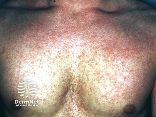
Clinical photography of the chest shows a diffuse erythematous maculopapular eruption with a rough, sandpaper-like texture covering the anterior trunk. The rash appears blanching with uniform distribution, without discrete vesicles, and involves the chest and upper abdomen. This exanthem is classically seen in scarlet fever, a streptococcal pharyngitis–associated illness, and is characterized by a diffuse erythematous rash, circumoral pallor, and sometimes strawberry tongue; desquamation can occur after several days. The image supports dermatologic assessment of an acute febrile illness with a prominent cutaneous eruption and provides a visual reference for exanthematous patterns. Differential considerations include measles, rubella, drug eruption, erythema marginatum, and, less often, Kawasaki disease, each with distinct clinical cues. Clinical correlation with fever, sore throat, tender or enlarged lymph nodes, and tongue findings is essential for accurate diagnosis. This photograph is valuable for educational purposes in dermatology and infectious disease, aiding recognition of exanthematous patterns, triage decisions, and patient counseling. Ideal applications include exam preparation, visual differential diagnostic training, and algorithm development for fever with rash workflows. It reinforces the role of rapid streptococcal testing and timely antibiotic therapy when scarlet fever is suspected. Cross-disciplinary collaboration and follow-up testing optimize patient outcomes and reduce transmission risk. Clinical documentation should include lesion mapping and progression notes.
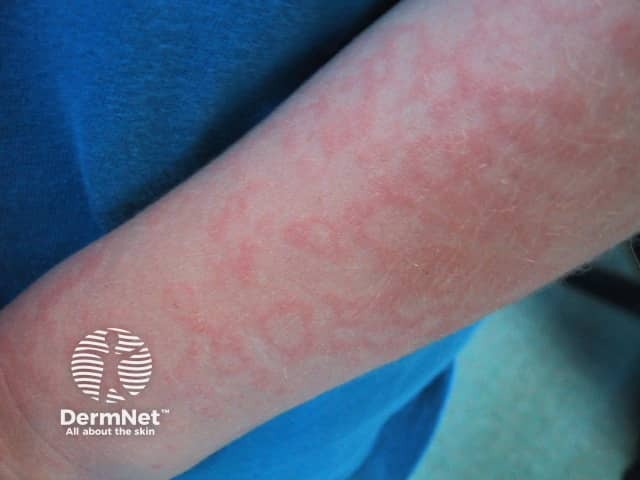
This clinical photograph depicts a skin eruption consistent with erythema infectiosum (fifth disease) on the forearm. Imaging modality: clinical photography in standard color profile; single anterolateral forearm view. The rash shows a fine, lacy or reticular erythema pattern distributed along the skin surface; pink to light red coloration with mild blanching on palpation. The epidermis is uninvolved to mildly accentuated by vascular dilation; there is no vesiculation or necrosis. The lesion tracks a superficial dermal capillary network, producing a net-like appearance that is more conspicuous in lighter skin tones. The pattern may begin subtely and intensify during febrile illness, then evolve to a reticular exanthem that spares the face early in disease. In erythema infectiosum, parvovirus B19 infection is the etiologic agent; the clinical correlation includes low-grade fever, malaise, and sometimes pruritus. Differential considerations include other viral exanthems (rubella, roseola), pityriasis rosea, drug eruption, and contact dermatitis. The diagnostic significance lies in recognizing the lace-like rash in children, which is typically self-limited and resolves without scarring within days to weeks. This image is relevant for dermatology education, infectious disease references, and clinical case repositories focusing on pediatric exanthems and viral rash patterns. Image assists in differential diagnosis and education globally.
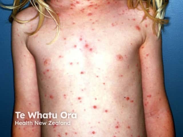
Imaging modality: Clinical photography of a pediatric patient with varicella (chickenpox) rash. Anterior frontal full-body view documenting widespread vesicular lesions on an erythematous base, most prominent on the trunk with scattered involvement of the limbs and face. The lesions appear in crops and at different stages, including macules, pruritic papules, clear-vesicles (dew drop on a rose petal), pustules, and crusted crusts. The distribution is generalized and centripetal, with higher density on the trunk and chest relative to the peripheral extremities. Skin texture shows mild erythema surrounding vesicles; some lesions show crusting as healing begins. No mucosal involvement visible in this image. Lighting is even and natural, color rendition preserved to aid lesion characterization. This dermatologic eruption is characteristic of varicella-zoster virus infection: contagious, self-limited in healthy children, typically presenting with fever, malaise, and pruritic vesicular rash that evolves over several days. Clinically, the image demonstrates classic features used for diagnosis: vesicles at multiple stages on erythematous base, cropped distribution, and trunk predominance. The image is useful for educational purposes, differential diagnosis training (including disseminated herpes simplex, pityriasis rosea-like viral exanthem), and patient counseling about contagion control and rash progression. It may guide teledermatology triage and documentation of clinical course in dermatology and pediatrics.
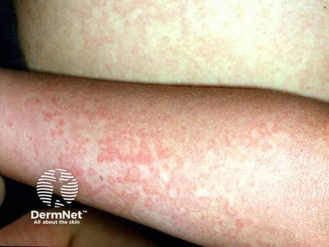
Clinical photography, dermatology assessment of a forearm reveals a lace-like, reticular erythematous rash consistent with Fifth disease (erythema infectiosum) due to parvovirus B19. The image shows a close-up, high-resolution view of cutaneous vascular dilation forming a net-like pattern across the affected skin with fine blanching and even distribution. Surface integrity remains preserved, with flat, non-vesicular erythematous patches that accentuate the interlacing lines. This dermatosis classically occurs in children and can accompany mild fever or malaise; in many cases facial erythema (slapped cheeks) is prominent, though not always visible in a limb-focused photograph. The rash tends to appear on the trunk and proximal limbs and may spare mucosa. Resolution is typically spontaneous within 1–3 weeks, with possible mild pigmentary after-effects. Clinically, the image supports recognition of a viral exanthem with the characteristic reticular pattern, assisting in differential diagnosis from pityriasis rosea, rubella, roseola, drug eruptions, and other viral exanthems. Parvovirus B19 serology or PCR can confirm infection if clinical suspicion is high. This image is valuable for education in pediatric dermatology, infectious disease, and primary care, enabling pattern-based learning, case-based discussion, and image database curation. High-quality imaging enhances diagnostic confidence and education, especially useful for telemedicine and remote consultation.
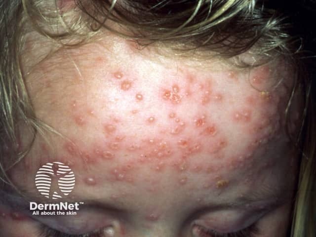
Clinical photography of active varicella (chickenpox) rash on the frontal scalp and forehead of a pediatric patient. Modality: digital color photograph taken under ambient lighting; standard clinical technique without dermoscopy or fluorescence. The image shows numerous small vesicles on an erythematous, slightly raised base distributed across the forehead and extending toward the temple. Lesions are in multiple evolutionary stages, with some vesicles capturing clear serous content, others showing crusted surfaces, and crusts around the margins. The pattern is characteristic of varicella: centripetal distribution with trunk and face involvement, crops of lesions appearing sequentially, and a tendency for vesicles to be more numerous on exposed skin areas. The epidermis and superficial dermis contain superficial fluid-filled vesicles, each about 2-4 mm in diameter, some conforming to the classic dew drop on a rose petal description. Facial involvement may accompany fever and malaise in pediatric patients. Although lesions may be pruritic, Nikolsky sign is typically absent; scarring is uncommon in children. Diagnosis is clinical, supported by history of varicella exposure or vaccination status; differential includes disseminated herpes zoster, pityriasis rosea, and vesicular dermatitis. Recognizing multistage lesions in a contagious exanthem is clinically significant for isolation, supportive therapy, and public health reporting as Varicella-zoster virus infection.
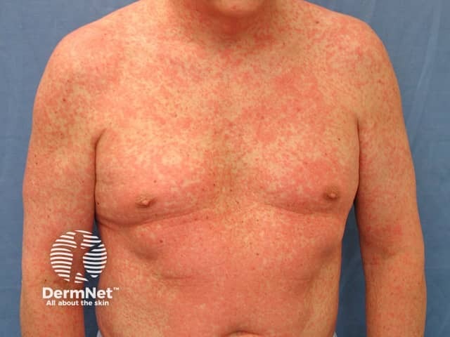
Clinical photography of an adult male presenting with a diffuse morbilliform eruption involving the trunk, shoulders, and proximal upper limbs. The image, obtained in frontal/anterior view under standard white‑light illumination, shows numerous erythematous macules and papules that are closely spaced and blanch with pressure. The rash is symmetric on the chest, upper abdomen, and the upper arms, with minimal confluent areas on the torso. No vesicles, purpura, or necrosis are evident, and there is no obvious mucosal involvement in the visible portions of the face or oral cavity. The morphology is characteristic of a morbilliform (measles‑like) exanthem, typically arising from either a viral infection or a drug reaction. The color is uniform red, and the lesions appear non-desquamating with intact epidermal surface in this field of view. The patient's age and body habitus are consistent with adults who can develop drug eruptions or viral rashes, though vaccination history and recent medications are critical for interpretation. Clinically, this pattern warrants evaluation for infectious etiologies (measles, rubella, parvovirus B19) and common drug eruptions (antibiotics, anticonvulsants, NSAIDs). Management considerations include symptom control, identification of potential triggers, and monitoring for evolution to fever, systemic involvement, or mucosal lesions. Documentation supports differential diagnosis, and education for patient counseling and follow-up.
Kawasaki disease rash children strawberry tongue
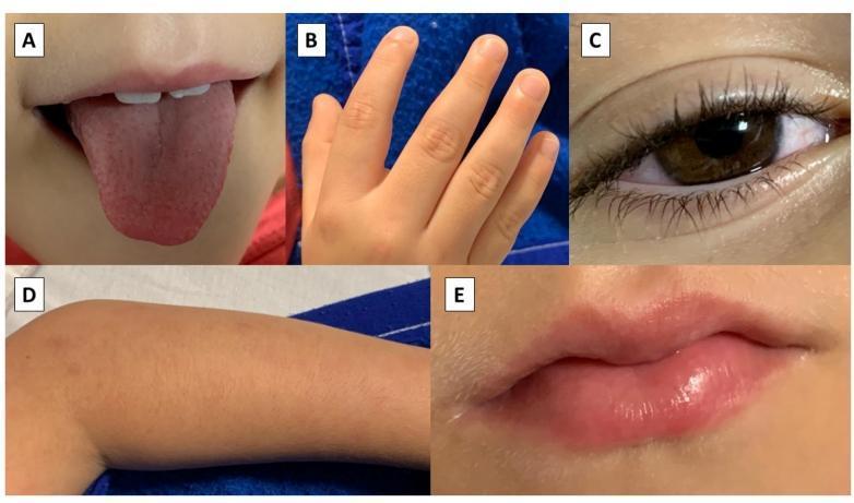
A composite of five clinical photographs (labeled A-E) illustrating the diagnostic features of Multisystem Inflammatory Syndrome in Children (MIS-C) and Kawasaki-like disease. Panel A shows a 'strawberry tongue' with prominent, inflamed papillae and diffuse erythema. Panel B demonstrates edema of the digits on the hand, characterized by swelling and loss of normal skin creases. Panel C displays bilateral bulbar conjunctival injection, notably sparing the limbus and presenting without exudate. Panel D shows a diffuse maculopapular cutaneous rash on the extremity. Panel E highlights oral mucositis, specifically erythematous, swollen, and cracked (fissured) lips. These visual markers are critical for the clinical identification of systemic inflammatory responses in pediatric patients, particularly those temporally associated with COVID-19 infection.
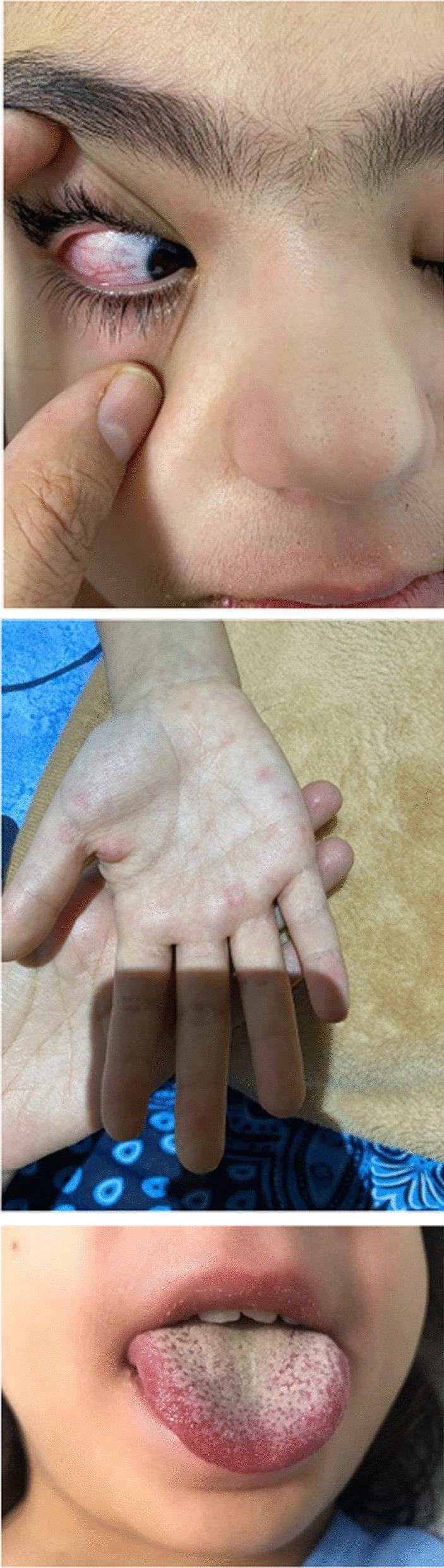
A composite of three clinical photographs illustrating diagnostic signs of Kawasaki Disease in a pediatric patient. The top image shows the right eye with bilateral, nonpurulent bulbar conjunctival injection; the sclera is markedly erythematous without exudate or discharge. The middle image displays the palmar surface of the hand, exhibiting a maculopapular rash characterized by discrete, erythematous lesions on the palm and fingers. The bottom image shows an oropharyngeal examination revealing a classic 'strawberry tongue,' featuring a bright red, edematous appearance with prominent, hypertrophied fungiform papillae against a white-coated dorsal background. Together, these visual findings—conjunctival sparing, peripheral extremity changes, and mucosal inflammation—serve as key clinical criteria for diagnosing Kawasaki Disease or Multisystem Inflammatory Syndrome in Children (MIS-C).
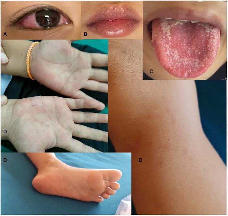
A composite of clinical photographs illustrating the classic mucocutaneous manifestations of Multisystem Inflammatory Syndrome in Children (MIS-C) and Kawasaki disease. Panel A shows bilateral non-exudative conjunctival injection, sparing the limbus. Panel B demonstrates erythematous, fissured, and swollen lips. Panel C displays a classic 'strawberry tongue' characterized by prominent lingual papillae and diffuse erythema with a patchy white coating. Panel D contains several images of the extremities, showing a faint, blanching maculopapular rash on the palms and soles, as well as an erythematous patch on the knee. These findings collectively represent systemic inflammation and vasculitis of the small and medium-sized vessels. The clinical significance of these visual signs is to aid in the prompt diagnosis of inflammatory syndromes following pediatric viral infections, specifically SARS-CoV-2. The target audience includes pediatricians and emergency medicine residents learning to recognize patterns of pediatric inflammatory conditions.
meningococcal purpuric rash petechiae child
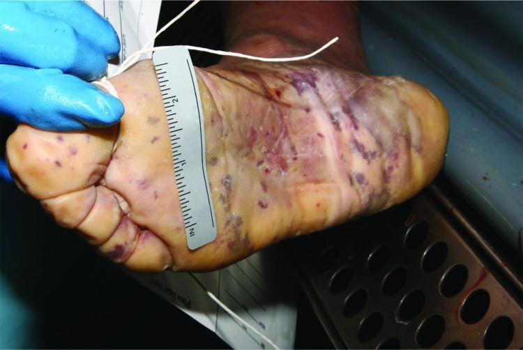
Clinical photograph of the plantar surface (sole) of a human foot during postmortem examination, demonstrating classic dermatological manifestations of invasive meningococcal disease. The image shows a widespread purpuric rash consisting of pinpoint petechiae and larger, confluent ecchymoses. These lesions are dark purple to blue-black in color and are most concentrated on the toes, heel, and lateral borders of the foot. Linear streaks of hemorrhage are visible across the arch. The surrounding skin exhibits postmortem changes, including pallor, mild maceration, and loss of turgor (wrinkling). A forensic ruler and identifying tags are present for scale and documentation. This visual serves as a critical educational example of fulminant meningococcemia (purpura fulminans) and its persistence in forensic pathology settings. The presentation highlights the importance of recognizing skin signs in the diagnosis of Neisseria meningitidis infection, even in cases of sudden death where clinical history may be absent.
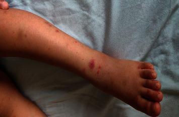
This clinical photograph displays a pediatric patient's lower extremity, showing a characteristic non-blanching purpuric rash. The lesions are distributed along the lower leg and dorsal aspect of the foot, exhibiting an ascending pattern. The morphology of the rash includes individual red-to-purple petechiae (pinpoint lesions) and larger, palpable purpura that have become confluent in areas above the ankle. The distribution appears scattered but predominantly involves the extensor surfaces. This visual finding is highly suggestive of immunoglobulin A (IgA) vasculitis, formerly known as Henoch-Schönlein purpura (HSP), particularly when associated with the clinical context of joint pain, abdominal symptoms, or hematochezia. The image serves as an educational example of cutaneous small-vessel vasculitis in a child, emphasizing the recognition of palpable purpura in dependent areas as a primary diagnostic sign.
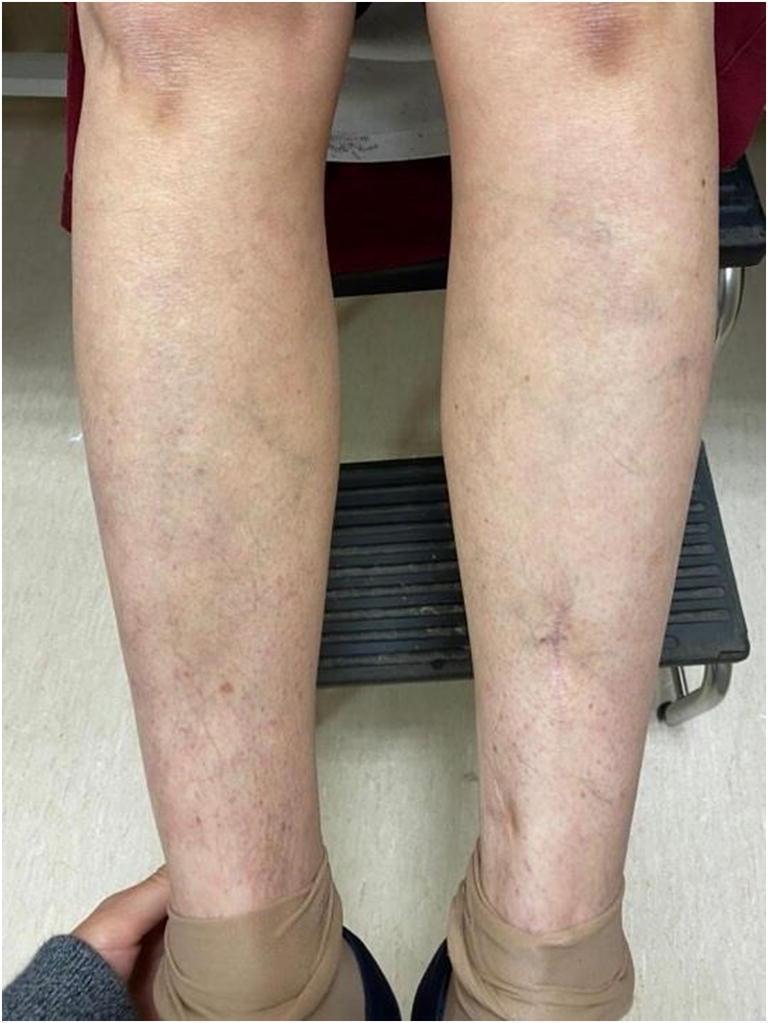
A clinical photograph showing the anterior view of both lower legs of a 62-year-old patient, demonstrating the healing phase of a petechial purpuric rash following 7 days of corticosteroid therapy. The image reveals scattered, fading purplish-red macules and fine petechiae, predominantly distributed on the distal shins and ankles. There is evidence of post-inflammatory hyperpigmentation and subtle residual ecchymosis. Visible vascular markings including superficial veins are apparent. The distribution is roughly symmetrical, showing a marked reduction in lesion density and inflammatory signs compared to an acute purpuric presentation. A small, faint surgical scar is visible on the pretibial area of the right leg. This visual serves to illustrate the clinical progression and therapeutic response of cutaneous vasculitis or a similar systemic inflammatory condition (e.g., associated with elevated rheumatoid factor and ANA) to systemic prednisone.
scarlet fever rash sandpaper skin child
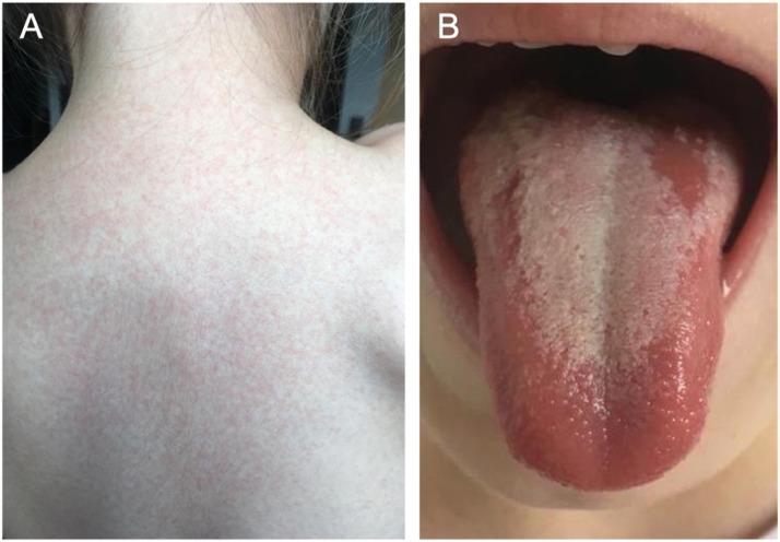
A composite of two clinical photographs (A and B) demonstrating classic physical manifestations of scarlet fever (Scarlatina) in a pediatric patient. Image A displays a characteristic scarlatiniform rash on the posterior trunk and neck. The rash is composed of diffuse, fine, erythematous maculopapular lesions that are closely spaced, giving the skin a 'sandpaper' texture. Image B shows an intraoral view of the tongue, illustrating 'white strawberry tongue.' The dorsal surface is covered by a thick, yellowish-white coating through which enlarged, hyperemic fungiform papillae project as prominent red dots. The lateral borders and the tip of the tongue appear intensely erythematous. These findings are hallmarks of a Group A Streptococcus infection. This educational material is suitable for illustrating systemic pediatric infections and dermatological-oral correlations in infectious diseases.
Clinical photography of the chest shows a diffuse erythematous maculopapular eruption with a rough, sandpaper-like texture covering the anterior trunk. The rash appears blanching with uniform distribution, without discrete vesicles, and involves the chest and upper abdomen. This exanthem is classically seen in scarlet fever, a streptococcal pharyngitis–associated illness, and is characterized by a diffuse erythematous rash, circumoral pallor, and sometimes strawberry tongue; desquamation can occur after several days. The image supports dermatologic assessment of an acute febrile illness with a prominent cutaneous eruption and provides a visual reference for exanthematous patterns. Differential considerations include measles, rubella, drug eruption, erythema marginatum, and, less often, Kawasaki disease, each with distinct clinical cues. Clinical correlation with fever, sore throat, tender or enlarged lymph nodes, and tongue findings is essential for accurate diagnosis. This photograph is valuable for educational purposes in dermatology and infectious disease, aiding recognition of exanthematous patterns, triage decisions, and patient counseling. Ideal applications include exam preparation, visual differential diagnostic training, and algorithm development for fever with rash workflows. It reinforces the role of rapid streptococcal testing and timely antibiotic therapy when scarlet fever is suspected. Cross-disciplinary collaboration and follow-up testing optimize patient outcomes and reduce transmission risk. Clinical documentation should include lesion mapping and progression notes.
roseola infantum rash infant exanthem subitum
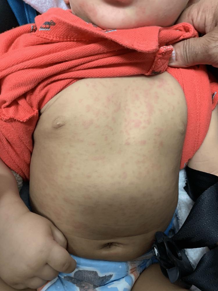
A clinical photograph of an infant's torso and lower face demonstrating a classic exanthem. The image shows a diffuse, erythematous, macular rash consisting of small, discrete, pale-pink macules. The lesions are primarily distributed centrally on the chest and abdomen with extension towards the neck and proximal extremities. The rash appears non-scaling and non-confluent, with clear areas of skin visible between the lesions. This presentation is characteristic of the defervescence rash associated with Roseola Infantum (Exanthema Subitum), typically caused by Human Herpesvirus 6 (HHV-6). The educational focus is on identifying the morphology and centrifugal distribution of viral exanthems in the pediatric population following a febrile episode.
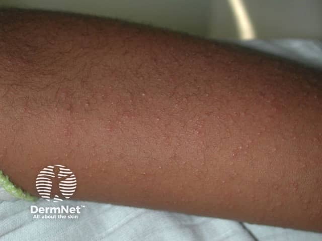
Imaging modality: Clinical photography of cutaneous surface. The image shows a close-up, anterior forearm view with diffuse, erythematous papules arranged in a maculopapular pattern. Lesions are small (roughly 1–3 mm), non-vesicular, non-follicular, and slightly raised, with even distribution across the exposed skin. The surrounding skin demonstrates a uniform complexion with no necrosis or purpura. Lighting is diffuse and neutral, suitable for dermatologic photography; no magnification or dermatoscopic features are evident. The findings are consistent with a viral exanthem—a contagious, self-limited dermatologic eruption seen in primary viral infections. The pattern is nonspecific but typical of pediatric viral illnesses and presents as a diffuse rash that may accompany fever or systemic symptoms. Potential etiologies include measles (rubeola), rubella, roseola (exanthem subitum), parvovirus B19, enteroviruses, and adenovirus infections. Clinically, the exanthem may precede or follow other signs such as cough, conjunctivitis, pharyngitis, lymphadenopathy, or malaise. Diagnostic significance lies in correlating clinical history, vaccination status, exposure risk, and evolution of the rash; laboratory tests or serology can aid confirmation when indicated. Differential considerations encompass drug eruption and pityriasis rosea. This image is valuable for dermatology education and clinical exemplars of viral rashes.
Fever with Rash in Paediatric Patients
Step 1 - Characterise the Rash Morphology
| Morphology | Key Examples |
|---|---|
| Maculopapular (morbilliform) | Measles, rubella, roseola, drug reaction, EBV |
| Vesicular | Varicella, HSV, hand-foot-mouth disease |
| Petechial / Purpuric | Meningococcaemia, IgA vasculitis (HSP), ITP |
| Sandpaper / Scarlatiniform | Scarlet fever |
| Polymorphous (non-vesicular) | Kawasaki disease |
| Targetoid | Erythema multiforme |
| Lacy / Reticular | Erythema infectiosum (Fifth disease) |
Major Causes: Diagnostic Summary
1. Measles (Rubeola)
- Begins behind ears/hairline, spreads caudally (face → trunk → arms → legs)
- Erythematous, maculopapular, semiconfluent
- Appears at day 14 post-exposure; fades in same progression over 3-4 days
- May be followed by desquamation (especially in malnourished children)
Harrison's Principles of Internal Medicine 22E (2025)
2. Varicella (Chickenpox)
- Centripetal distribution (trunk > face > limbs)
- Lesions in multiple stages simultaneously: macules → papules → vesicles ("dew drop on a rose petal") → pustules → crusts
- Intensely pruritic
- Scalp and mucous membranes involved
3. Roseola Infantum (Exanthem Subitum) - HHV-6/HHV-7
- High fever (39-40°C) for 3-5 days in a well-appearing child
- Fever abruptly defervesces
- Rash appears as fever breaks - rose-pink discrete macules/papules on trunk, spreads to neck/proximal extremities
4. Erythema Infectiosum (Fifth Disease) - Parvovirus B19
- "Slapped cheek" - bright red erythema on both cheeks, circumoral pallor
- Lacy reticular rash on trunk and extremities
- Rash may recur with heat, exercise, sunlight
5. Scarlet Fever
- Fine, erythematous, "sandpaper" texture (papular)
- Begins on trunk and neck, spreads to extremities
- Pastia's lines (petechiae in skin folds - axillae, antecubital fossa)
- Circumoral pallor (perioral sparing)
- "White strawberry tongue" initially (white coat with red papillae projecting through)
- Later "red strawberry tongue" (coat sheds, red tongue with prominent papillae)
- Pharyngeal erythema
Sleisenger & Fordtran's GI and Liver Disease
6. Kawasaki Disease
| Feature | Detail |
|---|---|
| Bilateral conjunctival injection | Non-exudative; perilimbic sparing |
| Oral/mucosal changes | Red cracked lips, pharyngeal erythema, strawberry tongue |
| Cervical lymphadenopathy | At least one node >1.5 cm |
| Peripheral extremity changes | Diffuse erythema/oedema of hands/feet (acute); periungual desquamation (convalescent) |
| Polymorphous rash | Non-vesicular, non-bullous; typically starts on trunk |
Rosen's Emergency Medicine; Goldman-Cecil Medicine
7. Meningococcaemia
- Initially non-specific maculopapular → petechial → purpuric/ecchymotic (non-blanching)
- Rapidly evolving; can progress to purpura fulminans
- Distribution: trunk, lower limbs
- High fever + meningism (neck stiffness, photophobia, Kernig's/Brudzinski's)
- Child may appear very unwell/toxic
- Non-blanching rash = DO NOT WAIT for investigations - give IV/IM benzylpenicillin immediately
8. Rubella (German Measles)
- Fine maculopapular, begins on face, spreads downward rapidly (24h to cover body, gone in 3 days)
- Milder than measles, no cough
- Posterior auricular/suboccipital lymphadenopathy (characteristic)
- Forchheimer spots (petechiae on soft palate)
- Low-grade fever
9. IgA Vasculitis (Henoch-Schönlein Purpura)
- Palpable purpura (lower limbs/buttocks) - non-thrombocytopenic
- Arthritis/arthralgia
- Abdominal pain (colicky)
- Renal involvement (haematuria, proteinuria)
10. Hand-Foot-Mouth Disease (HFMD)
- Oval greyish vesicles on palms, soles, and between fingers/toes
- Oral ulcers (painful)
- Fever + malaise precede rash
Quick Diagnostic Comparison Table
| Disease | Fever-Rash Timing | Rash Type | Location | Key Clue |
|---|---|---|---|---|
| Measles | Rash at peak fever | Maculopapular, confluent | Cephalocaudal | Koplik's spots |
| Roseola | Rash after fever resolves | Discrete macules, rose-pink | Trunk first | Well-appearing child with high fever |
| 5th disease | Minimal/no fever at rash | Lacy reticular | Arms/trunk; slapped cheek | "Slapped cheek" |
| Scarlet fever | Rash with fever | Sandpaper, blanching | Trunk, flexures | Pastia's lines, strawberry tongue |
| Varicella | Rash with mild fever | Vesicular (multi-stage) | Centripetal | All lesion stages simultaneously |
| Rubella | Rash with fever | Fine maculopapular | Cephalocaudal (fast) | Posterior auricular LN |
| Kawasaki | Rash during fever (day 2-5) | Polymorphous, non-vesicular | Trunk first | Strawberry tongue + conjunctivitis |
| Meningococcaemia | Rash with fever (rapid) | Petechiae → purpura | Trunk, lower limbs | Non-blanching, toxic child |
| HFMD | Rash with fever | Oval vesicles | Palms, soles, mouth | Oral ulcers |
| HSP | With/after URTI | Palpable purpura | Below waist | Non-thrombocytopenic |
Red Flags - Urgent Action Required
- Non-blanching rash (petechiae/purpura) + fever → rule out meningococcaemia; give antibiotics empirically
- Toxic/septic-appearing child regardless of rash type
- Kawasaki criteria met → echocardiogram + IVIG within first 10 days of fever
- Haemodynamic instability
- Altered consciousness + rash
Approach to Investigation
| Suspected Diagnosis | First-Line Tests |
|---|---|
| Measles | Measles IgM serology |
| Meningococcaemia | Blood culture, FBC, CRP, LP (if stable) |
| Kawasaki | FBC (elevated WBC, platelet), ESR, CRP, echo |
| Scarlet fever | Throat swab + RADT, ASO titre |
| Varicella | Clinical; PCR of lesion fluid if immunocompromised |
| Parvovirus B19 | Parvovirus IgM/IgG; blood film |
| HSP | Urinalysis, IgA level |
Sources: Harrison's Principles of Internal Medicine 22E (2025); Rosen's Emergency Medicine; Goldman-Cecil Medicine; Tintinalli's Emergency Medicine; Sherris & Ryan's Medical Microbiology