
Sub mandibular gland Its complete anatomy , including imp landmarks and blood and nerve supply Submandibular sailadenitis , its mc cause , complications , etiology , clinical features and management Explain sub mandibular gland excision steps , post op And intera complications
-
Submandibular gland anatomy
- Parts, relations, duct, surface anatomy and key surgical landmarks
- Arterial, venous, lymphatic, parasympathetic, sympathetic and sensory nerve supply
- Facial artery, lingual nerve, hypoglossal nerve, and marginal mandibular branch of facial nerve
-
Submandibular sialadenitis
- Acute versus chronic disease
- Most common cause: duct obstruction by a submandibular stone (sialolithiasis)
- Etiology, clinical features, investigations, complications, and medical/interventional management
-
Submandibular gland excision
- Indications and preoperative planning
- Stepwise operative technique
- Intraoperative complications and their prevention
- Postoperative care, complications, and follow-up
1 anatomy
anatomy diagram submandibular gland relations Wharton duct lingual nerve hypoglossal nerve facial artery
"submandibular gland" AND anatomy
submandibular gland anatomy relations duct nerve blood supply anatomy
https://www.ncbi.nlm.nih.gov/books/NBK542272
Submandibular gland: applied surgical anatomy
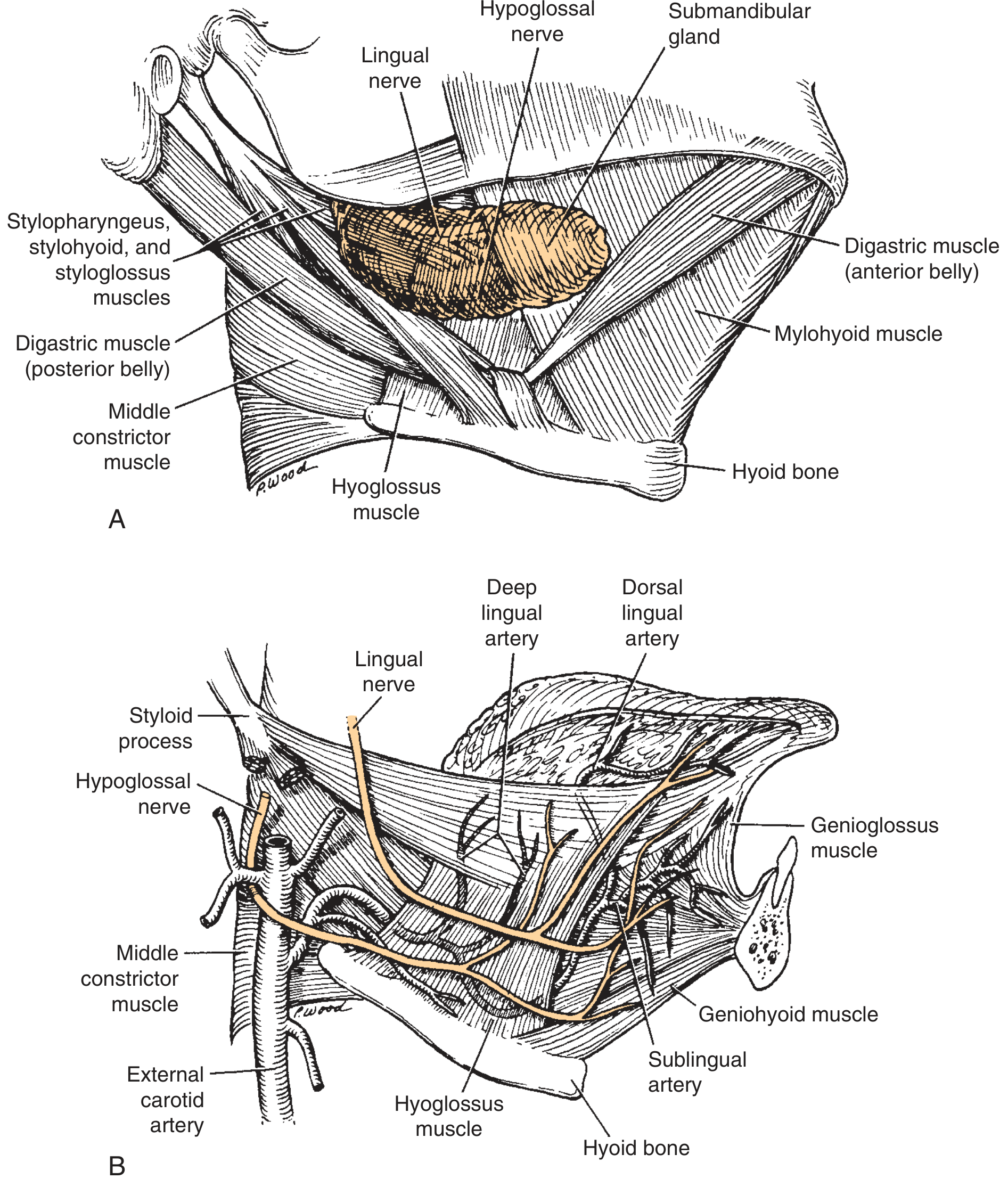
1. Overview
- A paired major salivary gland, second largest after parotid.
- Produces predominantly mixed saliva, with serous and mucous acini. It provides much of the resting, unstimulated salivary flow.
- Situated in the submandibular (digastric) triangle and floor of mouth.
- Enclosed by a capsule derived from the investing layer of deep cervical fascia.
- Has:
- A large superficial part
- A small deep part
- The two parts are continuous by wrapping around the posterior free border of mylohyoid.
2. Submandibular triangle
Boundaries
| Boundary | Structure |
|---|---|
| Superior / base | Inferior border of body of mandible |
| Anteroinferior | Anterior belly of digastric |
| Posteroinferior | Posterior belly of digastric |
| Floor | Mylohyoid anteriorly; hyoglossus and middle constrictor more posteriorly |
| Roof | Skin, superficial fascia with platysma, investing deep cervical fascia |
3. Parts and relations
A. Superficial part
Relations of superficial part
| Surface / relation | Structures |
|---|---|
| Superficial or lateral | Skin, platysma, investing fascia, facial vein, submandibular lymph nodes; marginal mandibular branch of facial nerve lies superficial to the gland |
| Superior | Submandibular fossa on medial surface of mandible |
| Inferior | Posterior belly of digastric, stylohyoid |
| Medial / deep | Mylohyoid anteriorly; hyoglossus posteriorly; posteriorly related to styloglossus |
| Posterolateral | Facial artery may groove, traverse, or lie closely applied to the posterior part of gland |
B. Deep part
Relations of deep part
| Direction | Important relation |
|---|---|
| Lateral | Mylohyoid |
| Medial | Hyoglossus and posteriorly styloglossus |
| Superior | Lingual nerve and submandibular ganglion |
| Inferior | Hypoglossal nerve and accompanying lingual veins |
| Anterior | Sublingual gland and submandibular duct |
In the floor of mouth, the structures from superior to inferior on hyoglossus are usually:
- Lingual nerve
- Submandibular duct
- Hypoglossal nerve
- Lingual veins
4. Submandibular duct, Wharton duct
Origin and course
- About 5 cm long.
- Arises from the anterior aspect of the deep lobe or gland hilum.
- Hooks around the posterior border of mylohyoid.
- Runs forward in the floor of mouth, between:
- Mylohyoid laterally
- Hyoglossus and genioglossus medially
- It then passes forwards between the sublingual gland and genioglossus.
Opening
- Opens at the sublingual papilla (caruncle) on either side of the lingual frenulum.
Critical relation with lingual nerve
5. Arterial supply
-
Facial artery
- Gives direct glandular branches.
- Passes deep to posterior belly of digastric and stylohyoid.
- May pass through, groove, or run close to the posterior portion of gland.
- Curves around the lower border of mandible, just anterior to masseter.
-
Submental artery
- Branch of facial artery.
- Supplies inferior part of gland and adjacent floor of mouth.
-
Sublingual artery
- Branch of lingual artery.
- Contributes particularly to deep part and duct region.
6. Venous drainage
- Facial vein lies superficial and lateral to the gland.
- Venous drainage also communicates with lingual and sublingual veins.
- Ultimately drains into the internal jugular venous system, mainly via facial/common facial vein.
7. Lymphatic drainage
- Drains first to submandibular lymph nodes, which are mainly periglandular.
- Efferents drain to upper deep cervical lymph nodes, especially jugulo-omohyoid and jugulodigastric chains.
8. Nerve supply
A. Parasympathetic secretomotor supply
→ facial nerve, CN VII
→ chorda tympani
→ joins lingual nerve, V3
→ preganglionic fibers synapse in submandibular ganglion
→ postganglionic fibers supply submandibular and sublingual glands.
B. Sympathetic supply
- Postganglionic sympathetic fibers originate from the superior cervical sympathetic ganglion.
- They travel along periarterial plexuses around branches of the external carotid artery.
- They cause vasoconstriction and modify salivary secretion, making it relatively scant and viscous.
C. Sensory supply
- General sensation from the region is carried mainly through the lingual nerve, a branch of mandibular division of trigeminal nerve, V3.
- The gland does not have a major independent sensory nerve that is routinely identified.
9. Important nerves in relation to the gland
1. Marginal mandibular branch of facial nerve, CN VII
- Runs in the superficial fascia near the lower border of mandible.
- It may lie below the inferior border of mandible, especially posterior to the facial vessels.
- Supplies muscles of lower lip, particularly depressor anguli oris, depressor labii inferioris, and mentalis.
2. Lingual nerve, V3
- Closely related to deep lobe, submandibular ganglion, and Wharton duct.
- Carries:
- General sensation from anterior two-thirds of tongue
- Taste fibers from chorda tympani
- Preganglionic parasympathetic fibers to submandibular ganglion
3. Hypoglossal nerve, CN XII
- Lies inferior to the duct, on the surface of hyoglossus.
- Motor nerve to intrinsic and extrinsic tongue muscles, except palatoglossus.
4. Mylohyoid nerve
- Branch of inferior alveolar nerve, V3.
- Runs in the mylohyoid groove on medial mandible.
- Supplies mylohyoid and anterior belly of digastric.
- Less commonly injured in standard gland excision but relevant during dissection near the mandible.
10. High-yield surgical landmarks
| Landmark | Why it matters |
|---|---|
| Inferior border of mandible | Reference for skin incision and marginal mandibular nerve protection |
| Facial artery pulse at mandibular border | Identifies facial artery; it is closely related to posterior gland |
| Posterior belly of digastric | Forms posterior boundary of triangle and helps locate facial artery |
| Mylohyoid posterior border | Point where gland turns from superficial to deep lobe |
| Wharton duct | Identify before division during excision |
| Lingual nerve | Lies superior to duct distally and loops under it |
| Hypoglossal nerve | Inferior to duct, superficial to hyoglossus |
| Hyoglossus | Lingual artery lies deep to it; hypoglossal nerve lies superficial to it |
| Facial vein | Usually superficial to gland and often encountered early |
11. One-minute viva answer
2 sailadenitis
"sialadenitis" AND management
submandibular sialadenitis diagnosis treatment complications guideline
ultrasound submandibular sialadenitis sialolithiasis stone Wharton duct
https://pmc.ncbi.nlm.nih.gov/articles/PMC10426371
Submandibular sialadenitis
Most common cause
- About 80% to 90% of salivary stones occur in the submandibular system.
- This predisposition results from the long, tortuous, upward course of Wharton duct and the relatively viscous, calcium-rich submandibular saliva.
- In acute bacterial sialadenitis, the commonest organism is Staphylococcus aureus. Streptococci and anaerobic oral flora may also contribute. K J Lee's Essential Otolaryngology, p. 648.
| Question | Best answer |
|---|---|
| Commonest cause of submandibular sialadenitis | Obstruction by a submandibular sialolith |
| Commonest pathogen in acute suppurative sialadenitis | Staphylococcus aureus |
Etiology and predisposing factors
1. Obstructive
- Sialolithiasis: most important cause.
- Ductal stricture or stenosis.
- Duct trauma, foreign body, or mucus plug.
- External compression by a mass, rarely.
2. Infective
- Bacterial: S. aureus, streptococci, anaerobic oral flora, and occasionally gram-negative organisms in frail or hospitalized patients.
- Viral: mumps, influenza, cytomegalovirus, coxsackievirus, HIV.
- Specific chronic infections: tuberculosis, actinomycosis, cat-scratch disease.
3. Reduced salivary flow or salivary stasis
- Dehydration
- Poor oral intake, fasting, postoperative state
- Fever and severe systemic illness
- Xerostomia due to anticholinergic drugs, antidepressants, antihistamines, diuretics, etc.
- Diabetes mellitus, renal failure, malnutrition
- Previous head and neck radiotherapy
4. Autoimmune or inflammatory disease
- Sjögren disease
- IgG4-related disease, including chronic sclerosing sialadenitis or Küttner tumour
- Sarcoidosis
Pathogenesis
→ salivary stagnation
→ duct dilatation and glandular oedema
→ retrograde bacterial ascent through Wharton duct
→ acute inflammation, pus formation, and possible abscess.
- Ductal stenosis
- Sialectasis, or dilated ductal system
- Acinar atrophy
- Fibrosis
- Reduced gland function
Clinical features
A. Acute bacterial submandibular sialadenitis
- Sudden painful swelling below the mandible
- Tender, warm, firm or indurated gland
- Erythema of overlying skin in more severe cases
- Pain aggravated during meals, especially with a stone
- Fever, malaise, dehydration
- Reduced salivary flow
- Foul taste in mouth
- Pus expressed from Wharton duct orifice at the sublingual caruncle after bimanual gland massage
- Trismus or dysphagia if infection is severe or spreads to deep neck spaces
- Cervical lymphadenopathy may occur
B. Chronic obstructive sialadenitis
- Recurrent swelling and discomfort, classically during meals
- “Salivary colic”: pain and swelling after gustatory stimulation, settling after the meal
- Gland may become firm, enlarged, or fibrotic
- Recurrent episodes of infection and purulent duct discharge
- A stone may be palpable bimanually in the floor of mouth, usually along Wharton duct
- Between episodes, the patient may be asymptomatic
C. Viral or autoimmune disease
- Viral disease is often bilateral and associated with systemic features.
- Sjögren disease typically produces chronic dry mouth and dry eyes, often with bilateral gland enlargement.
- A unilateral hard persistent gland needs evaluation to exclude neoplasm, particularly if accompanied by fixation, facial nerve symptoms, neck nodes, or weight loss.
Investigations
Clinical examination
- Inspect and palpate the submandibular region.
- Examine the floor of mouth.
- Palpate Wharton duct bimanually, one finger intraorally and the other externally.
- Milk the gland from posterior to anterior and observe ductal flow.
- Send expressed pus for Gram stain, culture, and antibiotic sensitivity when present.
Imaging
- Ultrasound: first-line for gland enlargement, stones, duct dilatation, and abscess.
- Non-contrast CT: best when a stone is suspected but not visible on ultrasound, or when complications/deep neck infection are suspected.
- Contrast CT: useful for abscess, cellulitis, deep-neck extension, or tumour differential.
- Sialendoscopy: diagnostic and therapeutic for ductal stones and stenosis.
- Sialography is avoided in acute infection, as it may worsen inflammation. K J Lee's Essential Otolaryngology, p. 648.
Management
- Treat the acute infection and restore flow.
- Treat the cause of obstruction to prevent recurrence.
A. Acute uncomplicated bacterial sialadenitis
- M - Massage: gentle gland massage from posterior to anterior, toward Wharton duct opening.
- A - Antibiotics: empirical therapy should cover S. aureus, streptococci, and oral anaerobes; modify according to culture results and local antibiotic guidance.
- S - Sialogogues: lemon drops, sour sugar-free sweets, or chewing gum if safe and not completely obstructed.
- H - Heat: warm compresses.
- H - Hydration: oral or intravenous hydration, depending on severity.
- Analgesia and antipyretics
- Good oral hygiene and chlorhexidine mouth care where appropriate
- Stop or modify xerostomia-causing medicines if clinically feasible
- Treat contributing systemic illness, dehydration, or diabetes
B. When to admit and give intravenous treatment
- Toxic appearance, high fever, sepsis, or immunocompromise
- Inability to swallow, dehydration, or failed oral treatment
- Trismus, tongue or floor-of-mouth elevation
- Rapidly increasing neck swelling
- Suspected abscess or deep neck-space extension
- Any airway concern
C. Treating the obstruction
| Situation | Usual management |
|---|---|
| Small distal duct stone | Hydration, massage, sialogogues; sometimes transoral duct incision and stone removal |
| Mobile intraductal stone / stenosis | Sialendoscopy with basket retrieval, duct dilatation, or irrigation |
| Larger proximal or hilar stone | Sialendoscopy-assisted transoral stone removal, sometimes lithotripsy depending on expertise |
| Recurrent infection, intraglandular stones, fibrosed non-functioning gland, failed minimally invasive management | Submandibular gland excision |
D. Abscess management
- Confirm with ultrasound or contrast CT.
- Drain by image-guided aspiration or surgical incision and drainage, depending on site and extent.
- Send pus for culture.
- Continue antibiotics and manage the underlying obstruction once infection is controlled.
Complications
Local complications
- Submandibular gland abscess
- Recurrent or chronic sialadenitis
- Fibrosis, acinar atrophy, and reduced salivary function
- Ductal stenosis and sialectasis
- Persistent stone disease
- Salivary fistula, rarely
Severe infective complications
- Spread to submandibular, sublingual, parapharyngeal, or other deep neck spaces
- Ludwig angina
- Floor-of-mouth oedema and tongue elevation
- Airway obstruction
- Septicaemia
- Rarely thrombophlebitis, osteomyelitis, or mediastinal spread
Viva-style short answer
3 excusion Its steps, most common complications
"submandibular gland excision" AND complications
submandibular gland excision operative steps complications marginal mandibular lingual hypoglossal nerve
https://www.ncbi.nlm.nih.gov/books/NBK568740
Submandibular gland excision
Structures at risk
- Marginal mandibular branch of facial nerve
- Lingual nerve
- Hypoglossal nerve
- Facial artery and facial vein
- Wharton duct and submandibular ganglion
Steps of external submandibular gland excision
This is an exam-oriented outline for trained surgical practice, not a substitute for operative supervision.
1. Preparation and positioning
- General anaesthesia, usually with oral endotracheal intubation.
- Supine position, shoulder roll, neck extended, and head turned to the opposite side.
- Prepare neck and oral cavity.
- Give perioperative antibiotics when indicated, particularly if active or recent infection is present.
- Palpate the lower border of mandible, gland, and stone if present.
2. Skin incision
- Make a transverse incision in a natural neck crease, at least two fingerbreadths, approximately 2 cm, below the lower border of mandible.
- This lower placement helps avoid the marginal mandibular branch of facial nerve and yields a more acceptable scar. Bailey and Love’s Short Practice of Surgery, 28th ed., p. 857.
3. Raise skin-subplatysmal flap
- Incise skin and platysma.
- Raise a superior subplatysmal flap toward the inferior border of mandible.
- Identify and protect the marginal mandibular nerve. It is commonly protected by elevating the fascia over the gland upward with the flap.
- Avoid blind clamping, excessive traction, or dissection directly on the undersurface of the mandible.
4. Expose superficial surface of gland
- Identify the inferior border of the gland.
- Divide or retract fascia and expose the superficial lobe.
- The facial vein, which lies superficial/lateral to the gland, is identified and ligated or divided as necessary.
- Identify the facial artery at the posterior or superior aspect of the gland.
5. Control facial vessels
- Preserve the facial artery if feasible by ligating its small glandular branches.
- Alternatively, ligate and divide facial artery and vein if necessary for safe removal or oncological clearance.
- Secure haemostasis carefully.
6. Mobilise gland from inferior and posterior attachments
- Identify posterior belly of digastric and separate the gland from it.
- Continue dissection from the inferior pole upward and posteriorly.
- For malignant disease, remove Level Ib fibrofatty nodal tissue en bloc with the gland when indicated.
7. Dissect gland from mylohyoid and identify deep lobe
- Mobilise the gland from the mylohyoid muscle.
- Retract mylohyoid anteriorly to expose the deep part of the gland and floor-of-mouth structures.
8. Identify and preserve lingual and hypoglossal nerves
- Identify the lingual nerve superiorly, with the submandibular ganglion attached to it.
- Identify the hypoglossal nerve inferior to Wharton duct, superficial to hyoglossus.
- Divide the small ganglionic branches from submandibular ganglion to the gland while preserving the lingual nerve.
- Do not place blind clamps near the duct or deep gland attachments.
9. Ligate Wharton duct
- Identify Wharton duct and accompanying vessels.
- Ligate and divide the duct as far anteriorly as safely possible, especially in stone disease, to reduce risk of a retained distal duct stone.
- Preserve lingual and hypoglossal nerves.
10. Deliver gland
- Release residual attachments and remove gland.
- Send specimen for histopathology.
- If malignancy is suspected, maintain oncological margins and do not breach tumour capsule.
11. Haemostasis, drain, closure
- Inspect wound for bleeding, especially facial artery branches and venous ooze.
- Check integrity/function of marginal mandibular nerve if nerve monitoring is used.
- Insert a closed suction drain when required.
- Close platysma and skin in layers.
Important operative anatomy to state in viva
- Marginal mandibular nerve: superficial to the gland, near lower border of mandible and facial vessels.
- Facial vein: superficial/lateral to gland.
- Facial artery: closely applied to, grooves, or occasionally passes through posterior part of gland.
- Lingual nerve: superior to Wharton duct in the deep part/floor of mouth.
- Hypoglossal nerve: inferior to Wharton duct.
- Lingual artery: deep to hyoglossus.
Most common complication
Temporary marginal mandibular nerve neuropraxia
- Weakness of depressor muscles of lower lip
- Asymmetry while smiling, showing teeth, pouting, or depressing lower lip
- Lower lip may not be pulled downward on the affected side
Complications
A. Intraoperative complications
| Complication | Cause / consequence | Prevention |
|---|---|---|
| Marginal mandibular nerve injury | Lower lip weakness/asymmetry | Low incision, subplatysmal flap, elevate fascia with nerve, avoid traction and blind cautery |
| Lingual nerve injury | Numb anterior two-thirds of tongue, loss of taste, reduced salivary parasympathetic input | Identify nerve before dividing ganglion/duct, careful deep dissection |
| Hypoglossal nerve injury | Ipsilateral tongue weakness and tongue deviation toward affected side | Identify nerve below duct before duct ligation/division |
| Facial artery or vein bleeding | Haemorrhage, postoperative haematoma | Deliberate identification, ligation of branches, meticulous haemostasis |
| Lingual vessel bleeding | Haemorrhage in deep floor-of-mouth plane | Avoid blind clamping near hyoglossus and hypoglossal nerve |
| Duct injury or retained distal stone | Continued pain/swelling, recurrent infection | Ligate duct appropriately and assess for distal duct calculi |
B. Early postoperative complications
-
Haematoma or seroma
- Can cause painful swelling, wound tension and, rarely, airway concern.
- Prevent with meticulous haemostasis and selective use of suction drain.
-
Wound infection
- Higher risk with active infection, diabetes, poor oral hygiene, or contaminated surgery.
-
Temporary marginal mandibular weakness
- Commonest nerve complication.
-
Pain, neck stiffness, scar tenderness or numbness
- Usually improves over time.
-
Salivary leak, sialocele, or orocutaneous fistula
- Uncommon. May result from residual ductal secretion or wound problems.
C. Late complications
- Persistent marginal mandibular palsy
- Lingual nerve deficit:
- Tongue numbness
- Dysgeusia or loss of taste in anterior two-thirds
- Hypoglossal palsy:
- Tongue weakness, dysarthria, swallowing difficulty
- Hypertrophic or cosmetically unsatisfactory scar
- Retained stone in distal Wharton duct stump
- Recurrence or persistent symptoms if ductal obstruction remains
- Tumour recurrence if excision was inadequate in malignancy
Postoperative care
- Observe airway, wound swelling, drain output, pain, fever, and bleeding.
- Check:
- Lower lip movement for marginal mandibular nerve function
- Tongue protrusion and mobility for hypoglossal nerve
- Tongue sensation and taste symptoms for lingual nerve
- Analgesia, hydration, and soft diet as tolerated.
- Antibiotics only as indicated by contamination, active infection, or local policy.
- Remove suction drain when output is low, often within 24 to 48 hours depending on practice.
- Review wound, histopathology, and facial/tongue nerve function.
- Advise urgent review for rapidly enlarging neck swelling, breathing difficulty, fever, pus, worsening pain, or persistent saliva leak.
One-minute examination answer
Create a one-page PDF of submandibular gland anatomy
~/
mkdir -p "/tmp/workspace/submandibular-gland-anatomy/src" "/tmp/workspace/submandibular-gland-anatomy/output" && python - <<'PY' from reportlab.lib.pagesizes import A4 from reportlab.lib import colors from reportlab.lib.styles import getSampleStyleSheet, ParagraphStyle from reportlab.lib.units import mm from reportlab.platypus import SimpleDocTemplate, Paragraph, Spacer, Table, TableStyle, KeepTogether from reportlab.pdfbase.ttfonts import TTFont from reportlab.pdfbase import pdfmetrics from reportlab.lib.enums import TA_CENTER from reportlab.pdfbase.pdfmetrics import stringWidth out='/tmp/workspace/submandibular-gland-anatomy/output/Submandibular_Gland_Anatomy_One_Page.pdf' page_w,page_h=A4 margin=10*mm styles=getSampleStyleSheet() styles.add(ParagraphStyle(name='TitleX', parent=styles['Title'], fontName='Helvetica-Bold', fontSize=16, leading=18, alignment=TA_CENTER, textColor=colors.HexColor('#123B57'), spaceAfter=3)) styles.add(ParagraphStyle(name='SubX', parent=styles['Normal'], fontName='Helvetica', fontSize=7.8, leading=9, alignment=TA_CENTER, textColor=colors.HexColor('#4A5D68'), spaceAfter=5)) styles.add(ParagraphStyle(name='HeadX', parent=styles['Heading2'], fontName='Helvetica-Bold', fontSize=9, leading=10, textColor=colors.white, backColor=colors.HexColor('#176B87'), borderPadding=(2.3,4,2.3,4), spaceBefore=3, spaceAfter=3)) styles.add(ParagraphStyle(name='BodyX', parent=styles['Normal'], fontName='Helvetica', fontSize=7.25, leading=8.45, spaceAfter=1.5)) styles.add(ParagraphStyle(name='SmallX', parent=styles['Normal'], fontName='Helvetica', fontSize=6.65, leading=7.7, spaceAfter=0.8)) styles.add(ParagraphStyle(name='BoxX', parent=styles['Normal'], fontName='Helvetica-Bold', fontSize=7.15, leading=8.4, textColor=colors.HexColor('#542B0A'))) P=lambda text, style='BodyX': Paragraph(text, styles[style]) doc=SimpleDocTemplate(out,pagesize=A4,rightMargin=margin,leftMargin=margin,topMargin=8*mm,bottomMargin=8*mm) story=[] story += [P('SUBMANDIBULAR GLAND: SURGICAL ANATOMY','TitleX'),P('One-page revision sheet | relations, duct, blood supply, innervation and operative landmarks','SubX')] left=[] left += [P('1. POSITION, PARTS & CAPSULE','HeadX'), P('<b>Paired mixed major salivary gland</b>, second largest after parotid. It lies chiefly in the <b>submandibular (digastric) triangle</b> and produces much of resting saliva.'), P('<b>Capsule:</b> investing layer of deep cervical fascia splits to enclose it.'), P('<b>Parts:</b> a large <b>superficial lobe</b> and smaller <b>deep lobe</b>, continuous around the posterior free border of <b>mylohyoid</b>.')] left += [P('2. SUBMANDIBULAR TRIANGLE','HeadX'), P('<b>Boundaries:</b> superior - lower border of mandible; anteroinferior - anterior belly of digastric; posteroinferior - posterior belly of digastric. <b>Floor:</b> mylohyoid anteriorly, hyoglossus posteriorly.'), P('<b>Superficial relations:</b> skin, platysma, fascia, facial vein, submandibular nodes and marginal mandibular branch of facial nerve.'), P('<b>Deep/medial relations:</b> mylohyoid; posteriorly hyoglossus and styloglossus. The facial artery closely grooves, traverses, or runs alongside the posterior gland.')] left += [P('3. WHARTON DUCT','HeadX'), P('<b>Origin:</b> from deep lobe/hilum. <b>Course:</b> turns around posterior border of mylohyoid and runs forward in floor of mouth between mylohyoid laterally and hyoglossus/genioglossus medially. <b>Opening:</b> sublingual caruncle, beside frenulum linguae.'), P('<b>Key relationship:</b> lingual nerve starts lateral to duct, loops <b>under</b> it, then lies medial to it. The duct is above the hypoglossal nerve.')] right=[] right += [P('4. “TOP TO BOTTOM” ON HYOGLOSSUS','HeadX'), P('<b>Lingual nerve → Wharton duct → hypoglossal nerve → lingual veins.</b><br/>The <b>lingual artery</b> lies deep to hyoglossus.'), P('<b>Clinical importance:</b> identify lingual and hypoglossal nerves before dividing submandibular ganglion or duct during excision.')] right += [P('5. VASCULAR & LYMPHATIC SUPPLY','HeadX'), P('<b>Arteries:</b> glandular branches of facial artery, submental branch of facial artery, and sublingual branch of lingual artery.'), P('<b>Veins:</b> facial vein is superficial/lateral to gland; drainage also to lingual/sublingual veins, then internal jugular system.'), P('<b>Lymphatics:</b> chiefly submandibular (periglandular) nodes → upper deep cervical nodes.')] right += [P('6. AUTONOMIC INNERVATION','HeadX'), P('<b>Parasympathetic secretomotor:</b> superior salivatory nucleus → facial nerve (VII) → chorda tympani → joins lingual nerve (V3) → synapse in <b>submandibular ganglion</b> → gland.'), P('<b>Sympathetic:</b> postganglionic fibers from superior cervical ganglion travel on periarterial plexuses of external carotid branches.'), P('<b>Sensory relation:</b> lingual nerve carries general sensation from anterior 2/3 of tongue and taste fibers from chorda tympani.')] right += [P('7. SURGICAL DANGER POINTS','HeadX'), P('<b>Marginal mandibular nerve:</b> superficial, near mandibular border/facial vessels. Injury → lower-lip asymmetry.<br/><b>Lingual nerve:</b> injury → numbness and taste loss in anterior 2/3 tongue.<br/><b>Hypoglossal nerve:</b> injury → ipsilateral tongue weakness; tongue deviates toward injured side.'), P('<b>External excision landmark:</b> make incision in a neck crease about 2 cm (two fingerbreadths) below mandible; elevate subplatysmal flap and protect nerve with fascia.')] colw=(page_w-2*margin-5*mm)/2 T=Table([[left,right]], colWidths=[colw,colw], hAlign='CENTER') T.setStyle(TableStyle([('VALIGN',(0,0),(-1,-1),'TOP'),('LEFTPADDING',(0,0),(-1,-1),2*mm),('RIGHTPADDING',(0,0),(-1,-1),2*mm),('LINEBEFORE',(1,0),(1,0),0.3,colors.HexColor('#A7C7D5'))])) story.append(T) story.append(Spacer(1,3*mm)) box=Table([[P('<b>Viva answer:</b> The submandibular gland has superficial and deep lobes that wrap around the posterior border of mylohyoid. Wharton duct crosses the floor of mouth and is crossed inferiorly by the lingual nerve. The three nerves at risk in gland surgery are marginal mandibular, lingual and hypoglossal.','BoxX')]],colWidths=[page_w-2*margin]) box.setStyle(TableStyle([('BACKGROUND',(0,0),(-1,-1),colors.HexColor('#FFF3D8')),('BOX',(0,0),(-1,-1),0.5,colors.HexColor('#D99A2B')),('LEFTPADDING',(0,0),(-1,-1),3*mm),('RIGHTPADDING',(0,0),(-1,-1),3*mm),('TOPPADDING',(0,0),(-1,-1),2.2*mm),('BOTTOMPADDING',(0,0),(-1,-1),2.2*mm)])) story.append(box) story.append(Spacer(1,2*mm)) story.append(P('Source basis: K J Lee’s Essential Otolaryngology, p. 643; Bailey & Love’s Short Practice of Surgery, 28th ed., p. 857; Cummings Otolaryngology Head and Neck Surgery, submandibular region anatomy.','SmallX')) doc.build(story) print(out) PY python - <<'PY' from pypdf import PdfReader p='/tmp/workspace/submandibular-gland-anatomy/output/Submandibular_Gland_Anatomy_One_Page.pdf' r=PdfReader(p) print('pages=',len(r.pages),'bytes=',__import__('os').path.getsize(p)) print(r.pages[0].extract_text()[:500]) PY
Submandibular Gland Anatomy One Page
PDF Document · PDF
Create odf of all the 3 responses
Yes
~/
mkdir -p "/tmp/workspace/submandibular-gland-complete-notes/output" "/tmp/workspace/submandibular-gland-complete-notes/src" && python - <<'PY' from reportlab.lib.pagesizes import A4 from reportlab.lib.styles import getSampleStyleSheet, ParagraphStyle from reportlab.lib import colors from reportlab.lib.units import mm from reportlab.platypus import SimpleDocTemplate, Paragraph, Spacer, Table, TableStyle, PageBreak, KeepTogether from reportlab.lib.enums import TA_CENTER from reportlab.pdfbase.pdfmetrics import stringWidth from odf.opendocument import OpenDocumentText from odf.text import P as OdfP, H as OdfH, List, ListItem from odf.style import Style, TextProperties, ParagraphProperties from odf.table import Table as OdfTable, TableRow, TableCell from odf import teletype import os base='/tmp/workspace/submandibular-gland-complete-notes/output' pdf_path=base+'/Submandibular_Gland_Complete_Notes.pdf' odt_path=base+'/Submandibular_Gland_Complete_Notes.odt' # Structured content derived from the three prior responses sections = [ ('1. Submandibular gland anatomy', [ ('Overview', ['Paired mixed major salivary gland, second largest after parotid. It produces a major proportion of unstimulated saliva.', 'Located in the submandibular (digastric) triangle and floor of mouth. It is enclosed in a capsule formed by the investing layer of deep cervical fascia.', 'It has a large superficial lobe and smaller deep lobe, continuous around the posterior free border of mylohyoid.']), ('Submandibular triangle', ['Boundaries: superior - lower border of mandible; anteroinferior - anterior belly of digastric; posteroinferior - posterior belly of digastric.', 'Floor: mylohyoid anteriorly, hyoglossus posteriorly. Roof: skin, superficial fascia, platysma and investing deep cervical fascia.']), ('Relations', ['Superficial/lateral: skin, platysma, facial vein, submandibular lymph nodes and marginal mandibular branch of facial nerve.', 'Superior: submandibular fossa on medial surface of mandible. Inferior/posterior: posterior belly of digastric and stylohyoid.', 'Deep/medial: mylohyoid anteriorly; hyoglossus and styloglossus posteriorly. The deep lobe lies on hyoglossus.']), ('Wharton duct', ['About 5 cm long. It arises from the deep lobe/hilum, hooks around the posterior border of mylohyoid, then runs forward in the floor of mouth.', 'It lies between mylohyoid laterally and hyoglossus/genioglossus medially, and opens at the sublingual caruncle on either side of the lingual frenulum.', 'Key relation: lingual nerve passes from lateral to medial by looping inferior to the duct.']), ('Vessels, lymphatics and nerves', ['Arterial supply: glandular branches of facial artery, submental branch of facial artery, and sublingual branch of lingual artery.', 'Venous drainage: facial vein is superficial/lateral to gland; drainage also reaches lingual/sublingual veins and then internal jugular system.', 'Lymphatic drainage: submandibular (mainly periglandular) nodes, then upper deep cervical nodes.', 'Parasympathetic secretomotor pathway: superior salivatory nucleus → facial nerve → chorda tympani → lingual nerve → submandibular ganglion → gland.', 'Sympathetic fibers: postganglionic fibers from superior cervical ganglion travel on periarterial plexuses.', 'On hyoglossus, from superior to inferior: lingual nerve, Wharton duct, hypoglossal nerve, lingual veins. Lingual artery is deep to hyoglossus.']), ('Surgical landmarks', ['Marginal mandibular nerve: superficial near mandibular border and facial vessels. Injury causes lower-lip weakness/asymmetry.', 'Lingual nerve: superior/closely related to duct and ganglion. Injury causes numbness and taste loss in anterior two-thirds of tongue.', 'Hypoglossal nerve: inferior to duct. Injury causes ipsilateral tongue weakness, with deviation toward the injured side.'])]), ('2. Submandibular sialadenitis', [ ('Definition and most common cause', ['Inflammation, with or without infection, of the submandibular gland.', 'Most common underlying cause: obstruction by a sialolith in Wharton duct. This causes stasis and permits retrograde infection from oral flora.', 'Most common bacterial pathogen in acute suppurative disease: Staphylococcus aureus.']), ('Etiology and risk factors', ['Obstructive: sialolith, ductal stricture/stenosis, mucus plug, trauma or foreign body.', 'Infective: Staphylococcus aureus, streptococci and anaerobic oral flora; viral causes include mumps, influenza, CMV and coxsackievirus.', 'Salivary stasis: dehydration, poor oral intake, postoperative state, xerostomic drugs, diabetes and prior radiotherapy.', 'Autoimmune/inflammatory: Sjögren disease, IgG4-related disease/Küttner tumour, and sarcoidosis.']), ('Clinical features', ['Acute bacterial disease: sudden painful, tender, warm, firm submandibular swelling; fever, malaise, foul taste and reduced salivary flow.', 'Purulent discharge may be expressed from Wharton duct after bimanual massage.', 'Obstruction: recurrent painful swelling, classically worse during meals (salivary colic); a duct stone may be palpable bimanually.', 'Severe disease may produce trismus, dysphagia, floor-of-mouth swelling or neck swelling.']), ('Investigations', ['Examine gland and floor of mouth; palpate bimanually; massage gland and culture any expressed pus.', 'Ultrasound is first-line for stones, duct dilatation and abscess. Non-contrast CT helps detect calculi; contrast CT assesses abscess/deep-neck spread.', 'Sialendoscopy is diagnostic and therapeutic for stone and stenosis. Avoid sialography during acute infection.']), ('Management', ['Acute uncomplicated disease: MASHH - Massage posterior to anterior, Antibiotics for suspected bacterial infection, Sialogogues, Heat, Hydration. Also give analgesia and improve oral hygiene.', 'If systemically unwell, immunocompromised, dehydrated, failing oral treatment, or if there is deep-neck/airway concern: urgent hospital assessment, airway evaluation, IV fluids, IV antimicrobial therapy per local policy and CT imaging.', 'Treat obstruction after infection settles: sialendoscopy with basket retrieval/dilatation, transoral stone removal, or selected lithotripsy. Excision is reserved for recurrent, refractory, intraglandular stone disease or a fibrosed non-functioning gland.']), ('Complications and red flags', ['Abscess, recurrent/chronic sialadenitis, duct stenosis, sialectasis, gland fibrosis and reduced secretion.', 'Deep neck-space spread, Ludwig angina, sepsis and airway obstruction.', 'Urgent red flags: rapidly increasing swelling, tongue/floor-of-mouth elevation, trismus, dysphagia, breathing difficulty, toxicity or failure to improve.'])]), ('3. Submandibular gland excision', [ ('Indications', ['Recurrent or chronic sialadenitis, recurrent stones not amenable to gland-preserving treatment, a fibrosed non-functioning gland, tumour, and selected ranula cases.', 'For malignancy, gland excision may include Level Ib nodal tissue and a neck dissection as indicated.']), ('External transcervical excision: operative steps', ['1. General anaesthesia; supine position, neck extended and head turned to opposite side. Prepare neck and oral cavity.', '2. Make a transverse skin-crease incision about 2 cm (two fingerbreadths) below the lower border of mandible.', '3. Incise platysma and raise a superior subplatysmal flap. Identify/protect marginal mandibular nerve, usually by elevating fascia with the flap.', '4. Expose superficial gland. Identify and control facial vein; identify facial artery and ligate glandular branches or divide vessel if required.', '5. Mobilise gland from digastric and mylohyoid. Retract mylohyoid anteriorly to expose deep lobe.', '6. Identify lingual nerve and submandibular ganglion superiorly. Identify hypoglossal nerve inferior to Wharton duct and preserve both nerves.', '7. Divide ganglionic branches, ligate and divide Wharton duct as far anteriorly as safely possible, then release remaining attachments and remove gland.', '8. Secure haemostasis, place a closed suction drain when required, and close in layers. Send specimen for histopathology.']), ('Postoperative care', ['Observe airway, wound swelling, drain output, pain, bleeding and fever.', 'Check lower-lip movement for marginal mandibular nerve function, tongue protrusion for hypoglossal function, and tongue sensation/taste symptoms for lingual nerve function.', 'Provide analgesia, hydration and soft diet as tolerated. Remove drain when output is low, commonly within 24 to 48 hours depending on practice.', 'Review wound and histopathology. Urgent review is needed for rapidly enlarging swelling, breathing difficulty, fever, pus, worsening pain or persistent salivary leak.']), ('Complications', ['Most common significant complication after external excision: temporary neuropraxia of marginal mandibular branch of facial nerve, causing lower-lip asymmetry. One published series reported 15.6% temporary and 2.2% permanent weakness; incidence varies by pathology and extent of surgery.', 'Bleeding, haematoma or seroma; facial vessel injury; wound infection; painful/hypertrophic scar or numbness.', 'Lingual nerve injury: numbness and taste loss in anterior two-thirds of tongue. Hypoglossal injury: ipsilateral tongue weakness/deviation. These are less common than marginal mandibular injury.', 'Salivary fistula/sialocele, retained distal duct stone, persistent symptoms and tumour recurrence when relevant.'])]) ] # PDF styles=getSampleStyleSheet() styles.add(ParagraphStyle(name='DocTitle',parent=styles['Title'],fontName='Helvetica-Bold',fontSize=19,leading=23,alignment=TA_CENTER,textColor=colors.HexColor('#123B57'),spaceAfter=5)) styles.add(ParagraphStyle(name='Intro',parent=styles['Normal'],fontSize=9,leading=12,alignment=TA_CENTER,textColor=colors.HexColor('#415A6B'),spaceAfter=10)) styles.add(ParagraphStyle(name='H1x',parent=styles['Heading1'],fontName='Helvetica-Bold',fontSize=14,leading=17,textColor=colors.white,backColor=colors.HexColor('#176B87'),borderPadding=5,spaceBefore=8,spaceAfter=6)) styles.add(ParagraphStyle(name='H2x',parent=styles['Heading2'],fontName='Helvetica-Bold',fontSize=10.5,leading=13,textColor=colors.HexColor('#123B57'),spaceBefore=5,spaceAfter=3)) styles.add(ParagraphStyle(name='Bx',parent=styles['Normal'],fontSize=9,leading=11.3,leftIndent=8,firstLineIndent=-7,spaceAfter=2)) styles.add(ParagraphStyle(name='Footx',parent=styles['Normal'],fontSize=7.3,leading=9,textColor=colors.HexColor('#52636D'))) def footer(canvas, doc): canvas.saveState(); canvas.setStrokeColor(colors.HexColor('#A9C6D2')); canvas.line(14*mm,12*mm,196*mm,12*mm) canvas.setFont('Helvetica',7.3); canvas.setFillColor(colors.HexColor('#52636D')) canvas.drawString(14*mm,7.5*mm,'Submandibular Gland: Anatomy, Sialadenitis & Excision') canvas.drawRightString(196*mm,7.5*mm,f'Page {doc.page}') canvas.restoreState() doc=SimpleDocTemplate(pdf_path,pagesize=A4,leftMargin=15*mm,rightMargin=15*mm,topMargin=13*mm,bottomMargin=17*mm,title='Submandibular Gland Complete Notes') story=[Paragraph('Submandibular Gland',styles['DocTitle']),Paragraph('Complete notes: anatomy, sialadenitis and surgical excision',styles['Intro'])] for sec_title, subs in sections: story.append(Paragraph(sec_title,styles['H1x'])) for subtitle, bullets in subs: story.append(Paragraph(subtitle,styles['H2x'])) for b in bullets: story.append(Paragraph('• '+b,styles['Bx'])) story.append(Spacer(1,6)) story.append(Paragraph('Educational revision notes. Treatment choices, antibiotics and operative decisions must follow local protocols and specialist supervision. Source basis: K J Lee’s Essential Otolaryngology; Bailey & Love’s Short Practice of Surgery; Cummings Otolaryngology Head and Neck Surgery; cited clinical summaries in the original responses.',styles['Footx'])) doc.build(story,onFirstPage=footer,onLaterPages=footer) # ODT odt=OpenDocumentText() title=Style(name='Title',family='paragraph'); title.addElement(TextProperties(fontsize='18pt',fontweight='bold',color='#123B57')); title.addElement(ParagraphProperties(textalign='center',spaceafter='0.15in')); odt.styles.addElement(title) subtitle=Style(name='Subtitle',family='paragraph'); subtitle.addElement(TextProperties(fontsize='10pt',color='#415A6B')); subtitle.addElement(ParagraphProperties(textalign='center',spaceafter='0.2in')); odt.styles.addElement(subtitle) h1=Style(name='H1Custom',family='paragraph'); h1.addElement(TextProperties(fontsize='14pt',fontweight='bold',color='#123B57')); h1.addElement(ParagraphProperties(spacebefore='0.18in',spaceafter='0.08in')); odt.styles.addElement(h1) h2=Style(name='H2Custom',family='paragraph'); h2.addElement(TextProperties(fontsize='11pt',fontweight='bold',color='#176B87')); h2.addElement(ParagraphProperties(spacebefore='0.1in',spaceafter='0.04in')); odt.styles.addElement(h2) body=Style(name='BodyCustom',family='paragraph'); body.addElement(TextProperties(fontsize='9.5pt')); body.addElement(ParagraphProperties(spaceafter='0.035in')); odt.styles.addElement(body) small=Style(name='SmallCustom',family='paragraph'); small.addElement(TextProperties(fontsize='8pt',color='#52636D')); odt.styles.addElement(small) odt.text.addElement(OdfP(stylename=title,text='Submandibular Gland')) odt.text.addElement(OdfP(stylename=subtitle,text='Complete notes: anatomy, sialadenitis and surgical excision')) for sec_title,subs in sections: odt.text.addElement(OdfH(outlinelevel=1,stylename=h1,text=sec_title)) for subtitle,bullets in subs: odt.text.addElement(OdfH(outlinelevel=2,stylename=h2,text=subtitle)) lst=List() for b in bullets: it=ListItem(); it.addElement(OdfP(stylename=body,text=b)); lst.addElement(it) odt.text.addElement(lst) odt.text.addElement(OdfP(stylename=small,text='Educational revision notes. Treatment choices, antibiotics and operative decisions must follow local protocols and specialist supervision. Source basis: K J Lee’s Essential Otolaryngology; Bailey & Love’s Short Practice of Surgery; Cummings Otolaryngology Head and Neck Surgery; cited clinical summaries in the original responses.')) odt.save(odt_path) print(pdf_path);print(odt_path) PY python - <<'PY' from pypdf import PdfReader import os, zipfile p='/tmp/workspace/submandibular-gland-complete-notes/output/Submandibular_Gland_Complete_Notes.pdf' o='/tmp/workspace/submandibular-gland-complete-notes/output/Submandibular_Gland_Complete_Notes.odt' r=PdfReader(p) print('PDF pages:',len(r.pages),'size:',os.path.getsize(p),'text:',len(''.join(x.extract_text() or '' for x in r.pages))) print('ODT valid:',zipfile.is_zipfile(o),'size:',os.path.getsize(o)) PY
python - <<'PY' from odf.opendocument import OpenDocumentText from odf.text import P as OdfP, H as OdfH, List, ListItem from odf.style import Style, TextProperties, ParagraphProperties import os, zipfile path='/tmp/workspace/submandibular-gland-complete-notes/output/Submandibular_Gland_Complete_Notes.odt' sections = [ ('1. Submandibular gland anatomy', [('Overview',['Paired mixed major salivary gland, second largest after parotid. It produces a major proportion of unstimulated saliva.','Located in the submandibular (digastric) triangle and floor of mouth. It is enclosed in a capsule formed by the investing layer of deep cervical fascia.','It has a large superficial lobe and smaller deep lobe, continuous around the posterior free border of mylohyoid.']),('Submandibular triangle',['Boundaries: superior - lower border of mandible; anteroinferior - anterior belly of digastric; posteroinferior - posterior belly of digastric.','Floor: mylohyoid anteriorly, hyoglossus posteriorly. Roof: skin, superficial fascia, platysma and investing deep cervical fascia.']),('Relations',['Superficial/lateral: skin, platysma, facial vein, submandibular lymph nodes and marginal mandibular branch of facial nerve.','Superior: submandibular fossa on medial surface of mandible. Inferior/posterior: posterior belly of digastric and stylohyoid.','Deep/medial: mylohyoid anteriorly; hyoglossus and styloglossus posteriorly. The deep lobe lies on hyoglossus.']),('Wharton duct',['About 5 cm long. It arises from the deep lobe/hilum, hooks around the posterior border of mylohyoid, then runs forward in the floor of mouth.','It lies between mylohyoid laterally and hyoglossus/genioglossus medially, and opens at the sublingual caruncle on either side of the lingual frenulum.','Key relation: lingual nerve passes from lateral to medial by looping inferior to the duct.']),('Vessels, lymphatics and nerves',['Arterial supply: glandular branches of facial artery, submental branch of facial artery, and sublingual branch of lingual artery.','Venous drainage: facial vein is superficial/lateral to gland; drainage also reaches lingual/sublingual veins and then internal jugular system.','Lymphatic drainage: submandibular (mainly periglandular) nodes, then upper deep cervical nodes.','Parasympathetic secretomotor pathway: superior salivatory nucleus → facial nerve → chorda tympani → lingual nerve → submandibular ganglion → gland.','Sympathetic fibers: postganglionic fibers from superior cervical ganglion travel on periarterial plexuses.','On hyoglossus, from superior to inferior: lingual nerve, Wharton duct, hypoglossal nerve, lingual veins. Lingual artery is deep to hyoglossus.']),('Surgical landmarks',['Marginal mandibular nerve: superficial near mandibular border and facial vessels. Injury causes lower-lip weakness/asymmetry.','Lingual nerve: superior/closely related to duct and ganglion. Injury causes numbness and taste loss in anterior two-thirds of tongue.','Hypoglossal nerve: inferior to duct. Injury causes ipsilateral tongue weakness, with deviation toward the injured side.'])]), ('2. Submandibular sialadenitis',[('Definition and most common cause',['Inflammation, with or without infection, of the submandibular gland.','Most common underlying cause: obstruction by a sialolith in Wharton duct. This causes stasis and permits retrograde infection from oral flora.','Most common bacterial pathogen in acute suppurative disease: Staphylococcus aureus.']),('Etiology and risk factors',['Obstructive: sialolith, ductal stricture/stenosis, mucus plug, trauma or foreign body.','Infective: Staphylococcus aureus, streptococci and anaerobic oral flora; viral causes include mumps, influenza, CMV and coxsackievirus.','Salivary stasis: dehydration, poor oral intake, postoperative state, xerostomic drugs, diabetes and prior radiotherapy.','Autoimmune/inflammatory: Sjögren disease, IgG4-related disease/Küttner tumour, and sarcoidosis.']),('Clinical features',['Acute bacterial disease: sudden painful, tender, warm, firm submandibular swelling; fever, malaise, foul taste and reduced salivary flow.','Purulent discharge may be expressed from Wharton duct after bimanual massage.','Obstruction: recurrent painful swelling, classically worse during meals (salivary colic); a duct stone may be palpable bimanually.','Severe disease may produce trismus, dysphagia, floor-of-mouth swelling or neck swelling.']),('Investigations',['Examine gland and floor of mouth; palpate bimanually; massage gland and culture any expressed pus.','Ultrasound is first-line for stones, duct dilatation and abscess. Non-contrast CT helps detect calculi; contrast CT assesses abscess/deep-neck spread.','Sialendoscopy is diagnostic and therapeutic for stone and stenosis. Avoid sialography during acute infection.']),('Management',['Acute uncomplicated disease: MASHH - Massage posterior to anterior, Antibiotics for suspected bacterial infection, Sialogogues, Heat, Hydration. Also give analgesia and improve oral hygiene.','If systemically unwell, immunocompromised, dehydrated, failing oral treatment, or if there is deep-neck/airway concern: urgent hospital assessment, airway evaluation, IV fluids, IV antimicrobial therapy per local policy and CT imaging.','Treat obstruction after infection settles: sialendoscopy with basket retrieval/dilatation, transoral stone removal, or selected lithotripsy. Excision is reserved for recurrent, refractory, intraglandular stone disease or a fibrosed non-functioning gland.']),('Complications and red flags',['Abscess, recurrent/chronic sialadenitis, duct stenosis, sialectasis, gland fibrosis and reduced secretion.','Deep neck-space spread, Ludwig angina, sepsis and airway obstruction.','Urgent red flags: rapidly increasing swelling, tongue/floor-of-mouth elevation, trismus, dysphagia, breathing difficulty, toxicity or failure to improve.'])]), ('3. Submandibular gland excision',[('Indications',['Recurrent or chronic sialadenitis, recurrent stones not amenable to gland-preserving treatment, a fibrosed non-functioning gland, tumour, and selected ranula cases.','For malignancy, gland excision may include Level Ib nodal tissue and a neck dissection as indicated.']),('External transcervical excision: operative steps',['1. General anaesthesia; supine position, neck extended and head turned to opposite side. Prepare neck and oral cavity.','2. Make a transverse skin-crease incision about 2 cm (two fingerbreadths) below the lower border of mandible.','3. Incise platysma and raise a superior subplatysmal flap. Identify/protect marginal mandibular nerve, usually by elevating fascia with the flap.','4. Expose superficial gland. Identify and control facial vein; identify facial artery and ligate glandular branches or divide vessel if required.','5. Mobilise gland from digastric and mylohyoid. Retract mylohyoid anteriorly to expose deep lobe.','6. Identify lingual nerve and submandibular ganglion superiorly. Identify hypoglossal nerve inferior to Wharton duct and preserve both nerves.','7. Divide ganglionic branches, ligate and divide Wharton duct as far anteriorly as safely possible, then release remaining attachments and remove gland.','8. Secure haemostasis, place a closed suction drain when required, and close in layers. Send specimen for histopathology.']),('Postoperative care',['Observe airway, wound swelling, drain output, pain, bleeding and fever.','Check lower-lip movement for marginal mandibular nerve function, tongue protrusion for hypoglossal function, and tongue sensation/taste symptoms for lingual nerve function.','Provide analgesia, hydration and soft diet as tolerated. Remove drain when output is low, commonly within 24 to 48 hours depending on practice.','Review wound and histopathology. Urgent review is needed for rapidly enlarging swelling, breathing difficulty, fever, pus, worsening pain or persistent salivary leak.']),('Complications',['Most common significant complication after external excision: temporary neuropraxia of marginal mandibular branch of facial nerve, causing lower-lip asymmetry. One published series reported 15.6% temporary and 2.2% permanent weakness; incidence varies by pathology and extent of surgery.','Bleeding, haematoma or seroma; facial vessel injury; wound infection; painful/hypertrophic scar or numbness.','Lingual nerve injury: numbness and taste loss in anterior two-thirds of tongue. Hypoglossal injury: ipsilateral tongue weakness/deviation. These are less common than marginal mandibular injury.','Salivary fistula/sialocele, retained distal duct stone, persistent symptoms and tumour recurrence when relevant.'])])] odt=OpenDocumentText() def addstyle(name,size,bold=False,color=None,align=None): s=Style(name=name,family='paragraph'); s.addElement(TextProperties(fontsize=size,fontweight='bold' if bold else 'normal',color=color if color else '#000000')) if align: s.addElement(ParagraphProperties(textalign=align)) odt.styles.addElement(s); return s title=addstyle('TitleCustom','18pt',True,'#123B57','center'); sub=addstyle('SubtitleCustom','10pt',False,'#415A6B','center'); h1=addstyle('H1Custom','14pt',True,'#123B57'); h2=addstyle('H2Custom','11pt',True,'#176B87'); body=addstyle('BodyCustom','9.5pt'); small=addstyle('SmallCustom','8pt',False,'#52636D') odt.text.addElement(OdfP(stylename=title,text='Submandibular Gland')) odt.text.addElement(OdfP(stylename=sub,text='Complete notes: anatomy, sialadenitis and surgical excision')) for st, subs in sections: odt.text.addElement(OdfH(outlinelevel=1,stylename=h1,text=st)) for title2, bullets in subs: odt.text.addElement(OdfH(outlinelevel=2,stylename=h2,text=title2)) lst=List() for b in bullets: it=ListItem();it.addElement(OdfP(stylename=body,text=b));lst.addElement(it) odt.text.addElement(lst) odt.text.addElement(OdfP(stylename=small,text='Educational revision notes. Treatment choices, antibiotics and operative decisions must follow local protocols and specialist supervision. Source basis: K J Lee’s Essential Otolaryngology; Bailey & Love’s Short Practice of Surgery; Cummings Otolaryngology Head and Neck Surgery; cited clinical summaries in the original responses.')) odt.save(path) print('exists:',os.path.exists(path),'valid zip:',zipfile.is_zipfile(path),'size:',os.path.getsize(path)) PY python - <<'PY' from pypdf import PdfReader import os, zipfile p='/tmp/workspace/submandibular-gland-complete-notes/output/Submandibular_Gland_Complete_Notes.pdf' o='/tmp/workspace/submandibular-gland-complete-notes/output/Submandibular_Gland_Complete_Notes.odt' r=PdfReader(p) print('PDF pages=',len(r.pages),'size=',os.path.getsize(p)) print('ODT valid=',zipfile.is_zipfile(o),'size=',os.path.getsize(o)) PY
Submandibular Gland Complete Notes
PDF Document · PDF
Submandibular Gland Complete Notes
ODT File · ODT
Pdf?
Submandibular Gland Complete Notes
PDF Document · PDF