Good, found a relevant bone age X-ray image. I now have sufficient physiology, laboratory-medicine, and clinical evidence to build the complete CBL analysis. Let me finalize.# CBL Analysis: 10-Year-Old Girl with Growth Arrest (Army Medical College, Biochemistry CBL 2026)
Transcribed case data (from the attached CBL sheet):
A 10-year-old girl was referred to a pediatrician due to concerns about delayed growth. Her parents reported that she had experienced growth arrest over the past four years, as evidenced by her continuing to fit into the same size clothing. Despite the growth concerns, she appeared to be highly intelligent and had no history of academic difficulties. There was no history of headaches, vomiting, or visual disturbances. A growth hormone stimulation test was recommended. She was ultimately diagnosed with dwarfism.
| Investigation | Result | Normal range |
|---|
| GH Stimulation Test | 4 ng/mL | Normal peak ≥10 ng/mL; Indeterminate 5-10 ng/mL; Subnormal <5 ng/mL |
| X-ray both wrists (bone age) | 8 years | (chronological age = 10 years) |
| Serum IGF-1 | 20 nmol/L | 6.5-59.08 nmol/L |
1. Case Overview
A prepubertal 10-year-old girl presents with a 4-year history of markedly reduced growth velocity (functionally evidenced by static clothing size), normal cognition, and no signs of raised intracranial pressure or chiasmal compression. Work-up shows a subnormal peak GH on stimulation testing (4 ng/mL), a delayed bone age (8 years vs chronological 10 years), and a serum IGF-1 that is technically within the (very wide) normal reference range (20 nmol/L). She was labeled as having "dwarfism" - the case explanation frames this as growth hormone (GH) deficiency, one of many causes of pathological short stature. As we'll see, the data are strongly suggestive but not fully "textbook-clean" (the normal IGF-1 is a genuine point of discussion), and this is exactly the kind of nuance a CBL session should surface rather than gloss over.
2. Line-by-Line Analysis
"A 10-year-old girl"
- Relevance: Age tells us she is almost certainly prepubertal (average female puberty onset ~10-11 years), meaning her epiphyseal growth plates are still open and sex-steroid-driven plate fusion has not yet begun. This is diagnostically important because it means she should still have substantial growth potential, and any hormone deficiency affecting growth would show up as a widening gap between her height and peers rather than a plateau caused by plate closure.
- Sex matters because in any short girl, Turner syndrome (45,X) must always be on the differential list, regardless of how the story otherwise "fits" GH deficiency.
"referred to a pediatrician due to concerns about delayed growth"
- This is a growth velocity complaint, not a single-point-in-time height complaint. Referral pattern implies the parents/previous caretakers plotted her on a growth chart (or simply observed clothing) and saw her falling away from her expected trajectory - the single most sensitive clinical sign of a pathological growth disorder.
"growth arrest over the past four years"
- Growth arrest = a substantial, sustained fall in growth velocity (not necessarily zero growth, but "arrest" implies clinically obvious). Four years is a chronic, slowly evolving process, not acute. This argues against an acute event (acute trauma, acute infection, sudden hemorrhage into a pituitary lesion) and favors either:
- a congenital/genetic defect in the GH/IGF-1 axis that has been present from early life but only becomes clinically obvious once the normal growth curve should be climbing, or
- a slow-growing acquired lesion (e.g., a benign, slow-enlarging craniopharyngioma) or
- a static postnatal insult (e.g., perinatal hypoxic pituitary injury) whose growth-suppressing effect simply accumulates visibly over years.
- Four years back from age 10 places onset around age 6 - worth noting in a real classroom that congenital GH deficiency classically manifests once growth becomes GH/IGF-1-dependent, i.e., after the first 6-12 months of life (fetal and early infant growth is largely GH/IGF-1-independent, driven instead by placental factors, insulin, and IGF-2), so an onset at ~6 years without earlier severe growth failure is more compatible with either an evolving/partial deficiency or an acquired cause than with a severe congenital defect (which usually declares itself much earlier).
"as evidenced by her continuing to fit into the same size clothing"
- This is a practical surrogate for growth velocity, not an investigation, but clinically it is a very telling detail: clothing size tracks height (and to a lesser extent weight) — if a child hasn't outgrown clothes in years, her growth velocity is almost certainly well below the normal ~5-6 cm/year expected at this age (textbook cutoff: growth velocity <4-5 cm/year is considered pathological and warrants endocrine work-up, versus ≥4-5 cm/year, which favors familial short stature or constitutional delay). This detail is what converts "she's just short" into "something is pathologically wrong with her growth."
"Despite the growth concerns, she appeared to be highly intelligent and had no history of academic difficulties."
- This is a crucial negative finding used to narrow the differential:
- It argues against untreated congenital hypothyroidism (cretinism), which causes both growth failure and significant intellectual disability if untreated - GH deficiency and hypothyroidism are the two classic "short + delayed bone age" endocrinopathies, and normal intellect helps separate them.
- It argues against several syndromic causes of short stature that carry cognitive impairment (some skeletal dysplasias, storage disorders, severe chromosomal syndromes).
- It is consistent with isolated GH/IGF-1 deficiency, Turner syndrome (usually normal IQ), and familial/constitutional short stature - none of which typically impair cognition.
- Directly supported by case: yes. Interpretation ("excludes hypothyroidism/cretinism") is an inference, not a proven fact - we still need TSH/free T4 to formally exclude hypothyroidism, since mild/subclinical hypothyroidism can cause growth failure without overt cognitive impairment.
"There was no history of headaches, vomiting, or visual disturbances."
- This triad is the classic red-flag symptom complex for a mass lesion in the sellar/suprasellar region (e.g., craniopharyngioma, optic glioma, large pituitary adenoma):
- Headache and vomiting → raised intracranial pressure or hydrocephalus (craniopharyngiomas can obstruct the third ventricle/foramen of Monro).
- Visual disturbance → compression of the optic chiasm from above, classically causing bitemporal hemianopia.
- Their absence lowers the probability of a large space-occupying lesion but does not exclude a small lesion, a congenital structural anomaly (pituitary hypoplasia, ectopic posterior pituitary, absent/thin infundibulum), or an isolated genetic/idiopathic GH deficiency. This is why, despite reassuring negatives, an MRI of the hypothalamic-pituitary region is still a standard part of the work-up once biochemical GH deficiency is suspected - the physical exam being unremarkable does not replace imaging.
"A growth hormone stimulation test was recommended."
- Why a stimulation (provocative) test and not a random serum GH? Because GH is secreted in a pulsatile fashion, with the largest surges tied to slow-wave (deep) sleep; between pulses, serum GH can be near zero even in a perfectly normal child. A single random GH level is therefore useless for diagnosing deficiency. Stimulation tests use a pharmacologic or physiologic stimulus (insulin-induced hypoglycemia, glucagon, arginine, clonidine, L-dopa, GHRH+arginine, or exercise) to drive maximal somatotroph output, and the peak GH value achieved is what is interpreted against the cutoffs shown in the table.
- Note the deliberate contrast with the OGTT GH-suppression test, which is used for the opposite problem - suspected GH excess (acromegaly/gigantism) - where a normal pituitary suppresses GH after glucose, but an autonomous somatotroph adenoma does not. This case correctly uses a stimulation test because deficiency, not excess, is suspected.
"She was ultimately diagnosed with dwarfism."
- "Dwarfism" is a broad, older clinical umbrella term for markedly short stature from any cause (GH deficiency, achondroplasia, hypothyroidism, Turner syndrome, etc.). In this case, given the topic title of the CBL ("Deficiency of Growth Hormone... short stature"), the intended diagnosis is specifically pituitary dwarfism due to GH deficiency. As a matter of clinical reasoning, however, "dwarfism" by itself is not a final biochemical diagnosis - we should be precise and say short stature due to (suspected) growth hormone deficiency.
GH Stimulation Test = 4 ng/mL (Normal ≥10, indeterminate 5-10, subnormal <5)
- Falls into the subnormal category → consistent with an inadequate maximal secretory reserve of somatotrophs. This is the single strongest biochemical data point favoring GH deficiency.
- Caveat for CBL discussion: current diagnostic standards (Growth Hormone Research Society consensus) generally require an abnormal peak on two separate stimulation tests using two different provocative agents before confirming the diagnosis, because a single test has a meaningful false-positive rate (a healthy child can occasionally have a "bad day" on one test). The case as presented only shows one test - a real point worth raising in class.
X-ray both wrists for bone age = 8 years (chronological age 10 years)
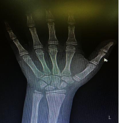
- A ~2-year delay in skeletal (bone) age relative to chronological age. Bone age reflects the cumulative growth-promoting hormonal drive (GH/IGF-1 plus thyroid hormone and sex steroids) on epiphyseal ossification centers (read via the Greulich-Pyle atlas from a hand/wrist film).
- This is a classic feature of GH deficiency and helps differentiate it from familial short stature (bone age = chronological age, normal growth velocity) and, to a lesser extent, from constitutional delay of growth and puberty (bone age delayed similarly, but growth velocity is usually near-normal and the child eventually catches up - which does not match this patient's described "growth arrest").
- Clinically it also matters for prognosis: because growth plates are still open (bone age 8, not fused), there is remaining growth potential if the underlying deficiency is treated promptly with recombinant human GH (rhGH).
Serum IGF-1 = 20 nmol/L (normal range 6.5-59.08 nmol/L)
- This is the most important nuance in the case. IGF-1 is squarely within the normal reference range, even though it sits on the lower half of a very wide range. IGF-1 is used clinically as a stable surrogate marker of average 24-hour GH secretion (long half-life due to IGF binding proteins, unlike pulsatile GH).
- In "textbook" GH deficiency, IGF-1 is expected to be low. Here it is not clearly low. Possible explanations to raise in discussion:
- IGF-1 reference ranges are wide and age/pubertal-stage dependent; a value that is "normal" for the general population may still be relatively low for this specific child.
- Partial or mild GH deficiency can have preserved (low-normal) IGF-1.
- The single stimulation test showing 4 ng/mL could be a false positive (test artifact, timing, assay variability), and the normal IGF-1 argues for confirmatory testing before finalizing the diagnosis.
- This is exactly why case-based teaching emphasizes that no single test is diagnostic in isolation - GH deficiency is a composite clinical + biochemical + radiological diagnosis.
- This should be explicitly flagged as an area of diagnostic tension in the case, not smoothed over.
Explanation paragraphs (given in the case)
- These summarize normal physiology (somatotroph origin, GHRH/somatostatin control, pulsatility, sleep-related surges, puberty-related peak, and the causes of GH deficiency: congenital hypothalamic/pituitary damage, acquired postnatal damage, or gene mutations affecting GH synthesis/secretion). These are covered comprehensively in Section 3 below.
3. Relevant Normal Physiology
Hypothalamic control:
- GHRH (growth hormone-releasing hormone), a 44-amino-acid hypothalamic peptide, is secreted in discrete pulses and stimulates GH synthesis and release from somatotrophs via a Gs-protein-coupled receptor → increased cAMP.
- Somatostatin (SRIF), from the periventricular/medial preoptic hypothalamus, inhibits GH release and sets the basal (inter-pulse) tone; GHRH pulses ride on top of this suppressed baseline, producing the characteristic pulsatile pattern.
- Ghrelin, a gastric-derived peptide, also stimulates GH release (and stimulates GHRH indirectly), linking nutritional/energy status to GH secretion.
- This dual push-pull system explains why random GH sampling is useless diagnostically and why provocative testing is required (as discussed above).
Pulsatility and sleep:
- GH surges occur mainly after the onset of slow-wave (deep) sleep, and these surges are especially prominent around puberty, coinciding with (and partly driven by) rising sex steroids that amplify GH pulse amplitude - this is part of why GH (and height velocity) peaks during the pubertal growth spurt.
Anterior pituitary somatotrophs:
- Somatotrophs make up up to ~50% of anterior pituitary cells, making GH the most abundant anterior pituitary hormone. Their differentiation and GH gene transcription depend on the Pit-1 transcription factor - mutations here (or in PROP1) cause combined pituitary hormone deficiencies, which is why anterior pituitary panels matter when GH deficiency is suspected.
- GH itself is synthesized as a larger prohormone, processed to the predominant 22-kDa, 191-amino-acid peptide.
GH receptor and signaling (JAK2-STAT5):
- The GH receptor is a single-transmembrane-domain receptor (cytokine receptor superfamily) without intrinsic enzymatic activity. GH binding causes receptor dimerization, which activates the associated cytoplasmic tyrosine kinase JAK2 (Janus kinase 2).
- JAK2 phosphorylates STAT5 (signal transducer and activator of transcription 5), which dimerizes, translocates to the nucleus, and drives transcription of GH-responsive genes - most importantly the IGF-1 gene. A MAPK/ERK pathway is also activated in parallel, contributing to some of GH's mitogenic/metabolic effects.
- This is the molecular basis of Laron dwarfism (GH receptor mutations → GH cannot signal → low IGF-1 despite normal/high GH), a useful contrast case for this patient (see differentials).
GH → IGF-1 axis:
- The liver is the major source of circulating (endocrine) IGF-1 in response to GH. IGF-1 is also produced locally in cartilage/growth plate and other tissues (paracrine/autocrine action) - the "dual effector" hypothesis: GH acts directly on the growth plate's resting-zone chondrocytes to stimulate their differentiation into proliferating chondrocytes, and it also stimulates local as well as hepatic IGF-1 production, which then drives chondrocyte proliferation and hypertrophy (via the IGF-1 receptor, a tyrosine-kinase receptor similar to the insulin receptor).
- IGF-1 circulates bound to IGF-binding proteins (especially IGFBP-3), which prolongs its half-life and makes it a stable, reliable integrated marker of average GH secretion - this is exactly why it is measured alongside (or instead of) pulsatile GH.
Negative feedback:
- IGF-1 feeds back at both the hypothalamus (stimulating somatostatin) and the pituitary (reducing somatotroph responsiveness to GHRH) to limit further GH release - a classic long-loop negative feedback circuit analogous to other hypothalamic-pituitary axes.
Direct metabolic effects of GH (independent of IGF-1):
- Protein: anabolic - increased amino acid uptake and protein synthesis in muscle and other tissues.
- Fat: lipolytic - increased free fatty acid mobilization from adipose tissue (used as fuel, sparing glucose/protein).
- Carbohydrate: "diabetogenic"/anti-insulin - GH reduces peripheral glucose uptake and increases hepatic gluconeogenesis, raising blood glucose; this is why chronic GH excess (acromegaly) can cause insulin resistance/diabetes, and why GH treatment is monitored for glycemic effects.
Growth plate and childhood vs adulthood effects:
- Longitudinal bone growth depends entirely on open epiphyseal growth plates. GH/IGF-1 drive chondrocyte proliferation and hypertrophy at the plate, followed by ossification. Once sex-steroid-driven plate fusion is complete (end of puberty), further GH excess causes acromegaly (soft tissue/bone thickening) rather than height gain, and GH deficiency in adulthood causes purely metabolic consequences (sarcopenia, central adiposity, dyslipidemia, osteoporosis, reduced quality of life) rather than height loss.
- This distinction is exactly why age and growth plate status are diagnostically decisive in this patient: at age 10, with bone age 8 (plates open), a GH deficiency is expected to manifest as height failure - which is precisely what we see - and remains treatable for height if caught now.
4. Symptom/Sign → Mechanism Analysis
| Finding | Mechanism chain |
|---|
| Growth arrest / static clothing size over 4 years | GH deficiency → reduced hepatic and local IGF-1 generation → reduced chondrocyte proliferation/hypertrophy at epiphyseal growth plates → reduced linear bone elongation → reduced height velocity → child does not outgrow clothing |
| Delayed bone age (8y vs 10y chronological) | Reduced GH/IGF-1 (and normally also thyroid hormone) drive on epiphyseal ossification centers → slower maturation of ossification centers → skeletal age lags chronological age on Greulich-Pyle wrist atlas |
| Subnormal GH stimulation test (4 ng/mL) | Deficient somatotroph secretory reserve (due to hypothalamic GHRH deficiency, pituitary somatotroph damage/hypoplasia, or a genetic defect in GH synthesis/secretion) → provocative stimulus (pharmacologic/physiologic) fails to elicit a normal peak GH surge → low measured peak |
| Normal-range (low-normal) IGF-1 | Because IGF-1 is a longer-acting integrated marker buffered by binding proteins, mild/partial reductions in GH secretion may not push IGF-1 clearly below the (wide) normal range → "discordant" or borderline picture, common in early/partial or single-test-positive GH deficiency |
| Preserved intelligence, no academic difficulty | GH/IGF-1 deficiency alone (unlike thyroid hormone deficiency) is not required for normal postnatal CNS/cognitive development → cognition preserved, arguing against hypothyroidism/cretinism or syndromes with intellectual disability |
| No headache/vomiting/visual disturbance | No mass effect on adjacent structures (ventricular system, optic chiasm) → argues against a large space-occupying sellar/suprasellar lesion (e.g., large craniopharyngioma) as the cause, though a small lesion or non-mass congenital anomaly is not excluded |
5. Differential Diagnosis
1. Growth hormone deficiency (isolated or as part of combined pituitary hormone deficiency) - the leading diagnosis
- Supports: subnormal stimulation peak (4 ng/mL), delayed bone age, chronic growth arrest with preserved cognition, no dysmorphism reported.
- Against/uncertain: IGF-1 is technically within normal range; only one stimulation test reported (guidelines usually want two).
- Expect if true: low/low-normal IGF-1 and IGFBP-3, delayed bone age, normal-to-low other pituitary axes (if isolated) or multiple low axes (if combined), possible structural pituitary/hypothalamic abnormality on MRI (or a completely normal MRI in idiopathic cases).
- Distinguishing test: second stimulation test with a different provocative agent + pituitary MRI + full anterior pituitary hormone panel.
- Likelihood: High - this is the working/most probable diagnosis given the data, though not yet fully confirmed by rigorous criteria.
2. Constitutional delay of growth and puberty
- Supports: delayed bone age is a hallmark of this condition too.
- Against: growth velocity in constitutional delay is usually near-normal (child tracks a lower but roughly parallel curve and eventually catches up at puberty), which does not match a described "growth arrest" with static clothing size for 4 years; also, GH stimulation test is typically normal in constitutional delay (delay is in puberty/bone maturation, not GH secretion).
- Distinguishing test: growth velocity charting over time, family/pubertal history (delayed puberty in parents), normal GH stimulation test.
- Likelihood: Low, given the abnormal stimulation test and described growth arrest.
3. Familial (genetic) short stature
- Supports: nothing specific in the case supports this.
- Against: bone age should equal chronological age (here it is delayed by 2 years), and GH stimulation test should be normal.
- Distinguishing test: parental height, bone age = chronological age.
- Likelihood: Low.
4. Hypothyroidism (acquired/juvenile)
- Supports: can cause delayed bone age and reduced growth velocity; can also blunt GH secretion/response secondarily.
- Against: no mention of cold intolerance, constipation, bradycardia, myxedematous features, delayed puberty, or (critically) any cognitive slowing - and juvenile-onset hypothyroidism, unlike congenital cretinism, doesn't have to impair intellect, so this can't be fully excluded on history alone.
- Distinguishing test: TSH and free T4 (not mentioned as done - should be ordered as standard short-stature work-up).
- Likelihood: Cannot be excluded without labs; moderate-low priority but must be tested.
5. Turner syndrome (45,X and variants)
- Supports: female sex, short stature, otherwise normal cognition (typical of Turner).
- Against: no dysmorphic features mentioned (webbed neck, cubitus valgus, widely spaced nipples, cardiac/renal anomalies) - though these can be subtle or absent.
- Distinguishing test: karyotype - standard recommendation in any girl with unexplained short stature, even without dysmorphism.
- Likelihood: Cannot be excluded from the data given; should be tested as a "do not miss" differential.
6. Structural hypothalamic-pituitary lesion (e.g., craniopharyngioma) causing acquired GH deficiency
- Supports: would explain both subnormal GH stimulation and delayed bone age.
- Against: absence of headache, vomiting, and visual disturbance makes a large mass less likely, though small lesions or congenital structural anomalies (pituitary hypoplasia, ectopic posterior pituitary, thin/absent infundibulum) can be silent clinically.
- Distinguishing test: MRI brain/pituitary with attention to the hypothalamic-pituitary region.
- Likelihood: Cannot be excluded without imaging; moderate priority.
7. Chronic systemic illness, malnutrition, or psychosocial (deprivation) short stature
- Supports: nothing specific.
- Against: no history suggestive of poor intake, GI symptoms, chronic disease, or neglect.
- Distinguishing test: basic screen (CBC, ESR/CRP, renal function, celiac serology) as part of standard short-stature work-up.
- Likelihood: Low based on history given, but standard screening still indicated.
8. Skeletal dysplasia (e.g., achondroplasia) or Laron-type GH insensitivity
- Against achondroplasia: would show disproportionate short stature (short limbs, normal trunk) and characteristic radiographic metaphyseal/epiphyseal changes, not a "delayed bone age" pattern; not suggested by case.
- Against Laron dwarfism: Laron syndrome shows elevated GH with low IGF-1 (receptor insensitivity); this case shows low stimulated GH with normal-range IGF-1 - the opposite pattern.
- Likelihood: Very low for both.
6. Differential Comparison
| Finding | GH deficiency | Constitutional delay | Familial short stature | Hypothyroidism | Turner syndrome |
|---|
| Growth arrest / static clothing (4y) | Strongly supportive | Against (velocity usually near-normal) | Against | Mildly supportive | Mildly supportive |
| Delayed bone age (8y vs 10y) | Strongly supportive | Strongly supportive | Against (should be equal) | Strongly supportive | Neutral/variable |
| Subnormal GH stimulation test (4 ng/mL) | Strongly supportive | Against (usually normal) | Against | Neutral (untested here) | Neutral (untested here) |
| Normal-range IGF-1 (20 nmol/L) | Mildly against / uncertain | Neutral | Neutral | Neutral | Neutral |
| Normal intelligence | Strongly supportive | Neutral | Neutral | Against overt cretinism, doesn't exclude juvenile-onset | Neutral |
| No headache/vomiting/visual loss | Mildly supportive (against large mass) | Neutral | Neutral | Neutral | Neutral |
| Female sex | Neutral | Neutral | Neutral | Neutral | Strongly relevant (must screen) |
The pattern of chronic growth arrest + delayed bone age + a subnormal (though single) GH stimulation peak, in a cognitively normal child without mass-effect symptoms, converges most strongly on GH deficiency, while the normal-range IGF-1 is the one piece of evidence that keeps this from being an open-and-shut, textbook-perfect case - which is exactly the kind of realistic ambiguity a CBL discussion should highlight.
7. Required Investigations
- GH stimulation (provocative) test (already done): measures the somatotrophs' maximal secretory capacity using a pharmacologic/physiologic stimulus, because random GH sampling is meaningless given pulsatile secretion. Result 4 ng/mL = subnormal, supporting GH deficiency. A normal result would essentially exclude GH deficiency (except in rare neurosecretory dysfunction). Best practice: repeat with a second, different provocative agent before finalizing diagnosis.
- Serum IGF-1 (already done): integrated, stable surrogate of average GH secretion over the preceding days. Expected low in GH deficiency; here it is within (low-normal) reference range - if normal, it should prompt confirmatory retesting rather than immediately confirming or excluding the diagnosis, especially when combined with a single positive stimulation test.
- Serum IGFBP-3: less nutrition-dependent than IGF-1, sometimes used as an adjunct, though evidence suggests it is not clearly superior to IGF-1 alone as a screening test.
- X-ray both wrists for bone age (already done): assesses skeletal maturation against chronological age using the Greulich-Pyle atlas. A delay (as found, 8y vs 10y) supports an organic cause of growth failure (GH deficiency or hypothyroidism) over familial short stature (bone age = chronological age).
Representative PA hand-wrist radiograph used for bone age assessment - delayed ossification relative to chronological age is classic for growth hormone deficiency or hypothyroidism.
- MRI of the hypothalamic-pituitary region: essential once biochemical GH deficiency is suspected, to look for a structural cause (craniopharyngioma, pituitary hypoplasia, ectopic posterior pituitary, empty sella, infiltrative disease). Absence of headache/vomiting/visual symptoms lowers but does not eliminate the pretest probability of a lesion - a normal MRI supports idiopathic/isolated GH deficiency; an abnormal MRI identifies a treatable structural cause and often predicts combined pituitary hormone deficiency.
- Full anterior pituitary hormone panel (TSH, free T4, ACTH/cortisol or morning cortisol, LH/FSH as age-appropriate, prolactin): needed to exclude combined pituitary hormone deficiency (CPHD), since isolated GH deficiency management/prognosis differs from panhypopituitarism, and untreated central hypothyroidism itself would also impair growth and confound interpretation.
- Visual field examination/formal ophthalmology assessment: low yield here given no visual complaints, but standard if any structural lesion is found on MRI or if suspicion for a suprasellar mass remains.
- Karyotype: recommended in any girl with unexplained short stature to exclude Turner syndrome, even without overt dysmorphic features.
- Baseline metabolic/chronic disease screen (CBC, ESR/CRP, renal and liver function, celiac serology): to exclude chronic systemic disease or malnutrition as an alternative cause of growth failure and delayed bone age.
8. Interpretation of Expected Results
- GH stimulation test 4 ng/mL → below the subnormal cutoff of 5 ng/mL → consistent with impaired somatotroph reserve. Physiologically, this means the hypothalamic-pituitary axis cannot mount a normal GH surge even under maximal pharmacologic/physiologic stimulation, implicating a defect anywhere from GHRH production/action to somatotroph synthesis/release of GH.
- Bone age 8 years vs chronological 10 years → 2-year delay → consistent with chronically reduced growth-promoting hormonal drive on the growth plates; a normal bone age would argue strongly against GH deficiency and favor familial short stature.
- IGF-1 20 nmol/L (within 6.5-59.08) → does not clearly support GH deficiency on its own; if IGF-1 had been low, it would reinforce the diagnosis very strongly (per the Tietz textbook of laboratory medicine, "if IGF-1 is squarely within its reference interval for age and sex, GH deficiency is excluded" in general screening use - so a normal IGF-1 here is a genuine reason to insist on a confirmatory second stimulation test and imaging rather than close the case).
- If MRI is normal: supports idiopathic/isolated GH deficiency (possibly genetic, e.g., GHRH receptor or GH1 gene mutation) as the working diagnosis.
- If MRI shows a mass or structural anomaly: confirms an acquired or congenital structural cause and often predicts additional pituitary hormone deficits.
- If TSH/free T4, cortisol, or gonadotropins are abnormal: reclassifies this as combined pituitary hormone deficiency, changing management (multi-hormone replacement) and often making a structural/genetic pituitary cause more likely.
- If karyotype is abnormal: reclassifies the case entirely as Turner syndrome-associated short stature rather than isolated GH deficiency (though some Turner patients also have blunted GH response and benefit from GH therapy).
9. Final Diagnosis
Based on the data provided, the most probable diagnosis is growth hormone deficiency (pituitary dwarfism), presenting as pathological short stature with growth arrest in a prepubertal girl.
- Why: chronic (4-year) growth arrest with a functionally significant reduction in growth velocity, a subnormal peak GH on stimulation testing (4 ng/mL, below the 5 ng/mL subnormal cutoff), and a delayed bone age (8y vs 10y chronological) together form the classic clinical-biochemical-radiological triad of GH deficiency.
- Age/growth plate relevance: at 10 years chronological / 8 years bone age, her epiphyseal growth plates are still open, meaning height deficit is currently reversible with timely rhGH replacement - this is why early diagnosis matters enormously; once plates fuse (post-puberty), the window for height gain closes permanently, even though metabolic consequences of GH deficiency would persist into adult life (reduced lean mass, increased adiposity, dyslipidemia, osteoporosis risk).
- Caveat (must be stated honestly): the normal-range serum IGF-1 (20 nmol/L) is not fully typical of classic GH deficiency and represents genuine diagnostic uncertainty. Per standard practice, this diagnosis should be confirmed with a second stimulation test using a different provocative agent, and MRI plus a full pituitary hormone panel, before treatment is finalized. As presented, the case supports GH deficiency as the leading/working diagnosis, but is not proven to the level of rigorous diagnostic certainty from the data given alone.
- Distinguishing from related entities:
- Gigantism/acromegaly are conditions of GH excess, not relevant here except as the physiological "mirror image" for teaching contrast (gigantism = excess before plate fusion → tall stature; acromegaly = excess after fusion → bony/soft tissue thickening without height gain).
- Pituitary adenoma here would refer to a GH-secreting tumor causing excess, which is not this case; if any pituitary abnormality exists here, it would be a cause of deficiency (hypoplastic pituitary, non-secreting mass compressing normal tissue, or damage from another process), not a secretory adenoma.
10. Underlying Cause
The case explanation itself outlines the general causes of GH deficiency, and from the data provided we cannot definitively pinpoint which applies to this specific child, but the plausible categories are:
- Congenital GH deficiency: damage to the hypothalamus or pituitary during fetal development (structural malformations such as pituitary hypoplasia, ectopic posterior pituitary, septo-optic dysplasia) or a genetic mutation affecting GH synthesis/secretion (e.g., GH1 gene mutations, GHRHR mutations impairing the GHRH receptor, or transcription factor defects like PROP1/POU1F1 causing combined deficiencies). Congenital forms would typically declare themselves once GH-dependent growth begins.
- Acquired GH deficiency: postnatal damage to the hypothalamic-pituitary axis - e.g., a slow-growing tumor (craniopharyngioma being the classic pediatric example, though the absence of headache/vomiting/visual loss makes a large one less likely), cranial irradiation, head trauma, infection (meningitis/encephalitis), or infiltrative disease.
- Idiopathic isolated GH deficiency: no identifiable structural or genetic cause found even after full work-up - a common final "diagnosis by exclusion" in pediatric endocrinology when imaging and genetics are unrevealing.
Without birth history, family history, and (crucially) an MRI result, the specific underlying cause cannot be determined from this case alone - this is an important limitation to state explicitly rather than guess.
11. Complete Pathophysiological Chain
Underlying cause (congenital hypothalamic/pituitary defect, acquired postnatal insult, or gene mutation affecting GH synthesis/secretion - not yet determined from data given)
→ Hormonal abnormality: deficient GHRH-driven pulsatile GH release from anterior pituitary somatotrophs
→ Molecular/physiological mechanism: inadequate GH binding to peripheral GH receptors → reduced JAK2-STAT5 signaling → reduced hepatic and local (growth plate) IGF-1 gene transcription
→ IGF-1/GH effects: reduced IGF-1-mediated stimulation of chondrocyte proliferation and hypertrophy at the epiphyseal growth plate; reduced GH-driven protein anabolism, lipolysis, and gluconeogenic/anti-insulin metabolic effects
→ Tissue/organ effects: slowed linear bone growth (skeletal maturation lags), reduced growth velocity
→ Symptoms/signs: chronic growth arrest evidenced by unchanged clothing size over 4 years; delayed bone age on wrist X-ray (8y vs 10y); preserved intelligence (since cognition is not GH/IGF-1-dependent postnatally); absence of mass-effect symptoms (headache/vomiting/visual disturbance) if the cause is non-structural or a small lesion
→ Investigations: subnormal GH stimulation test (4 ng/mL); IGF-1 within normal range (a point of diagnostic tension); delayed bone age confirmed radiologically
→ Diagnosis: growth hormone deficiency (pituitary dwarfism), pending confirmatory second stimulation test, MRI, and pituitary hormone panel to define the exact cause and rule out combined deficiencies or structural lesions.
12. CBL Learning Objectives
- Structure, synthesis site, and secretion of GH (somatotrophs, Pit-1-dependent, 22-kDa/191-aa peptide).
- Hypothalamic regulation of GH: GHRH (stimulatory) vs somatostatin (inhibitory), ghrelin, pulsatility, sleep-related surges, pubertal amplification.
- GH receptor signaling: JAK2-STAT5 pathway and its role in IGF-1 gene transcription.
- The GH → IGF-1 axis: hepatic (endocrine) vs local (paracrine) IGF-1 production; the "dual effector" hypothesis at the growth plate.
- Direct (IGF-1-independent) metabolic effects of GH on protein, fat, and carbohydrate metabolism.
- Negative feedback regulation of the GH axis by IGF-1.
- Differences between childhood and adult consequences of GH deficiency/excess, tied to growth plate status.
- Clinical use and limitations of random GH, stimulation tests, IGF-1, IGFBP-3, and bone age in diagnosing pediatric GH deficiency.
- Differential diagnosis of short stature in children, and how bone age and growth velocity help distinguish the causes.
13. Likely Teacher/Class Questions
- Why can't we just measure a random serum GH level to diagnose GH deficiency?
- Why is IGF-1 considered a more reliable single measurement than GH itself?
- What is the physiological reason GH secretion peaks during puberty?
- Why does bone age lag in GH deficiency, and how is it measured?
- If this were an adult instead of a child, would GH deficiency present differently? How?
- What additional tests would you want before committing to a permanent diagnosis of GH deficiency in this specific case, given the normal IGF-1?
- How would the work-up change if this child also had polyuria/polydipsia, or diabetes insipidus symptoms?
- What is the difference between gigantism and acromegaly, and why does growth plate status determine which one occurs?
14. Viva Questions + Answers
Q: What secretes GH, and what percentage of anterior pituitary cells are these cells?
A: Somatotrophs, comprising up to ~50% of the anterior pituitary cell population.
Q: Name the two main hypothalamic regulators of GH and their effects.
A: GHRH (stimulates) and somatostatin/SRIF (inhibits).
Q: What is the intracellular signaling pathway activated by the GH receptor?
A: JAK2-STAT5 pathway (receptor dimerization → JAK2 activation → STAT5 phosphorylation/dimerization → nuclear translocation → IGF-1 gene transcription), with parallel MAPK/ERK signaling.
Q: Where is most circulating IGF-1 produced?
A: The liver (endocrine IGF-1); local paracrine IGF-1 is also produced in cartilage/growth plate and other tissues.
Q: Why is a single random GH level not useful diagnostically?
A: Because GH secretion is pulsatile, with very low/undetectable levels between pulses even in healthy individuals.
Q: What test would you use if you suspected GH excess instead of deficiency?
A: An oral glucose tolerance test (OGTT) with GH suppression - normal individuals suppress GH after glucose; autonomous somatotroph adenomas do not.
Q: Why does GH deficiency in adults not cause short stature?
A: Because epiphyseal growth plates have already fused after puberty; GH deficiency in adults causes metabolic effects (sarcopenia, central adiposity, dyslipidemia, osteoporosis) rather than height loss.
Q: What is Laron dwarfism, and how does it differ biochemically from classic GH deficiency?
A: A GH receptor insensitivity syndrome due to loss-of-function receptor mutations - GH levels are normal/elevated, but IGF-1 is low because the receptor cannot signal, unlike classic GH deficiency where GH itself is low.
Q: Why is bone age assessed via a hand/wrist X-ray?
A: It captures multiple ossification centers with well-established maturation atlases (Greulich-Pyle), allowing comparison of skeletal maturity to chronological age.
15. High-Yield Takeaways
- GH deficiency is diagnosed with a combination of clinical growth pattern, bone age, IGF-1, and confirmed stimulation testing - never on one test alone.
- Common misconception #1: "Low GH on one stimulation test = confirmed GH deficiency." In reality, guidelines typically require two abnormal provocative tests with different agents before confirming the diagnosis, because of a meaningful false-positive rate.
- Common misconception #2: "IGF-1 must always be low in GH deficiency." As this case shows, IGF-1 can sit within a wide normal reference range in partial/early/single-test-positive deficiency - a normal IGF-1 does not fully exclude GH deficiency, especially when other clinical/biochemical features fit.
- Common misconception #3: Confusing "dwarfism" (a broad descriptive term) with a specific biochemical diagnosis - always specify the underlying cause (GH deficiency, achondroplasia, hypothyroidism, Turner syndrome, etc.).
- Common misconception #4: Believing acromegaly and gigantism are the "same disease at different ages" without appreciating that the only reason they look different is epiphyseal growth plate status at the time GH excess begins.
- Common misconception #5: Assuming GH deficiency necessarily impairs intelligence - it typically does not (unlike hypothyroidism/cretinism), which is why the normal cognition in this case is a genuinely useful discriminating clue.
- Clinical pearl: Absence of headache/vomiting/visual disturbance lowers but never fully excludes an intracranial structural cause - MRI remains indicated once biochemical GH deficiency is suspected.
A brief literature check found no recent evidence contradicting this reasoning; recent systematic reviews (e.g., Mameli et al., Endocrine 2024, PMID 38498128, on GH deficiency epidemiology) and network meta-analyses on newer long-acting GH formulations (PMIDs 38580693, 39261416) relate mainly to treatment advances (weekly rhGH analogs) rather than changing diagnostic criteria, so the diagnostic approach outlined above remains current.