
from Guyton and Hall textbook of medical physiology, 4th South Asia edition, write a 10-mark long answer on the Gastric Juice-composition; HCl Secretion-mechanism, factors regulating
HCl secretion mechanism parietal cell hydrogen potassium ATPase proton pump
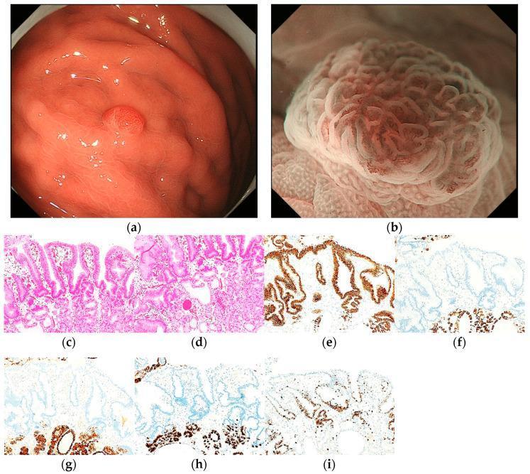
This composite figure illustrates the endoscopic and histopathological features of a proton pump inhibitor-related lesion (PPI-L) with low-grade dysplasia. (a) White-light imaging (WLI) reveals a 6 mm, homogenous reddish, protruded lesion located on the greater curvature of the upper gastric third. (b) Magnifying endoscopy with narrow-band imaging (ME-NBI) demonstrates a regular microvascular (MV) pattern and a regular microsurface (MS) pattern, bordered by a distinct demarcation line. (c, d) H&E stained sections at 100x magnification show parietal cell hyperplasia, foveolar epithelial dilation and elongation, and stromal vascularity. (e–i) Immunohistochemical analysis (100x) displays strong MUC5AC positivity in foveolar epithelial cells, while MUC6 (f), pepsinogen-I (g), and H+/K+-ATPase (h) are negative. (i) Ki-67 immunostaining reveals a high labeling index (80%), indicating significant cellular proliferation. The image serves as an educational reference for differentiating benign-appearing gastric protruded lesions from early gastric cancer using the MESDA-G diagnostic algorithm.
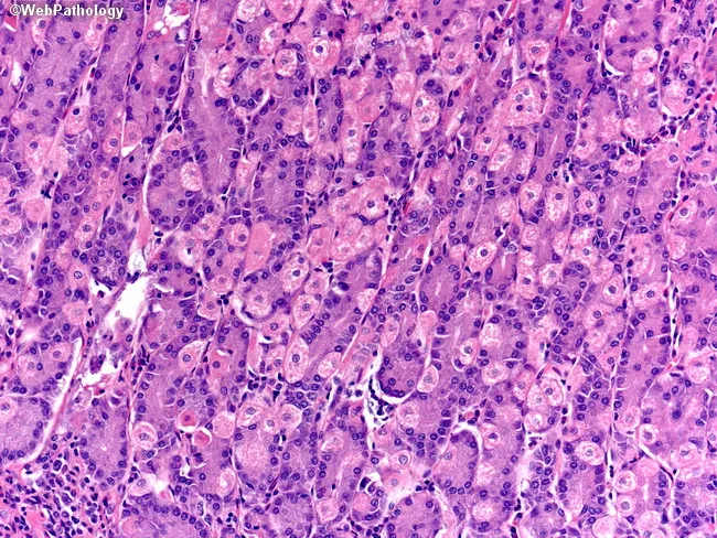
Gastric mucosal histology viewed by bright-field light microscopy on a Hematoxylin and Eosin (H&E) stained section of mucosa from the stomach, typically the oxyntic/fundic region. The glands are tubular and packed with two principal cell types: parietal (oxyntic) cells and chief (peptic) cells. Parietal cells display abundant eosinophilic (pink) cytoplasm and a central or slightly eccentric nucleus, giving a characteristic fried-egg appearance; they contribute acid secretion via gastric H+/K+-ATPase. Chief cells have basophilic (purple) cytoplasm with basal nuclei and apical zymogen granules, reflecting pepsinogen production. Neuroendocrine cells are present in minute numbers and are usually inconspicuous on routine H&E sections. Stem cells are scarce and not readily visible without special markers. The overall architecture shows intact gastric fundic glands with uniform cell density, minimal cytologic atypia, and preserved mucosal layering. The image emphasizes contrasts between cytoplasmic staining: eosinophilic parietal cells versus basophilic chief cells, as well as the densely staining nuclei. This morphology is essential for recognizing normal gastric mucosa, distinguishing parietal cell-rich areas, and identifying early metaplastic changes or inflammatory patterns in gastritis. Clinically, such images support reports of gastric biopsy evaluation, autoimmune gastritis assessment, and correlating acid-secreting cell distribution with disorders of digestion and nutrition and metabolic balance.
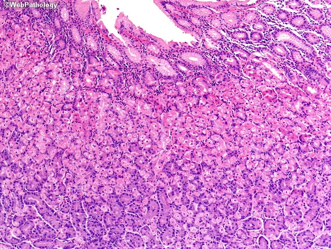
This is a bright-field light microscopy image of fixed gastric mucosa section stained with Hematoxylin and Eosin (H&E). The tissue shows gastric pits with an isthmus and neck populated by conspicuous parietal (oxyntic) cells with abundant eosinophilic cytoplasm and central nuclei. Parietal cells secrete hydrochloric acid and intrinsic factor, essential for digestion and B12 absorption. Deeper in the gland, chief (peptic) cells possess basophilic cytoplasm and secrete pepsinogen. Interspersed neuroendocrine cells release hormones; a stem cell zone resides at the neck, supporting constant epithelial renewal. The overall architecture corresponds to fundic-type oxyntic mucosa with tubular glands arranged in distinct isthmus/neck and base compartments. No overt inflammatory infiltrate or architectural distortion is evident, consistent with normal histology. This image is valuable for teaching gastric histology, differentiating mucous neck cells, parietal cells, chief cells, enteroendocrine cells, and stem cell niches, and for contextualizing pathologies affecting acid secretion, intrinsic factor production, or gastric carcinogenesis in educational and research settings.
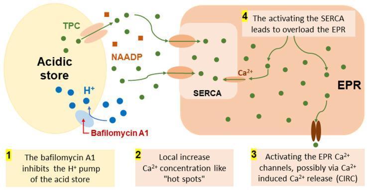
This pathophysiology diagram illustrates the hypothetical molecular mechanism of Bafilomycin A1's effect on cellular calcium (Ca2+) signaling and the cross-talk between acidic stores (lysosomes/endosomes) and the endoplasmic reticulum (EPR). The diagram follows a numbered four-step sequence: (1) Bafilomycin A1 inhibits the H+ pump on the acidic store membrane, disrupting the proton gradient. (2) This leads to Ca2+ release from the acidic store via TPC (Two-Pore Channels), which are also modulated by NAADP (nicotinic acid adenine dinucleotide phosphate), creating localized 'hot spots' of high Ca2+ concentration. (3) These hot spots activate SERCA (Sarco/Endoplasmic Reticulum Ca2+-ATPase) pumps, which transport Ca2+ into the EPR. (4) The resulting EPR overload triggers Ca2+ efflux through EPR Ca2+ channels, likely through calcium-induced calcium release (CICR). The visual uses color-coded symbols: green dots for Ca2+, blue dots for H+, and brown squares for NAADP molecules. This model explains how endo-lysosomal inhibitors can modulate global cellular calcium homeostasis and signaling in human tissues, such as the colon mucosa.
Gastric Juice - Composition; HCl Secretion - Mechanism, Factors Regulating
I. GASTRIC JUICE - COMPOSITION
A. Glands and their Secretory Cells
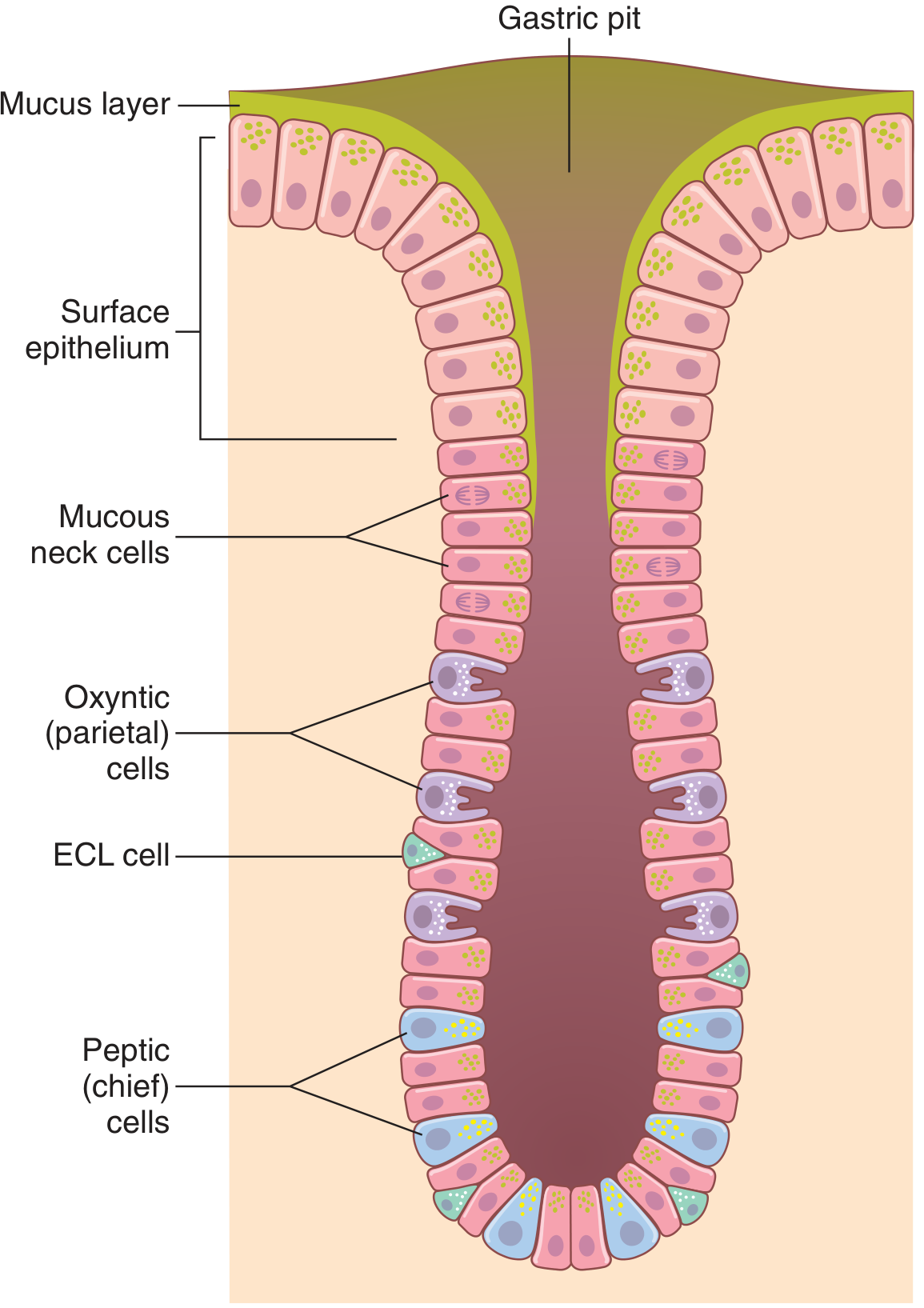
| Cell Type | Product Secreted |
|---|---|
| Mucous neck cells | Mucus |
| Peptic (chief) cells | Pepsinogen |
| Parietal (oxyntic) cells | Hydrochloric acid + Intrinsic factor |
| ECL cells (enterochromaffin-like) | Histamine |
B. Components of Gastric Juice
- Hydrochloric Acid (HCl) - Secreted by parietal cells; ~160 mmol/L; pH ~0.8
- Pepsinogen - Inactive precursor secreted by chief cells; activated to pepsin by HCl (pH 2-3)
- Intrinsic Factor - Secreted by parietal cells; essential for vitamin B12 absorption in the ileum
- Mucus - Secreted by mucous neck cells and surface epithelial cells; forms a protective layer that prevents self-digestion; alkaline in nature
- Gastrin - Hormone from G cells of pyloric glands; stimulates acid secretion
- Water - Forms the bulk of gastric juice
II. MECHANISM OF HCl SECRETION
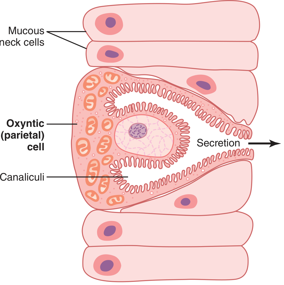
A. Structure of the Parietal Cell
B. Step-by-Step Mechanism (Fig. 65.6)
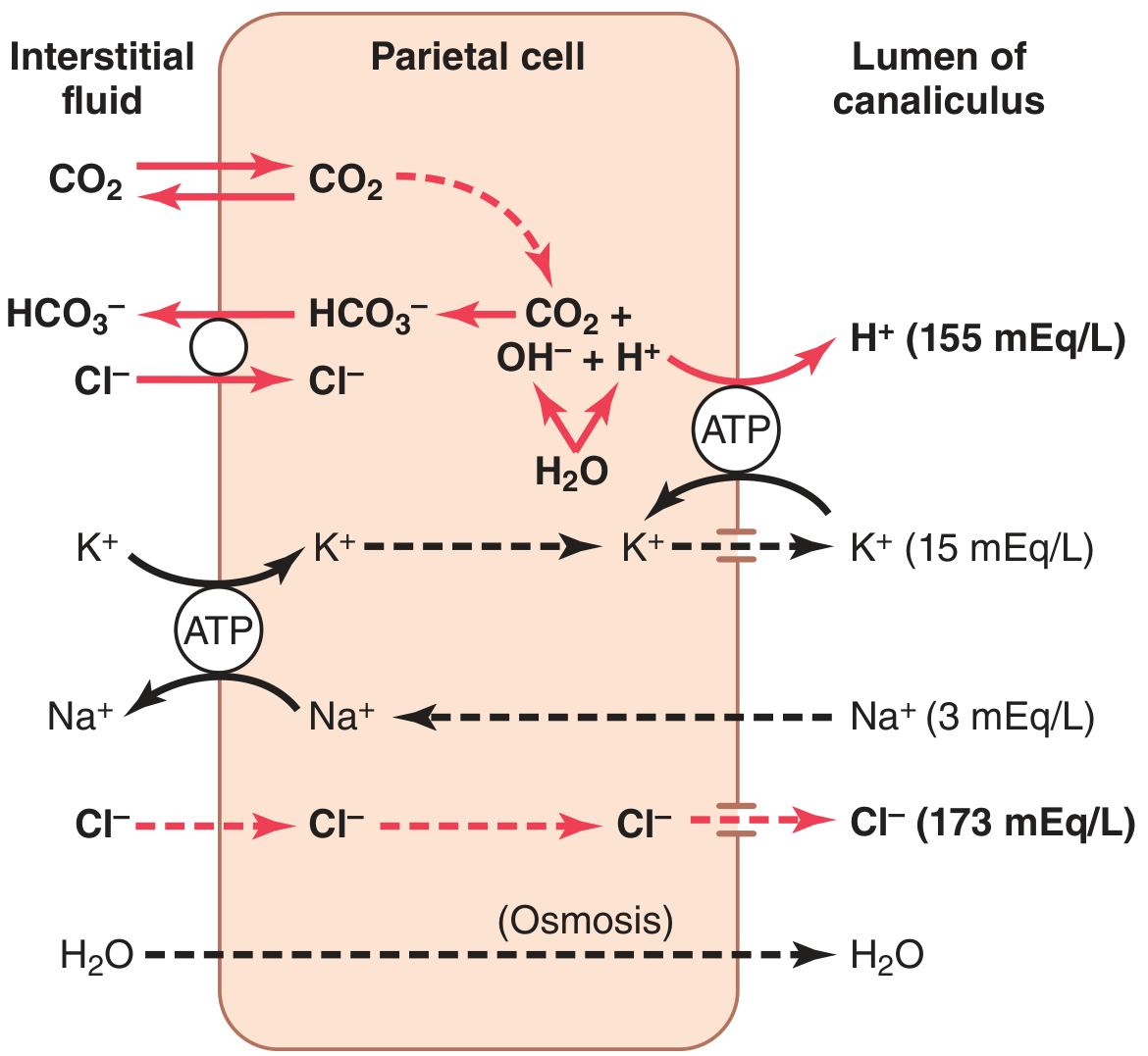
- Water inside the parietal cell dissociates into H⁺ and OH⁻ in the cytoplasm.
- H⁺ is actively pumped into the canalicular lumen in exchange for K⁺ by the H⁺-K⁺ ATPase (proton pump).
- K⁺ transported into the cell by the basolateral Na⁺-K⁺ ATPase tends to leak into the lumen but is recycled back via H⁺-K⁺ ATPase - this recycling keeps the H⁺-K⁺ pump running.
- The basolateral Na⁺-K⁺ ATPase creates low intracellular Na⁺, contributing to Na⁺ reabsorption from the canaliculus. Most K⁺ and Na⁺ is reabsorbed, and H⁺ takes their place.
- Pumping H⁺ out allows OH⁻ to accumulate in the cell.
- OH⁻ combines with CO₂ (from metabolism or blood) to form HCO₃⁻, catalyzed by carbonic anhydrase.
- HCO₃⁻ is transported across the basolateral membrane in exchange for Cl⁻ (chloride shift).
- Cl⁻ enters the cell and is secreted through chloride channels into the canaliculus.
- H⁺ and Cl⁻ combine in the canaliculus to form HCl at ~150-160 mEq/L.
- (Note: HCO₃⁻ diffusing into the blood causes gastric venous blood pH to be higher than arterial blood during active acid secretion - the "alkaline tide.")
- Water passes into the canaliculus by osmosis due to the high ion concentration.
- Final secretion: HCl ~150-160 mEq/L, KCl ~15 mEq/L, small amount of NaCl.
C. Gastric Mucosal Barrier
- Alkaline mucus layer covering the epithelium
- Tight junctions between epithelial cells
III. FACTORS REGULATING GASTRIC ACID SECRETION
A. Stimulatory Factors
- Acetylcholine (ACh): Released by parasympathetic (vagal) stimulation. Stimulates pepsinogen secretion by peptic cells, HCl by parietal cells, and mucus by mucous cells. Acts directly on parietal cells via muscarinic (M3) receptors.
- Gastrin: Hormone from G cells of pyloric glands. Strongly stimulates acid secretion by parietal cells. It primarily works by stimulating ECL cells to release histamine, which is the proximal stimulant of parietal cells.
- Histamine: Secreted by ECL cells. Directly stimulates parietal cells via H₂ receptors and is the most potent immediate stimulant of acid secretion. ECL cells are stimulated by gastrin and by the enteric nervous system.
B. Phases of Gastric Secretion
- Occurs before food enters the stomach
- Triggered by: sight, smell, thought, or taste of food
- Mediated by: vagal (parasympathetic) stimulation via the dorsal motor nucleus of the vagus
- Results in: ACh release → stimulates peptic and parietal cells and also causes G cells to secrete gastrin
- Occurs when food enters the stomach
- Three mechanisms:
- Vago-vagal reflexes - distension of the stomach stimulates stretch receptors → long vago-vagal reflexes to the brainstem → vagal efferents back to stomach
- Local enteric reflexes - distension activates local nerve plexuses → increased secretion
- Gastrin release - proteins and amino acids in food directly stimulate G cells in antrum to secrete gastrin → gastrin in blood → ECL cells → histamine → parietal cells → HCl
- Distension of the antrum is the most powerful stimulus during this phase.
- Occurs when chyme enters the duodenum
- Small intestinal mucosa contains G cells that secrete a small amount of gastrin in response to proteins entering the duodenum
- Maintains modest gastric acid secretion as food passes into the intestine
C. Inhibitory Factors
- When pH in the antrum falls to ~3.0, gastrin secretion is partially inhibited.
- When pH falls to ~1.0-1.5, gastrin secretion is almost completely blocked.
- This prevents dangerous over-acidification.
- Enterogastric nervous reflexes via the sympathetic chain - distension of duodenum, irritation of duodenal mucosa, or acidity of chyme all inhibit gastric secretion
- Hormonal inhibitors (enterogastrones):
- Secretin - released by duodenal mucosa in response to acid; inhibits gastric secretion and stimulates pancreatic bicarbonate
- Cholecystokinin (CCK) - released by jejunal mucosa; inhibits gastric secretion
- Gastric inhibitory peptide (GIP) / Glucose-dependent insulinotropic peptide - released when fat, protein, and carbohydrate are in the upper small intestine; moderately inhibits gastric acid secretion
- Vasoactive intestinal peptide (VIP) - inhibits gastric acid secretion
- These intestinal inhibitory mechanisms collectively prevent excessive acid from overwhelming the neutralizing capacity of the duodenum and damaging the small intestinal mucosa.
Summary Table: Factors Regulating Gastric Acid Secretion
| Factor | Type | Effect | Mechanism |
|---|---|---|---|
| Acetylcholine | Neural | Stimulation | Direct on parietal cells (M3 receptor) |
| Gastrin | Hormonal | Stimulation | Via ECL cells → histamine |
| Histamine | Paracrine | Stimulation | Direct on parietal cells (H2 receptor) |
| Low antral pH | Chemical | Inhibition | Suppresses gastrin release |
| Secretin | Hormonal | Inhibition | Released by duodenum in response to acid |
| CCK | Hormonal | Inhibition | Released by jejunum in response to fat/protein |
| GIP | Hormonal | Inhibition | Released by fat/CHO/protein in duodenum |
| Enterogastric reflex | Neural | Inhibition | Sympathetic; duodenal distension/acidity |
| Somatostatin | Paracrine | Inhibition | Inhibits parietal cells and G cells |