
Clinical features and management of orbital cellulitis
"orbital cellulitis" management
orbital cellulitis periorbital proptosis clinical signs
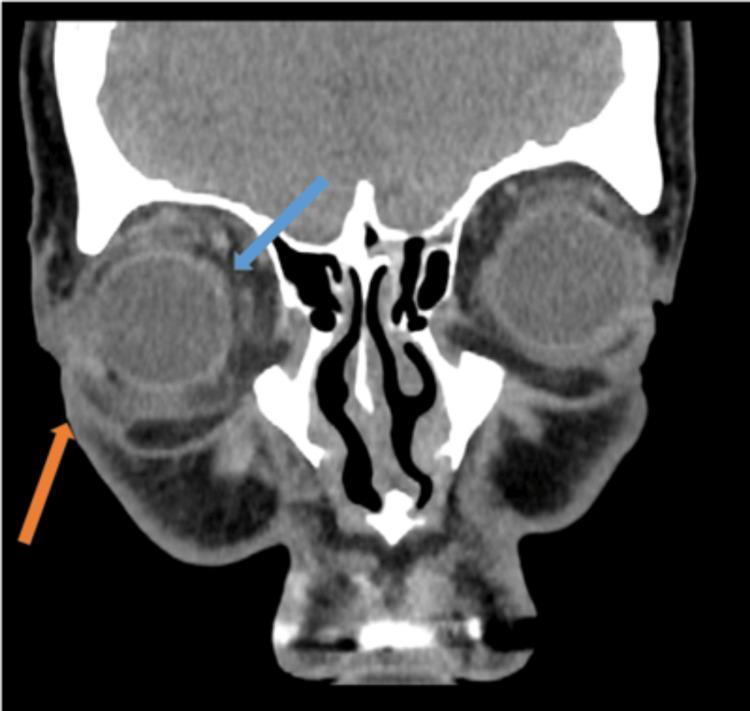
This coronal contrast-enhanced CT scan of the orbits demonstrates clinical features of orbital and periorbital cellulitis. The primary pathological findings include significant stranding of the post-septal orbital fat (indicated by the blue arrow), which appears as increased reticular density within the normally hypodense fat compartment. Additionally, there is marked thickening and increased volume of the periorbital soft tissues (indicated by the orange arrow), consistent with periorbital oedema. The globe is visible, showing signs of mild proptosis relative to the orbital rim. The extraocular muscles and the optic nerve complex are visualized within the inflammatory changes. Bony structures, including the ethmoid air cells and orbital walls, are also depicted. This imaging is characteristic of acute inflammatory or infectious processes within the orbital cavity, illustrating the key diagnostic features necessary to differentiate between pre-septal and orbital cellulitis in a clinical radiology context.
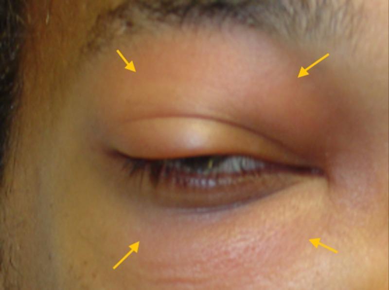
A clinical photograph of a patient's right eye and periorbital region illustrating acute inflammatory changes. The image demonstrates significant periorbital edema, characterized by prominent swelling of both the upper and lower eyelids, which narrows the palpebral fissure. Diffuse erythema is visible across the eyelid skin, extending towards the malar and frontal regions. There is a perceptible forward protrusion of the globe, consistent with proptosis. Yellow arrows are used to annotate the areas of maximal soft tissue swelling and inflammation. These clinical signs are hallmark indicators of orbital or preseptal cellulitis, frequently associated with underlying sinusitis or odontogenic infection. The image is intended for medical education to aid in the visual recognition of ocular emergencies and the differentiation between preseptal and orbital involvement based on the presence of proptosis.
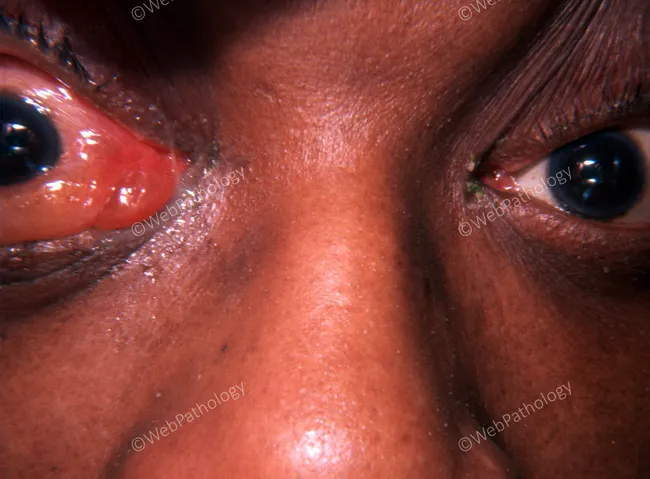
This is a clinical external photograph of the bilateral periorbital region demonstrating signs consistent with orbital cellulitis secondary to ethmoid sinusitis. Modality and technique: Clinical photography (external ocular/adnexal image), frontal view with close-up focus on the orbits, standard color photography under even lighting, with the patient seated. Anatomical context: Orbits and periorbital soft tissues, including eyelids and conjunctiva; medial orbital walls adjacent to the ethmoid sinuses. Visual features: pronounced bilateral periorbital edema and erythema of the eyelids, conjunctival injection, and mild chemosis; proptosis is not clearly evident in this view, though subtle posterior displacement may be present. Pathological interpretation: findings are compatible with orbital cellulitis due to contiguous spread from ethmoid sinus infection; inflammatory edema in the eyelids and orbital soft tissues, with mucosal involvement in the adjacent sinuses. Diagnostic relevance: this pattern signals urgent infectious process requiring prompt systemic antibiotics and imaging to delineate extent and exclude abscess; risk of vision loss and intracranial spread if untreated. Clinical correlation and use cases: useful for ophthalmology and radiology education, differential with preseptal cellulitis, dacryoadenitis, and orbital abscess, and for case-based teaching on sinus-origin orbital infections and emergency management. Early recognition facilitates timely multidisciplinary care and improves visual prognosis.
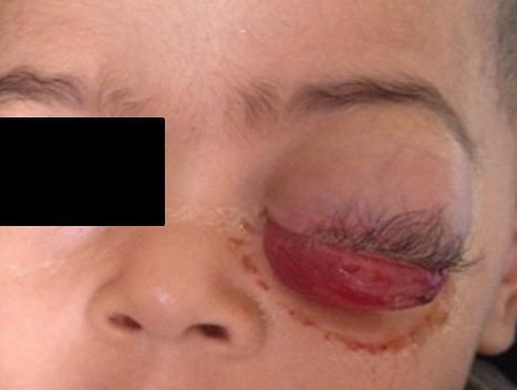
This clinical photograph displays a pediatric patient with severe unilateral ocular pathology of the right eye, characteristic of an orbital rhabdomyosarcoma. The image shows significant axial proptosis (exophthalmos), where the eyeball is markedly displaced forward from the orbit. Associated findings include severe mechanical ptosis due to the bulk of the tumor and inflammatory response. A prominent feature is the severe chemosis, with the bulbar conjunctiva appearing prolapsed, edematous, and erythematous, extending beyond the eyelid margins. The periorbital tissues show secondary signs of inflammation and irritation, with visible skin discoloration following the inferior orbital rim. The rapid progression indicated by these signs is a hallmark of pediatric orbital sarcomas. This visual serves as a critical educational example for distinguishing malignant orbital tumors from periorbital cellulitis or other inflammatory conditions in ophthalmology and pediatric oncology.
Orbital Cellulitis
Chandler Classification (Orbital Complications of Sinusitis)
| Stage | Name | Description |
|---|---|---|
| I | Inflammatory oedema (Preseptal cellulitis) | Infection anterior to orbital septum; no true orbital involvement |
| II | Orbital cellulitis | Diffuse infection of postseptal orbital fat; no abscess |
| III | Subperiosteal abscess | Extraconal pus collection between periorbita and sinus wall |
| IV | Orbital abscess | Intraconal pus collection within the muscle cone |
| V | Cavernous sinus thrombosis | Septic thrombosis of the cavernous sinus |
- Scott-Brown's Otorhinolaryngology Vol 2, p. 311
Aetiology and Pathogenesis
- Preseptal (periorbital) cellulitis spreading posteriorly
- Dacryocystitis, dacryoadenitis, infected hordeolum
- Facial cellulitis via valveless midface venous anastomoses
- Orbital/periorbital trauma (including retained foreign body - can present months later)
- Dental abscess (aggressive mixed aerobes/anaerobes)
- Post-operative (ocular, paranasal sinus, or orbital surgery)
- Haematogenous spread from distant bacteraemia
Causative Organisms
| Population | Organisms |
|---|---|
| All ages (most common) | S. pneumoniae, S. aureus (including MRSA), S. pyogenes, H. influenzae |
| Children (vaccinated era) | S. pneumoniae, M. catarrhalis, non-typeable H. influenzae; anaerobes (Bacteroides, Fusobacterium) |
| Adults | Staphylococcus spp., Streptococcus spp., Bacteroides spp. |
| Post-traumatic | Gram-negative rods |
| Dental source | Mixed aggressive aerobes and anaerobes |
| Immunocompromised / diabetic | Fungi: Mucor (zygomycosis), Aspergillus |
Clinical Features
Symptoms
- Rapid onset periorbital pain, worse on eye movement
- Eyelid swelling, malaise, and fever (often marked)
- Visual impairment and diplopia
- Nasal congestion/discharge; recent history of URTI or sinusitis
- Sinus headache, tooth pain (if dental source)
Signs
- Proptosis (often non-axial/dystopia if abscess is present; may be obscured by lid oedema)
- Painful ophthalmoplegia - restricted and painful extraocular movements
- Chemosis (conjunctival oedema) with injection, sometimes subconjunctival haemorrhage
- Tender, firm, erythematous, warm eyelids
- Reduced visual acuity and colour vision
- Relative afferent pupillary defect (RAPD) - almost certain sign of optic nerve compression
- Choroidal folds and optic disc swelling on fundoscopy
- Bilateral eyelid oedema/bilateral proptosis
- Complete ophthalmoplegia, cranial nerve palsies (III, IV, VI, V1)
- Papilloedema, meningeal signs, spiking fevers
Clinical Photo

Preseptal vs. Orbital Cellulitis - Key Differentiators
| Feature | Preseptal (Stage I) | Orbital (Stage II+) |
|---|---|---|
| Pain on eye movement | Absent | Present |
| Proptosis | Absent | Present |
| Restricted EOM | Absent | Present |
| Visual acuity | Normal | May be reduced |
| RAPD | Absent | May be present |
| Chemosis | Usually absent | Present |
| Fever | May be mild | Often marked |
Investigations
-
CT orbit + paranasal sinuses + brain (with contrast) - the most important investigation:
- Confirms diagnosis, delineates postseptal involvement
- Identifies subperiosteal or orbital abscess
- Evaluates intracranial extension
- Guides surgical planning
- MRI is complementary (better soft tissue resolution, detects early cavernous sinus thrombosis)
-
MR venogram - gold standard for cavernous sinus thrombosis (shows absence of venous flow)
-
Laboratory: FBC with differential, CRP, blood cultures, nasal/conjunctival swabs
-
Lumbar puncture: If headache, lethargy, neurological symptoms, or toxic appearance (to exclude meningitis) - after neuroimaging
-
Tetanus immunisation status in trauma cases
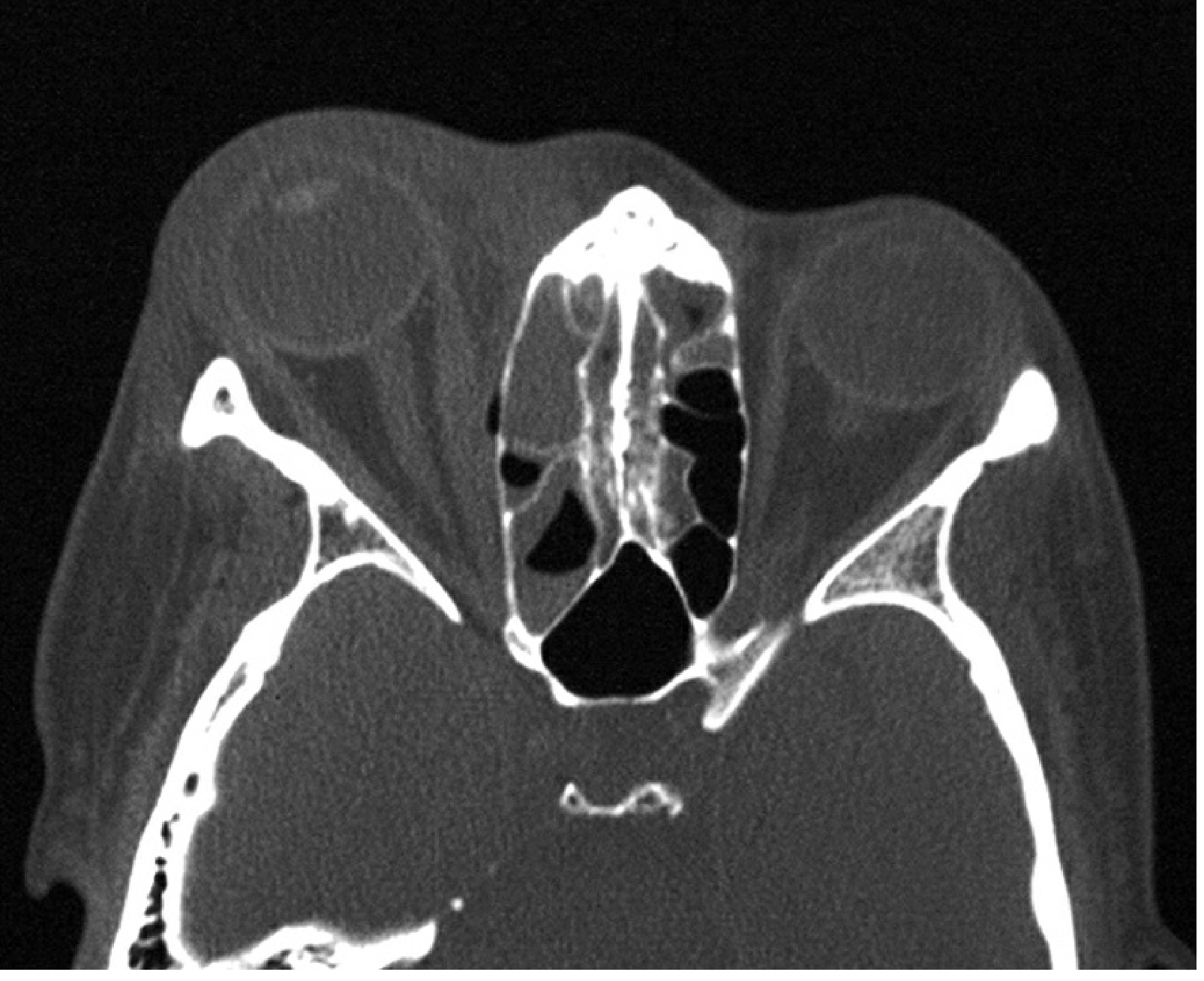
Management
1. Admission and Multidisciplinary Team
- Ophthalmology - optic nerve monitoring, surgical decompression
- Otolaryngology/ENT - sinus assessment and drainage
- Paediatrics (children), Neurosurgery (intracranial complications)
- Infectious disease (atypical, severe, or unresponsive cases)
- Oral and maxillofacial surgery (dental source - infections from this source are aggressive and may spread to cavernous sinus)
2. Optic Nerve Monitoring
- Visual acuity, colour vision, light brightness appreciation, pupillary reactions
- Any deterioration prompts consideration of surgical intervention
3. Antibiotic Therapy
| Scenario | First-Line Antibiotic(s) |
|---|---|
| Standard (community-acquired) | Ampicillin-sulbactam 3g IV q6h (adults); 300 mg/kg/day ÷4 doses (children) |
| Alternative standard | Piperacillin-tazobactam 4.5g IV q8h (adults) |
| Anaerobic suspected / ceftazidime used | Add metronidazole 500mg IV q8h or clindamycin 10mg/kg IV q6h |
| Suspected HA-MRSA / severe / skin source / meningitis | Add vancomycin 15-20mg/kg IV q8-12h (adults); 40-60mg/kg/day ÷3-4 doses (children) |
| Penicillin-allergic, tolerates cephalosporins | Vancomycin + ceftriaxone 2g IV daily + metronidazole |
| Penicillin/cephalosporin allergy | Fluoroquinolone (moxifloxacin 400mg IV daily) + metronidazole |
| Immunocompromised (fungal risk) | Antifungal cover for Mucor/Aspergillus; surgical debridement essential |
- IV antibiotics continued until apyrexial for 4 days
- Then oral antibiotics for 1-3 weeks (Tintinalli recommends a total 3-week course)
4. Surgical Indications
- Subperiosteal or orbital abscess confirmed on CT - drain at an early stage
- Failure to respond to IV antibiotics (typically 48-72 hours)
- Deterioration of vision or worsening optic nerve function
- Severe sinus disease requiring decompression
- RAPD present - indicates optic nerve compression → emergency lateral canthotomy/cantholysis for orbital decompression
- Endoscopic sinus surgery - drainage of infected sinuses (ethmoidectomy most commonly)
- Orbital abscess drainage - external (Lynch incision) or endoscopic
- Emergency canthotomy/cantholysis - for acute orbital compartment syndrome / severe optic nerve compression
Complications
Ocular
- Optic neuropathy - permanent visual loss (most feared)
- Exposure keratopathy
- Raised intraocular pressure
- Central retinal artery or vein occlusion
- Endophthalmitis, panophthalmitis
Intracranial (3-4% but extremely serious)
- Meningitis
- Brain abscess
- Cavernous sinus thrombosis - septic; associated with bilateral signs, CN palsies, spiking fever, papilloedema
- Epidural/subdural empyema
Subperiosteal Abscess
Differential Diagnosis of Acute Orbital Inflammation
| Category | Conditions |
|---|---|
| Infection | Preseptal cellulitis, dacryocystitis, dacryoadenitis, fungal orbital infection |
| Vascular | Cavernous sinus thrombosis, acute orbital haemorrhage, carotid-cavernous fistula |
| Neoplasia | Rhabdomyosarcoma (children), lymphoma, metastasis, rapidly progressive retinoblastoma |
| Inflammation | Idiopathic orbital inflammatory disease (orbital pseudotumour), Tolosa-Hunt syndrome, orbital myositis, GPA (Wegener's), scleritis, herpes zoster |
| Endocrine | Acute thyroid eye disease |
Special Considerations
-
Zygomycosis (mucormycosis): In diabetic or otherwise immunocompromised patients - presents with severe pain, external ophthalmoplegia, rapid profound visual loss, eschar in nasal passages. Associated with metabolic acidosis. Rapidly progressive and life-threatening; requires emergent surgical debridement + antifungal therapy.
-
MRSA: CA-MRSA is increasing, especially in urban areas. Cover empirically with vancomycin in severe cases, skin-source cases, failed conventional therapy, or high-incidence areas.
-
Retained foreign body: Cellulitis can develop months after apparently minor orbital trauma - always consider if no obvious precipitant.
Explain cavernous sinus thrombosis for ent subject of mbbs from basics explain like a top lecturer easy to understand
cavernous sinus anatomy diagram contents cranial nerves
cavernous sinus thrombosis bilateral proptosis ophthalmoplegia
cavernous sinus MRI venography thrombosis filling defect
🧠 Cavernous Sinus Thrombosis (CST)
ENT - MBBS | Explained from Basics
STEP 1: Understand the Anatomy First
What runs THROUGH the cavernous sinus?
LATERAL WALL (top to bottom):
  ├── CN III  (Oculomotor nerve)
  ├── CN IV   (Trochlear nerve)
  ├── CN V1   (Ophthalmic branch of Trigeminal)
  └── CN V2   (Maxillary branch of Trigeminal)

INSIDE THE SINUS ITSELF:
  ├── CN VI   (Abducens nerve) ← runs loose inside, most vulnerable!
  └── Internal Carotid Artery (with its sympathetic plexus)
When the cavernous sinus gets infected and swells, ALL these structures get compressed - and that explains every single clinical feature!
STEP 2: Why Does It Get Infected? (Pathogenesis)
The Valveless Venous System 🔑
The Danger Triangle of the Face
Route of Infection - Know These Paths:
SOURCE                          ROUTE TO CAVERNOUS SINUS
──────────────────────────────────────────────────────────
Furuncle of face/nose       → Facial vein → Ophthalmic veins → Cavernous sinus
Sinusitis (ethmoidal,       → Direct spread / emissary veins → Cavernous sinus
  sphenoidal - most common ENT cause)
Dental infection            → Pterygoid plexus → Cavernous sinus
Orbital cellulitis          → Ophthalmic veins → Cavernous sinus
Tonsil/pharyngeal abscess   → Haematogenous spread
Otitis media/mastoiditis    → Lateral sinus → Cavernous sinus (via petrosal sinus)
ENT exam point: Sphenoidal sinusitis is the single most direct and dangerous source because the sphenoid sinus is immediately adjacent to the cavernous sinus.
STEP 3: Causative Organisms
| Organism | Notes |
|---|---|
| Staphylococcus aureus | Most common - comes from facial furuncles, skin infections |
| Streptococcus species | From sinusitis, pharyngeal infections |
| Anaerobes | From dental sources |
| Gram-negative rods | Less common |
| Fungi (Mucor, Aspergillus) | In diabetics/immunocompromised |
STEP 4: Clinical Features - Understand, Don't Memorise
- Inflammation and thrombosis (clot formation) inside the sinus
- Compression of the structures running through and around it
A. General/Systemic Signs
- High spiking fever (septic, "picket fence" pattern)
- Severe headache (retro-orbital, behind the eye)
- Malaise, lethargy, nausea, vomiting
- Neck stiffness (meningeal irritation if meningitis develops)
- Altered consciousness in severe cases
B. Orbital Signs (Comes from venous congestion + CN compression)
| Feature | Why it happens |
|---|---|
| Proptosis (bulging eye) | Venous congestion → orbital oedema pushes eye forward |
| Chemosis (conjunctival swelling) | Venous congestion of conjunctival veins |
| Eyelid oedema + erythema | Venous backpressure |
| Periorbital oedema | Same mechanism |
| Congestion of conjunctival/retinal/facial veins | Obstructed drainage |
| Reduced vision | Optic nerve compression or retinal vein occlusion |
C. Cranial Nerve Signs (The Hallmark!)
| CN Affected | Feature Produced | Exam Point |
|---|---|---|
| CN III (Oculomotor) | Ptosis, dilated fixed pupil, eye looks "down and out" | Exits lateral wall |
| CN IV (Trochlear) | Cannot look downward-inward (diplopia on downward gaze) | Exits lateral wall |
| CN VI (Abducens) | Earliest and most common - cannot abduct eye (lateral gaze palsy) | Runs INSIDE the sinus - most vulnerable |
| CN V1 (Ophthalmic) | Periorbital pain and anaesthesia, impaired corneal reflex | Exits lateral wall |
| CN V2 (Maxillary) | Numbness of upper lip, cheek | Exits lateral wall |
| Sympathetic fibres (on ICA) | Horner syndrome: ptosis + miosis + anhidrosis | Travel with ICA |
MBBS key point: CN VI palsy (lateral rectus) is the FIRST and MOST COMMON nerve sign because it runs freely inside the sinus and is compressed earliest by even mild swelling.
D. THE MOST IMPORTANT CLINICAL SIGN - BILATERAL INVOLVEMENT
⚠️ BILATERAL proptosis + ophthalmoplegia
STEP 5: How to Remember All the Features - One Story
"A patient with a boil on their nose develops a terrible headache and spiking fever. Both eyes become red, swollen, and proptosed. They can't move their eyes (especially outward - CN VI goes first). Their eyelids droop (CN III). The area around the eye is numb (CN V1). They go drowsy - and now you know infection has reached the cavernous sinus."
STEP 6: Investigations
1. MRI Brain with Contrast + MR Venography (Gold Standard)
- Shows filling defect (absence of flow signal) in the cavernous sinus
- Dural enhancement around the cavernous sinus
- Shows associated sinusitis, orbital changes
- MR venography directly shows absence of venous flow in the thrombosed sinus
2. CT Brain with Contrast (Alternative - faster, widely available)
- Shows filling defects in cavernous sinus
- Identifies source (sinusitis, abscess)
- Less sensitive than MRI for early thrombosis
3. Other Investigations
- Full blood count (leukocytosis, elevated CRP/ESR)
- Blood cultures (identify organism)
- Lumbar puncture - to exclude meningitis (do after imaging to rule out raised ICP)
- Nasal swabs, culture of any discharge
- Blood glucose (check for diabetes)
STEP 7: Management
1. ICU / Critical Care Admission
2. IV Antibiotics (First and Most Important!)
- Must be broad-spectrum, covering Gram-positive, Gram-negative, and anaerobes
- Given for at least 2 weeks beyond clinical resolution - because bacteria hidden inside the thrombus are not killed until the sinus begins to recanalize
- Duration typically 4-6 weeks or until radiological resolution
- Be alert for relapse up to 6 weeks after stopping antibiotics
| Situation | Drug Choice |
|---|---|
| Standard | Ampicillin-sulbactam / Piperacillin-tazobactam IV |
| Add if anaerobes suspected | Metronidazole |
| Suspected MRSA / severe | Add Vancomycin |
| Penicillin-allergic | Fluoroquinolone + Metronidazole |
3. Anticoagulation (Controversial - know both sides!)
| For Anticoagulation | Against Anticoagulation |
|---|---|
| Prevents clot from growing | Thrombus "walls off" the infection - removing it may spread it |
| Reduces inflammation | Risk of intracranial haemorrhage |
| Helps recanalization |
4. Surgical Treatment
- Surgery is not the primary treatment for CST itself
- Surgery is indicated to:
- Drain any underlying sinusitis (especially sphenoidal, ethmoidal)
- Drain any orbital or intracranial abscess
- Endoscopic sinus surgery is preferred
- Follow-up for at least 6 months after recovery
5. Supportive Treatment
- IV fluids and resuscitation
- Treat exposure keratopathy (lubricating eye drops/ointment - eyes may not close due to proptosis)
- Treat secondary glaucoma
- Steroids: controversial; occasionally used with antibiotics
STEP 8: Complications
- Meningitis - infection breaks through to subarachnoid space
- Brain abscess
- Cerebral infarction - internal carotid artery compromise
- Pituitary abscess / hypopituitarism
- Blindness - optic nerve ischaemia or central retinal vein occlusion
- Death - mortality 30-40% even with treatment
STEP 9: Differential Diagnosis (Examiners Love This!)
| Condition | How to Differentiate |
|---|---|
| Orbital cellulitis | Unilateral; no CN palsies beyond orbit; no meningism |
| Carotid-cavernous fistula | Pulsatile proptosis, bruit, no fever |
| Tolosa-Hunt syndrome | Painful ophthalmoplegia but no fever, responds to steroids |
| Orbital apex syndrome | CN II-VI all affected; includes optic nerve = visual loss |
| Superior orbital fissure syndrome | CN III, IV, VI, V1 affected; optic nerve SPARED |
QUICK REVISION CARD
CAVERNOUS SINUS THROMBOSIS - MBBS ESSENTIALS

ANATOMY:   CN III, IV, V1, V2 (lateral wall) + CN VI + ICA (inside)
CAUSE:     Valveless veins → retrograde spread
           ENT source: Sphenoidal/ethmoidal sinusitis (most common)
           Skin source: Facial furuncle (danger triangle of face)
ORGANISM:  S. aureus (most common)

FEATURES:  Spiking fever + severe headache
           Bilateral proptosis + chemosis ← KEY SIGN
           CN VI palsy (FIRST) → all EOM palsies
           CN V1 → periorbital numbness
           Meningism, altered consciousness

INVESTIGATION:
           MRI + MR Venogram = Gold Standard
           (absent flow in cavernous sinus)
           CT contrast = alternative
           LP after imaging (exclude meningitis)

TREATMENT:
           ICU + IV Antibiotics (4-6 weeks)
           ± Heparin anticoagulation (controversial)
           Surgery for underlying sinus/abscess

MORTALITY: 30-40% even with treatment
           Up to 100% if untreated
Explain it like donkey
🫏 Cavernous Sinus Thrombosis — Explained Like You're a Donkey
First, What Even IS the Cavernous Sinus?
🏊 Pool (cavernous sinus)
     ↓
Wires running through it:
  • Wire 1 = moves your upper eyelid + most eye muscles (CN III)
  • Wire 2 = moves eye downward (CN IV)
  • Wire 3 = moves eye OUTWARD - the lateral rectus (CN VI) ← inside the pool!
  • Wire 4 = feeling around your eye (CN V1)
  • Wire 5 = feeling in your cheek (CN V2)
  • Pipe   = the internal carotid artery (blood supply to brain)
Now — Why Does It Get Infected?
"The veins draining into this pool have NO valves."
Where Does the Infection Come From?
| Drain (Source) | How dirty water reaches the pool |
|---|---|
| 😤 Sinuses (sphenoid, ethmoid) | Directly next door — nearest neighbours |
| 🦷 Dental abscess | Via pterygoid veins, backwards into pool |
| 🟥 Boil/pimple on face | Via facial/ophthalmic veins, backwards |
| 👁️ Orbital cellulitis | Via ophthalmic veins, backwards |
| 👂 Ear infection | Via lateral sinus, into cavernous sinus |
ENT exam: Sphenoidal sinusitis is the most direct ENT cause because the sphenoid sinus literally SITS next to the cavernous sinus. Neighbours who share a wall.
What Happens Inside the Pool When Infected?
- Full of pus and clot 🤢
- Swollen and putting pressure on all those wires passing through it
- Blocked, so blood backs up into the eye and face
Clinical Features — Just Think "What Gets Squashed?"
🔥 First — The Infection Symptoms (Boring but Important)
- Very high spiking fever (spikes up and down like a mountain range)
- Terrible headache behind the eye
- Nausea, vomiting, lethargy
- Patient looks VERY sick (toxic)
👁️ Then — The Eye Symptoms (Blood backing up)
- Both eyes bulge out (proptosis) — blood can't drain, so it pools in the orbit and pushes the eye forward like squeezing a tube of toothpaste 🪥
- Red, swollen, puffy conjunctiva (chemosis) — same reason
- Eyelids swollen and red
🔌 Then — The Wire Symptoms (Nerves getting squashed)
| Wire squashed | What the patient can't do |
|---|---|
| CN VI (inside pool — squashed FIRST and MOST) | Can't look sideways (outward). Eye stuck looking inward. This is the FIRST sign! |
| CN III | Eyelid droops (ptosis). Pupil dilates. Eye points down and out |
| CN IV | Can't look diagonally down. Double vision on stairs |
| CN V1 | Numb/painful skin around the eye |
| CN V2 | Numb cheek and upper lip |
| Sympathetic fibres | Horner syndrome — droopy lid + small pupil + dry face |
The ONE Sign That Tells You It's CST and NOT Just an Eye Infection
👀👀 BOTH eyes are involved
How Do You Diagnose It?
🥇 Gold Standard: MRI + MR Venography
- MR Venography shows no blood flowing in the cavernous sinus (because it's all clotted up)
- Like checking if water flows through a pipe — if it's blocked, nothing comes out
🥈 CT Brain with Contrast
- Faster, more available
- Shows a filling defect (dark hole where the pool should be bright with contrast dye)
- Also shows the sinus infection that caused it
🩸 Blood tests
- High white cells, high CRP — confirms infection
- Blood cultures — grow the bug
💉 Lumbar Puncture
- Do it AFTER imaging (to make sure there's no brain swelling that could cause herniation)
- Rules out meningitis
Treatment — 4 Things
1. 🏥 Admit to ICU Immediately
2. 💉 IV Antibiotics — The Most Important Treatment
- Broad spectrum — kills Gram+, Gram-, anaerobes
- Ampicillin-sulbactam or Piperacillin-tazobactam to start
- Add Metronidazole for anaerobes
- Add Vancomycin if MRSA is suspected
- Give for 4-6 weeks (long time! Because bacteria hide inside the clot and can't be killed until the clot starts dissolving)
- Watch for relapse up to 6 weeks after stopping
3. 🩸 Anticoagulation (Heparin) — Controversial, Exam Favourite
| Team Heparin says... | Team No-Heparin says... |
|---|---|
| Stops the clot from growing | The clot is actually helpful — it walls off the infection |
| Reduces inflammation | Heparin can cause brain bleeding |
| Helps the pool drain again |
4. 🔪 Surgery — Only to Drain the Source
- Surgery is NOT done ON the cavernous sinus itself
- Surgery drains the sinuses (sphenoid, ethmoid) that caused it
- Endoscopic sinus surgery (FESS) is preferred
Complications (What Happens if You Miss It)
- 🧠 Meningitis
- 🧠 Brain abscess
- 💀 Cerebral infarction (the internal carotid artery inside the pool gets involved)
- 👁️ Blindness (optic nerve, retinal vein)
- ☠️ Death (30-40% even with treatment; nearly 100% if missed)
The Whole Story in One Paragraph (Read This the Night Before Exam)
A boil on the nose / sinusitis → bacteria travel backwards through valveless veins → reach the cavernous sinus → form a septic clot → patient gets spiking fever + severe headache → clot swells the sinus → compresses CN VI first (can't look outward) then CN III (droopy lid, big pupil) then V1 (numb eye area) → blood backs up into orbit → BOTH eyes bulge out and turn red → MRI venogram shows no flow in the sinus → treat with IV antibiotics for 6 weeks ± heparin → mortality still 30-40%.
Memory Hook 🎯
- Sinus infection (source)
- Backward spread (valveless veins)
- Locks the cavernous sinus (thrombosis)
- Ophthalmoplegia - CN VI first (wire squashed)
- Chemosis + bilateral proptosis (blood backed up)
- Kills at 30-40% (mortality)
Atrophic rhinitis explain like donkey
atrophic rhinitis ozaena nasal cavity crusting wide nasal passage
🫏 Atrophic Rhinitis — Explained Like You're a Donkey
STEP 1: Understand the Normal Nose First
1. 🌡️  WARM the air you breathe in
2. 💧  MOISTEN the air (like a humidifier)
3. 🧹  FILTER dust and germs (the mucus + tiny hairs = cilia)
- Tiny hair-like cilia that sweep mucus + dirt out
- Mucous glands that keep everything wet
- Blood vessels that help warm the air
STEP 2: Now What Goes Wrong in Atrophic Rhinitis?
Normal nose:       Atrophic rhinitis nose:
Wet sponge 🧽  →   Dry, cracked desert 🏜️
Pink mucosa   →   Pale, thin, dead mucosa
Moist + soft  →   Dry + crusty + smelly
Big turbinates →   Shrunken / absent turbinates
Narrow airway →   Paradoxically WIDE empty airway
⚡ Exam Bomb: This is called "paradoxical nasal obstruction" - the nose is actually WIDE OPEN but the patient feels blocked. Why? Because the nervous system relies on the feeling of air hitting the turbinates to sense "open nose." No turbinates = no sensation = feels blocked.
STEP 3: The Two Types — Primary vs Secondary
🔴 Primary Atrophic Rhinitis (= Ozaena)
- No obvious cause — the nose just starts dying on its own
- More common in:
- Developing countries (subtropical + temperate climates - think India, Middle East, Africa)
- Women more than men
- Young adults (20s-30s)
- Associated with bacterial colonization (the bacteria don't cause it but make it way worse)
🟡 Secondary Atrophic Rhinitis
- Has a clear cause — something destroyed the turbinates
- More common in developed countries
- Less severe and less progressive than primary
| Letter | Cause |
|---|---|
| T | Trauma to the nose |
| I | Irradiation (radiotherapy near face/nose) |
| G | Granulomatous diseases (TB, leprosy, sarcoidosis, Wegener's) |
| E | Excessive surgery (too much turbinate removed - "empty nose syndrome") |
| R | Rhinitis medicamentosa (overuse of nasal decongestant sprays) |
| S | Specific infections (syphilis, etc.) |
STEP 4: The Bugs Involved 🦠
| Bug | Significance |
|---|---|
| Klebsiella ozaenae | Named after this disease! Most associated organism |
| Staphylococcus aureus | Common coloniser |
| Proteus mirabilis | Contributes to foul smell |
| Escherichia coli | Also found |
Important: These bugs are not necessarily the cause — they are colonisers taking advantage of dead, dry tissue. Like vultures arriving at a carcass 🦅 — they didn't kill it, but they make it worse.
STEP 5: What Actually Happens Inside — The Histology (Pathology)
1. 🔄 Squamous Metaplasia
- No cilia = mucus can't be swept out = it sits and dries into crusts
2. 🫙 Glandular Atrophy
- No glands = no mucus = dry as a desert
3. 🩸 Endarteritis Obliterans
- No blood supply = tissue dies = atrophy
- This is the KEY underlying change — kill the blood supply, kill the tissue.
Think of it this way: The garden (nasal mucosa) lost its water pipe (blood vessels), lost its gardeners (glands), and the grass (cilia + epithelium) all died. What's left? Dry, cracked, dead land.
STEP 6: Clinical Features — What Does the Patient Look Like?
Symptoms (What the Patient Tells You)
| Symptom | Why? |
|---|---|
| Foul smell from nose (OZAENA) | Dead tissue + bacteria = rotten smell. The patient CANNOT smell it themselves! Relatives/friends smell it first. |
| Anosmia (can't smell anything) | Olfactory nerve endings are dead too |
| Nasal crusting (yellow/green/brown hard crusts) | Dried mucus + dead tissue + bacteria |
| Nasal obstruction (blocked feeling) | Paradoxical obstruction — wide nose but no sensation of airflow |
| Nasal bleeding | Crusts peel off and take tissue with them |
| Headache | From sinus involvement |
| Post-nasal drip | Dry secretions trickle back |
Signs (What You See on Examination)
👃 Wide, spacious, empty nasal cavities
🟫 Green/yellow/brown thick crusty deposits covering everything
🌸 Pale, thin, atrophic, fragile mucosa underneath the crusts
🪨 Shrunken or absent turbinates (especially inferior turbinate)
🩸 Bleeding when crusts are removed
🌟 In advanced cases — can see all the way to the nasopharynx
   (normally can't do this — turbinates block the view)
Exam tip: The ENT surgeon says they can see the posterior wall of nasopharynx directly through the front of the nose. That's how big the cavity has become.
STEP 7: Investigations
1. Clinical Diagnosis Mostly
2. Nasal Swab + Culture
- Grows Klebsiella ozaenae, Proteus, Staph, E. coli
- Guides antibiotic treatment
3. CT Paranasal Sinuses
- Shows atrophy of turbinates
- Widened nasal cavity
- Associated sinusitis
- Rules out other causes (tumour, granulomatous disease)
4. Biopsy (if diagnosis unclear)
- Confirms: squamous metaplasia + glandular atrophy + endarteritis obliterans
5. Blood Tests
- Haemoglobin (anaemia can make things worse)
- ESR, ANCA (to rule out Wegener's granulomatosis if secondary suspected)
STEP 8: Treatment — The Most Exam-Heavy Part
🧴 Medical Treatment (Non-Surgical)
| Treatment | What it does |
|---|---|
| Nasal saline irrigation (large volume lavage) | Washes out crusts + bacteria. First line. Use daily. |
| Steam inhalation + humidification | Moistens the dry mucosa |
| 25% Glucose in Glycerine nasal drops | Restores moisture, inhibits proteolytic bacteria (like Klebsiella). Classic old remedy. |
| Topical antibiotics (e.g. Naseptin - chlorhexidine + neomycin) | Reduces bacterial colonisation |
| Systemic antibiotics (based on culture) | Ciprofloxacin active against Klebsiella; given in courses |
| Oestrogen nasal spray | Stimulates mucosal regrowth; used in some protocols |
| Placental extract injections | Old treatment; stimulates vasodilation + mucosal healing |
| Vitamins A + D (topical or systemic) | Promote mucosal regeneration |
⚠️ Tell the examiner: Treatment of atrophic rhinitis is mostly symptomatic and supportive. There is NO cure. You manage it long-term. It is a chronic, progressive, distressing condition.
🔪 Surgical Treatment
- Air moves slower → mucosa stays moist longer
- Less turbulence → less drying
- Less surface area exposed to bacteria
| Surgery | What it does |
|---|---|
| Young's operation (Closure of nostrils) | Completely sew the nostrils shut for 6 months. Yes, literally closed. Builds up humidity and mucosa regenerates. Opened again later. Most dramatic option. |
| Modified Young's operation | Leave a tiny hole (partial closure) |
| Submucosal injection of Teflon / fat / cartilage | Implants inserted under nasal mucosa to bulk up + narrow the nasal cavity |
| Turbinate reconstruction | Using autologous fat, cartilage, platelet-rich plasma to rebuild turbinates (newer techniques) |
| Sinus drainage surgery | If sinusitis is a complication |
🎯 Exam favourite: Young's operation = surgical closure of nostrils = treatment of primary atrophic rhinitis. The opposite of what most conditions need!
STEP 9: The PARADOX You Must Know
"The nose is WIDE but the patient feels BLOCKED"
STEP 10: Complications
- Ozaena - the unbearable foul smell — social isolation, depression
- Atrophic pharyngitis / laryngitis — the dryness spreads downward
- Sinusitis — dead mucosa can't fight infection
- Nasal septal perforation — from repeated crusting + picking + bleeding
- Secondary infection — abscess, bone involvement (rare)
THE WHOLE STORY IN ONE PARAGRAPH (Night Before Exam)
Atrophic rhinitis = the nasal turbinates slowly shrink and die → mucosa becomes dry, pale, and scarred (squamous metaplasia + glandular atrophy + endarteritis obliterans) → nose gets abnormally wide but patient feels blocked (paradoxical obstruction) → Klebsiella ozaenae colonises the dead tissue → thick green/yellow crusts form + unbearable foul smell (ozaena) that the patient CANNOT smell (anosmia) → treat with saline irrigation, glucose-glycerine drops, antibiotics → surgery = Young's operation (close the nostrils to narrow the space and let mucosa recover).
QUICK MEMORY TABLE 🎯
ATROPHIC RHINITIS - MBBS ESSENTIALS

TYPE:       Primary (ozaena) = women, developing countries, unknown cause
            Secondary = surgery / trauma / radiation / granulomas / drugs

BUGS:       Klebsiella ozaenae (most classic), Proteus, Staph, E. coli

HISTOLOGY:  3 changes (SMS):
            S = Squamous Metaplasia (no more cilia)
            M = Mucous gland atrophy (no moisture)
            S = Small vessel obliteration (endarteritis)

FEATURES:   Foul smell (OZAENA) - patient can't smell it
            Anosmia
            Paradoxical nasal obstruction (wide nose, feels blocked)
            Green/yellow crusts on rhinoscopy
            Visible nasopharynx (turbinates gone)

TREATMENT:  Saline wash + Glucose-glycerine drops
            Antibiotics (topical + systemic)
            YOUNG'S OPERATION = close the nostril shut!
Explain rhinitis sicca like donkey
rhinitis sicca ENT definition causes symptoms treatment MBBS
https://www.chirurgiadelnaso.it/media/news/pdf/rinite-secca-…
🫏 Rhinitis Sicca — Explained Like You're a Donkey
FIRST: What Does "Rhinitis Sicca" Even Mean?
- Rhinitis = inflammation of the nose (rhino = nose, itis = inflammation)
- Sicca = Latin for DRY
✅ Key point for exams: Rhinitis Sicca is also called "Dry Nose Syndrome." In textbooks it also appears as "Rhinitis Sicca Anterior" when the dryness is limited to the front part of the nose. And yes - KJ Lee's textbook lists it as another name for Atrophic Rhinitis (though strictly speaking, they are different - more on that below).
STEP 1: Understand the Normal Wet Nose
- Mucous glands → secrete sticky mucus (keeps things moist + traps dust)
- Blood vessels → warm the air + keep the mucosa pink and alive
- Cilia → tiny hairs that sweep mucus + dirt out
- A protective moisture layer → like the screen protector on your phone
STEP 2: What Goes Wrong in Rhinitis Sicca?
Normal nose:            Rhinitis Sicca nose:
Moist and pink    →    Dry, pale, cracked 🏜️
Smooth surface    →    Rough with thin crusts
No crusting       →    Thin yellow/brown scabs form
No bleeding       →    Bleeds easily (epistaxis)
No discomfort     →    Itching, burning, soreness
STEP 3: How is Rhinitis Sicca DIFFERENT from Atrophic Rhinitis?
| Feature | Rhinitis Sicca | Atrophic Rhinitis (Ozaena) |
|---|---|---|
| Area affected | Mainly FRONT of nose (anterior) | Whole nose - deep and widespread |
| Turbinate damage | Turbinates are mostly INTACT | Turbinates are DESTROYED / shrunken |
| Nasal cavity size | Normal or slightly wide | Very wide and empty |
| Smell (ozaena) | Usually NO foul smell | YES - horrible foul smell |
| Severity | Mild to moderate | Severe and progressive |
| Bone involvement | No | YES - bone atrophies too |
| Paradoxical obstruction | Less prominent | Classic feature |
One-line memory: Rhinitis Sicca = DRY FRONT of nose, intact structure, no foul smell. Atrophic Rhinitis = DEAD whole nose, destroyed turbinates, horrible smell.
STEP 4: Why Does the Nose Dry Out? (Causes)
🌬️ Environmental / External Causes
| Cause | How it dries the nose |
|---|---|
| Dry air (air conditioning, heaters, winter) | Literally evaporates nasal moisture |
| Hot, dusty, windy environment | Blows away moisture, irritates mucosa |
| High altitude | Air is thinner and drier |
| Occupational exposure | Dust, chemicals, fumes (bakers, miners, painters) |
💊 Drug-Related Causes
| Drug | Mechanism |
|---|---|
| Overuse of nasal decongestant sprays (oxymetazoline, xylometazoline) | Causes rhinitis medicamentosa → then rebound dryness |
| Antihistamines | Dry up all secretions including nasal ones |
| Anticholinergics | Block mucous gland secretion |
| Cocaine (intranasal use) | Destroys nasal mucosa directly |
| Isotretinoin (acne drug) | Dries all mucosal surfaces |
| Radiotherapy to head/neck | Destroys mucous glands |
🩺 Systemic / Medical Causes
| Disease | Why nose dries |
|---|---|
| Sjögren's syndrome | Autoimmune - destroys ALL secretory glands (eyes, mouth, nose) |
| Hypothyroidism | Reduces mucosal secretion |
| Diabetes | Microvascular damage to nasal mucosa |
| Anaemia | Mucosal dryness |
| Ageing (senile rhinitis sicca) | Mucous glands naturally atrophy with age |
| Granulomatous diseases (TB, leprosy, Wegener's, sarcoidosis) | Destroy nasal tissue |
👆 Mechanical / Local Causes
- Nose picking (digital trauma) - most common in children!
- Nasal surgery - especially if too much turbinate was removed
- Nasal septum deviation - alters airflow, dries out certain areas
- Persistent nasal discharge - paradoxically, constant flow irritates and dries the skin of the anterior nose
STEP 5: Where Exactly in the Nose?
👃 Anterior nasal septum
   ↓
The "Little's Area" (Kiesselbach's Plexus)
= Front bottom part of nasal septum
= Where 5 arteries meet (most vascular spot in nose)
= MOST COMMON site of epistaxis
= MOST COMMON site of rhinitis sicca dryness + crusting
🎯 Exam point: Little's Area / Kiesselbach's Plexus = the site of rhinitis sicca anterior AND the most common site of epistaxis (nosebleed). They are directly connected — rhinitis sicca → crust forms → crust peels off → blood vessel tears → epistaxis.
STEP 6: Clinical Features — What Does the Patient Complain About?
"Doctor, my nose feels dry and scratchy all the time. It burns. There are little scabs inside. And I keep getting nosebleeds when I pick at them."
Symptoms:
| Symptom | Why? |
|---|---|
| Sensation of nasal dryness | Obvious - no moisture |
| Itching and burning | Dry, inflamed mucosa = irritation |
| Nasal obstruction | Crusts physically block the passage |
| Thin yellow/brown crusting | Dried mucus + dead cells |
| Epistaxis (nosebleeds) | Dry mucosa cracks → blood vessels exposed → bleed |
| Mild unpleasant smell (sometimes) | Crusts + secondary bacterial colonisation |
| Slightly reduced sense of smell | Dry mucosa → olfactory dysfunction |
| No/minimal discharge | Opposite of a runny nose! |
On Anterior Rhinoscopy (Looking Inside the Nose):
🔴 Dry, pale, fragile anterior nasal mucosa
🟫 Thin yellowish-brown crusts/scabs on anterior septum
🩸 Raw, reddish, eroded areas under the crusts
   (superficial ulceration in some cases)
💧 NO watery discharge, NO polyps, NO pus
STEP 7: Diagnosis
- History - dry environment? decongestant overuse? systemic disease? nose-picking habit?
- Anterior rhinoscopy - see the dry mucosa and crusting at Little's Area
- Nasal endoscopy - for deeper assessment
- Rule out sinister causes:
- Biopsy if crusting is unilateral, severe, or associated with bone destruction (to rule out granulomatous disease, malignancy)
- Blood tests if systemic cause suspected (Sjögren's antibodies, TFTs, blood sugar, ESR/ANCA for Wegener's)
- CT sinuses if structural abnormality or atrophy suspected
STEP 8: Treatment — Keep It Simple, Keep It Moist!
🥇 First Line — Moisturise, Moisturise, Moisturise
| Treatment | How it helps |
|---|---|
| Nasal saline irrigation (large volume, hypertonic 3% saline) | Washes crusts away + hydrates the mucosa. First and most important. Use daily. |
| Nasal saline sprays / drops | Keeps mucosa moist between irrigations |
| Humidifier at home/work | Puts moisture back into the air before it enters your nose |
| Steam inhalation | Directly moistens nasal lining |
| Avoid triggers | Stop overusing decongestant sprays; use humidified air; wear a mask in dusty environments |
| Stop nose picking | Most obvious, hardest to enforce 😅 |
🥈 Topical Ointments / Lubricants
| Ointment | Use |
|---|---|
| Vaseline (petroleum jelly) applied inside nose | Simple, cheap, very effective moisturiser |
| Glucose in glycerine 25% drops | Restores moisture + creates hostile environment for bacteria |
| Vitamin A + D ointment | Promotes mucosal healing and regeneration |
| Sesame oil nasal drops | Natural lubricant, evidence-based in some trials |
| Naseptin cream (chlorhexidine + neomycin) | When secondary bacterial infection is present |
🥉 Treating the Underlying Cause
| Cause found | Treatment |
|---|---|
| Rhinitis medicamentosa | Stop the decongestant spray (gradually) |
| Sjögren's syndrome | Treat the autoimmune disease |
| Hypothyroidism | Thyroid replacement |
| Occupational exposure | Workplace modifications, mask use |
| Excessive turbinate removal | Turbinate reconstruction (surgery) |
🔪 Surgery (Only If Medical Treatment Fails)
- Turbinate reconstruction using submucosal fat, cartilage, or platelet-rich plasma (to bulk up the cavity and reduce airflow turbulence)
- Treatment of associated septal deviation
- Avoid removing more turbinate tissue — this will make it WORSE
⚠️ Exam warning: Over-aggressive turbinate surgery CAUSES secondary atrophic rhinitis and "Empty Nose Syndrome." Less is more when it comes to turbinate surgery.
STEP 9: The Spectrum — Where Does Rhinitis Sicca Fit?
MILDEST                                                    MOST SEVERE
   │                                                            │
   ▼                                                            ▼
Rhinitis Sicca     →    Secondary Atrophic     →    Primary Atrophic
 Anterior               Rhinitis                    Rhinitis (OZAENA)
   │                        │                            │
Front of nose only      Whole nose                  Whole nose + bone
Mild dryness +          Dry + moderately            Destroyed turbinates +
crusting                atrophic                    foul smell (ozaena) +
No foul smell           May have smell              anosmia + paradoxical
Turbinates intact       Turbinates reduced          obstruction
STEP 10: One Paragraph Night-Before-Exam Summary
Rhinitis Sicca = dry nose - the nasal mucosa (especially the anterior septum / Little's Area) loses its moisture due to dry air, decongestant overuse, Sjögren's syndrome, ageing, or nose-picking → mucosa becomes dry, cracked, and covered in thin yellow crusts → patient gets itching, burning, nasal obstruction, and nosebleeds (epistaxis) → NO foul smell, NO destroyed turbinates (unlike atrophic rhinitis) → diagnose clinically with anterior rhinoscopy → treat with nasal saline irrigation + Vaseline/glycerine drops + humidification + remove the cause → surgery rarely needed.
QUICK MEMORY CARD 🎯
RHINITIS SICCA - MBBS ESSENTIALS

NAME:       Sicca = DRY. Dry nose syndrome.
            Anterior form = Rhinitis Sicca Anterior

SITE:       Anterior nasal septum (Little's Area / Kiesselbach's plexus)

CAUSES:     Dry air, AC, heaters (most common environmental)
            Decongestant spray overuse (rhinitis medicamentosa)
            Sjögren's syndrome (systemic)
            Nose picking (mechanical)
            Excessive nasal surgery
            Ageing

FEATURES:   Dryness + itching + burning
            Thin crusts (yellow/brown) on anterior septum
            Epistaxis ← most important complication
            NO foul smell (unlike atrophic rhinitis)
            NO paradoxical obstruction (unlike atrophic rhinitis)

TREATMENT:  Saline irrigation (FIRST)
            Vaseline / Glucose-glycerine drops / sesame oil
            Humidifier
            Remove the cause
            NO surgery usually needed

KEY DIFFERENCE from atrophic rhinitis:
            Sicca = MILD, FRONT only, no turbinate destruction, no foul smell
            Atrophic = SEVERE, whole nose, destroyed turbinates, ozaena
Write in detail about hypersensitivity
Which subject/context is this for? · How do you want it explained?
For pathology mbbs explain its types with suitable example
Write in detail about hypersensitivity For pathology mbbs explain its types with suitable example
hypersensitivity reactions types Gell and Coombs classification diagram
🛡️ Hypersensitivity Reactions
Pathology MBBS - Complete Notes with Examples
WHAT IS HYPERSENSITIVITY? (Start From Basics)
Definition: Hypersensitivity is an exaggerated or augmented immune response that causes tissue damage and disease in the host.
- It requires prior sensitisation - first exposure sensitises, second exposure causes the reaction
- The damage is caused by the immune response itself, not by the antigen
CLASSIFICATION - Gell and Coombs (1963)
Types I, II, III  →  ANTIBODY-MEDIATED (Humoral)
Type IV           →  T-CELL MEDIATED (Cellular)
| Type | Name | Mediator | Speed |
|---|---|---|---|
| I | Immediate / Anaphylactic | IgE + Mast cells | Seconds to minutes |
| II | Antibody-mediated / Cytotoxic | IgG or IgM | Hours |
| III | Immune complex-mediated | IgG + Complement | Hours to days |
| IV | Delayed / Cell-mediated | T lymphocytes | 24-72 hours |
🔴 TYPE I — IMMEDIATE (ANAPHYLACTIC) HYPERSENSITIVITY
The Donkey Version First:
You eat a peanut 🥜 → your body freaks out → releases a tsunami of chemicals → blood vessels burst open + airways clamp shut → anaphylaxis. You could die in minutes.
Definition:
Mechanism - Step by Step
PHASE 1: Sensitisation (First Exposure - No Symptoms)
Allergen enters body (pollen, food, drug, venom)
        ↓
Antigen presenting cells process it
        ↓
Th2 cells activated → release IL-4, IL-5, IL-13
        ↓
B cells stimulated → produce IgE antibodies
        ↓
IgE binds to FcεRI receptors on MAST CELLS and BASOPHILS
        ↓
Person is now SENSITISED (no symptoms yet)
PHASE 2: Second Exposure - The Reaction
Same allergen enters again
        ↓
CROSS-LINKS two adjacent IgE molecules on mast cell surface
        ↓
MAST CELL DEGRANULATION → releases TWO waves of mediators
Mediators Released
Wave 1: PREFORMED (Released immediately from granules)
| Mediator | Effect |
|---|---|
| Histamine (most important) | Vasodilation + increased vascular permeability + smooth muscle contraction (bronchospasm) + mucus secretion |
| Heparin | Anticoagulant; helps package granule contents |
| Tryptase, chymase (proteases) | Tissue damage, activate complement (C3a) |
Wave 2: NEWLY SYNTHESISED (from arachidonic acid - takes minutes)
| Mediator | Effect |
|---|---|
| Prostaglandin D2 | Bronchospasm, vasodilation |
| Leukotriene C4, D4 (Slow Reacting Substance of Anaphylaxis - SRS-A) | Prolonged bronchospasm + vascular permeability - 1000x more potent than histamine! |
| Leukotriene B4 | Chemotaxis - recruits neutrophils, eosinophils |
| Platelet Activating Factor (PAF) | Bronchospasm, platelet aggregation |
| Cytokines (IL-4, IL-5, TNF-α) | Late phase reaction, eosinophil recruitment |
💡 Exam tip: SRS-A (Slow Reacting Substance of Anaphylaxis) = Leukotrienes C4, D4, E4. These are responsible for the late phase reaction (2-24 hours later) and are the target of montelukast (leukotriene receptor antagonist used in asthma).
The Two Phases of Type I Reaction
🔴 Immediate Phase (within minutes)
- Vasodilation, oedema, smooth muscle spasm, mucus secretion
- Caused by histamine and lipid mediators
🟠 Late Phase Reaction (2-24 hours later)
- Infiltration by eosinophils, neutrophils, basophils, Th2 cells
- More tissue damage and inflammation
- Why asthma gets worse hours after the initial attack
- Caused by leukotrienes + cytokines
Clinical Examples
| Disease | Allergen | Manifestation |
|---|---|---|
| Anaphylaxis | Penicillin, bee sting, peanut, contrast dye | Systemic shock, laryngeal oedema, death |
| Bronchial asthma (atopic) | Dust mite, pollen, animal dander | Wheeze, breathlessness, bronchospasm |
| Allergic rhinitis (Hay fever) | Pollen, dust | Sneezing, runny nose, watery eyes |
| Urticaria (Hives) | Food, drugs | Itchy wheals on skin |
| Atopic dermatitis (Eczema) | Environmental allergens | Itchy, inflamed skin |
| Food allergy | Peanuts, shellfish, milk | GI symptoms, urticaria, anaphylaxis |
Treatment
- Adrenaline (Epinephrine) - first line for anaphylaxis (reverses bronchospasm and vasodilation)
- Antihistamines (H1 blockers) - block histamine receptors
- Corticosteroids - suppress late phase reaction
- Montelukast - leukotriene antagonist (used in asthma)
- Immunotherapy (desensitisation) - gradually increase allergen dose → switch from IgE to IgG response
🟠 TYPE II — ANTIBODY-MEDIATED (CYTOTOXIC) HYPERSENSITIVITY
The Donkey Version:
Wrong blood group transfusion 🩸 → your antibodies see the foreign red blood cells → label them → your immune system destroys them all → haemolytic transfusion reaction.
Definition:
Mechanism - 3 Ways Antibodies Kill Cells
Mechanism 1: Opsonisation + Phagocytosis
IgG/IgM binds to cell surface antigen
        ↓
Complement activated → C3b deposited on cell surface
        ↓
Macrophages/neutrophils recognise C3b via Fc receptors
        ↓
Cell engulfed (phagocytosis) and destroyed
Mechanism 2: Complement-Mediated Lysis
IgG/IgM binds to cell surface
        ↓
Classical complement pathway activated
        ↓
Membrane Attack Complex (MAC) formed (C5b-9)
        ↓
"Drills holes" in cell membrane → cell lyses (osmotic death)
Mechanism 3: Antibody-Dependent Cellular Cytotoxicity (ADCC)
IgG coats the target cell
        ↓
NK cells + macrophages bind via Fc receptors
        ↓
Kill the cell WITHOUT phagocytosis
Mechanism 4 (Special): Antibody Against Receptors - No Killing, Just Dysfunction!
| Action | Example |
|---|---|
| Stimulates receptor | Graves disease - anti-TSH receptor antibody → thyroid overstimulated → hyperthyroidism |
| Blocks receptor | Myasthenia gravis - anti-ACh receptor antibody → receptor blocked → muscle weakness |
Clinical Examples
| Disease | Target Antigen | Mechanism |
|---|---|---|
| Autoimmune haemolytic anaemia | RBC surface antigens | Opsonisation → phagocytosis |
| Transfusion reaction (ABO mismatch) | Blood group antigens on RBCs | Complement lysis |
| Goodpasture syndrome | Type IV collagen (glomerular/alveolar basement membrane) | Complement + inflammation → nephritis + lung haemorrhage |
| Graves disease | TSH receptor on thyroid | Anti-receptor stimulation → hyperthyroidism |
| Myasthenia gravis | Acetylcholine receptor (NMJ) | Anti-receptor blocking → muscle weakness |
| Pemphigus vulgaris | Desmoglein (skin cell junctions) | Complement + destruction → skin blisters |
| Immune thrombocytopenia (ITP) | Platelet surface glycoproteins | Opsonisation → platelet destruction → bleeding |
| Rhesus haemolytic disease (HDN) | Rh antigen on foetal RBCs | Maternal IgG crosses placenta → foetal RBC destruction |
🎯 Exam memory: Type II = antibodies attack fixed targets (cells and tissues). Think "target practice" - antibody aims at a specific cell. Kills directly or blocks function.
🟡 TYPE III — IMMUNE COMPLEX-MEDIATED HYPERSENSITIVITY
The Donkey Version:
Antibody meets antigen → they form a complex → complex gets stuck in blood vessel walls → activates complement → huge inflammatory explosion right there in the vessel wall → vasculitis, nephritis, arthritis.
Definition:
- Type II → antibody attacks fixed, cell-bound antigen
- Type III → antibody + antigen form soluble complexes that travel in circulation and get trapped in tissues
Mechanism - Step by Step
Antigen + IgG → Immune complex formed in circulation
        ↓
Normally cleared by RES (liver, spleen)
        ↓
If too many complexes OR complement is defective → NOT cleared
        ↓
Complexes trapped in:
  → Glomeruli (kidney)
  → Synovium (joints)
  → Blood vessel walls
  → Skin
        ↓
Complement activated (C3a, C5a = anaphylatoxins)
        ↓
C5a = powerful chemotactic factor → attracts NEUTROPHILS
        ↓
Neutrophils try to phagocytose immune complexes
        ↓
Release lysosomal enzymes + reactive oxygen species
        ↓
TISSUE DAMAGE → vasculitis, glomerulonephritis, arthritis
Morphology (What It Looks Like Under Microscope)
- Blood vessels: Necrotising vasculitis with fibrinoid necrosis of vessel wall
- Kidney: Glomerulonephritis - on immunofluorescence = granular ("lumpy-bumpy") deposits of IgG + C3 along GBM
- Joints: Arthritis with neutrophilic infiltrate
- Serum complement levels are LOW (consumed by immune complexes) → marker of disease activity in SLE
Two Classic Forms
1. Systemic Immune Complex Disease (Serum Sickness)
- Large amounts of foreign protein injected
- Immune complexes formed and deposited throughout body
- Classic triad: Fever + Arthralgia + Urticaria + Proteinuria (10 days after antigen exposure)
- Example: Antithymocyte globulin therapy, horse serum (old diphtheria treatment)
2. Local Immune Complex Disease (Arthus Reaction)
- Low dose antigen injected intradermally into already immunised individual
- Local immune complex formation at injection site
- Localised vasculitis with oedema and haemorrhage at the site
- Example: Localised skin reaction at site of repeated vaccine injection
Clinical Examples
| Disease | Antigen | Main Features |
|---|---|---|
| Systemic Lupus Erythematosus (SLE) | Nuclear antigens (DNA, histones) | Nephritis, skin rash, arthritis, vasculitis |
| Poststreptococcal glomerulonephritis | Streptococcal cell wall antigens | Nephritis 2-3 weeks after throat/skin infection |
| Serum sickness | Foreign serum proteins | Fever, urticaria, arthralgia, nephritis |
| Polyarteritis nodosa | HBsAg (Hepatitis B) | Systemic vasculitis |
| Arthus reaction | Foreign proteins (local) | Localised skin vasculitis at injection site |
| Hypersensitivity pneumonitis | Fungal spores, bird proteins | Lung inflammation ("Farmer's lung") |
| Reactive arthritis | Bacterial antigens (Yersinia, Chlamydia) | Arthritis after infection |
🎯 Exam memory: Type III = "complex crime" - immune complexes commit crimes at their landing spots. GN (lumpy deposits) + Low C3 + preceding infection = classic Type III.
🟢 TYPE IV — CELL-MEDIATED (DELAYED-TYPE) HYPERSENSITIVITY (DTH)
The Donkey Version:
Tuberculin skin test (Mantoux) → inject TB antigen → NOTHING for 24 hours → 48-72 hours later → hard, red, swollen bump. Your T cells were slowly gathering, recognising the antigen, and starting a fight. No antibodies involved at all.
Definition:
- Travel to the site
- Recognise the antigen
- Proliferate
- Secrete cytokines
Cells Involved
| Cell | Role |
|---|---|
| CD4+ Th1 cells | Secrete IFN-γ → activates macrophages; IL-2 → T cell proliferation; TNF-α → inflammation |
| CD4+ Th17 cells | Secrete IL-17 → recruit neutrophils |
| CD8+ Cytotoxic T Lymphocytes (CTLs) | Directly kill antigen-bearing target cells by perforin/granzyme |
| Macrophages | Activated by IFN-γ → become "angry macrophages" → release more cytokines and enzymes → tissue damage |
Mechanism - Step by Step
Phase 1: Sensitisation (First Exposure)
Antigen processed by APCs → presented on MHC Class II to CD4+ T cells
(OR on MHC Class I to CD8+ T cells)
        ↓
T cells proliferate and form MEMORY T CELLS
(No visible reaction)
Phase 2: Elicitation (Second Exposure - 24-72 hours later)
Antigen re-enters → processed by local APCs
        ↓
Memory T cells recognised antigen → activated → proliferate
        ↓
CD4+ Th1 cells release:
  IFN-γ → activates macrophages
  IL-2  → T cell proliferation
  TNF-α → vascular changes, fever
        ↓
Macrophages activated → "angry macrophages"
  → Release: IL-1, IL-6, TNF, reactive oxygen species, lysosomal enzymes
        ↓
TISSUE DAMAGE + GRANULOMA FORMATION
Three Subtypes of Type IV Hypersensitivity
1. Classic Delayed-Type Hypersensitivity (DTH)
- CD4+ Th1 mediated
- 48-72 hour delay
- Features: induration (hard swelling), erythema, central necrosis in severe cases
- Prototype: Tuberculin (Mantoux) test - positive = induration ≥10mm at 48-72 hours
2. Contact Hypersensitivity
- Small molecules (haptens) penetrate skin → bind to proteins → become complete antigens
- Sensitisation via Langerhans cells in skin
- Re-exposure → CD4+ T cell-mediated skin reaction
- Features: Erythema, vesicles, intense itching (eczematous reaction)
- Examples:
- Nickel (jewellery, belt buckles)
- Poison ivy / poison oak (urushiol)
- Formaldehyde, rubber chemicals
- Topical medications (neomycin, sulfonamides)
- Hair dyes (PPD - paraphenylenediamine)
3. Granulomatous Hypersensitivity
- Most severe form of Type IV
- Occurs when macrophages cannot destroy the antigen (e.g., mycobacteria, fungi, foreign material)
- Frustrated macrophages fuse → Epithelioid cells + Giant cells
- Granuloma forms - a ball of activated macrophages walling off the antigen
- Examples: Tuberculosis, leprosy, sarcoidosis, Crohn's disease
Clinical Examples
| Disease | Antigen | Mechanism |
|---|---|---|
| Tuberculin test (Mantoux) | PPD (TB antigen) | Classic DTH - Th1 cells |
| Contact dermatitis | Nickel, poison ivy, rubber, cosmetics | Contact hypersensitivity - haptens |
| Tuberculosis (tissue lesions) | Mycobacterium tuberculosis | Granulomatous hypersensitivity |
| Leprosy | Mycobacterium leprae | Granulomatous (in tuberculoid leprosy) |
| Sarcoidosis | Unknown antigen | Granuloma formation |
| Type 1 Diabetes Mellitus | Pancreatic beta cell antigens | CD8+ CTL destruction of beta cells |
| Multiple Sclerosis | Myelin basic protein | CD4+ Th1 cells attack myelin |
| Hashimoto's thyroiditis | Thyroid antigens | T cell-mediated + antibody-mediated |
| Graft rejection (acute) | Foreign HLA antigens on transplant | CD4+ + CD8+ T cells destroy graft |
🏆 MASTER COMPARISON TABLE (The Most Exam-Important Table)
| Feature | Type I | Type II | Type III | Type IV |
|---|---|---|---|---|
| Mediator | IgE | IgG, IgM | IgG (complexes) | T cells |
| Cells involved | Mast cells, basophils, eosinophils | Complement, phagocytes, NK cells | Neutrophils, complement | CD4+, CD8+, macrophages |
| Antigen location | Soluble, environmental | Cell surface / ECM | Soluble (circulating) | Cell-associated / tissue |
| Onset | Seconds to minutes | Minutes to hours | Hours to days | 24-72 hours |
| Complement involved | NO (usually) | YES | YES | NO |
| Transfer by | Serum (IgE) | Serum (IgG/IgM) | Serum (IgG complexes) | Cells (T cells) |
| Skin test result | Wheal + flare (minutes) | - | Arthus (hours) | Induration (48-72h) |
| Classic example | Anaphylaxis, hay fever, asthma | Autoimmune haemolytic anaemia, Goodpasture, Graves, Myasthenia | SLE, serum sickness, post-strep GN | Contact dermatitis, TB test, granulomas |
| Histology | Oedema, eosinophils | Cell lysis, fibrinoid necrosis | Fibrinoid necrosis of vessels | Lymphocytes, macrophages, granulomas |
🎯 EXAM FAVOURITE QUESTIONS + ANSWERS
ONE PARAGRAPH SUMMARY (Night Before Exam)
Hypersensitivity = exaggerated immune response causing host tissue damage, requiring prior sensitisation. Gell and Coombs classified it into 4 types. Type I (immediate) = IgE + mast cell degranulation → histamine + leukotrienes → anaphylaxis, asthma, hay fever; treat with adrenaline + antihistamines. Type II (cytotoxic) = IgG/IgM against cell surface antigens → lysis, phagocytosis, ADCC, or receptor dysfunction (Goodpasture, AIHA, Graves, Myasthenia). Type III (immune complex) = soluble antigen-antibody complexes deposited in tissues → complement + neutrophils → fibrinoid necrosis + granular GBM deposits → SLE, serum sickness, post-strep GN. Type IV (delayed, cell-mediated) = T cells only, no antibodies, 24-72 hours → Th1 cytokines + macrophage activation OR CD8 killing OR granuloma formation → contact dermatitis, tuberculin test, tuberculosis.
Give answer not so lengthy give it in short
Hypersensitivity — Short & Sharp (Pathology MBBS)
Definition
Gell & Coombs Classification (1963)
TYPE I — Immediate / Anaphylactic
- Allergen → IgE produced → IgE binds to mast cells
- Re-exposure → allergen cross-links IgE → mast cell degranulation
- Releases histamine, leukotrienes (SRS-A), prostaglandins
- Anaphylaxis (penicillin, bee sting)
- Bronchial asthma (atopic)
- Hay fever (allergic rhinitis)
- Urticaria, food allergy
TYPE II — Antibody-Mediated / Cytotoxic
- Opsonisation + phagocytosis (IgG coats cell → macrophage eats it)
- Complement lysis (MAC drills holes → cell dies)
- ADCC (NK cells kill IgG-coated cells)
- Receptor dysfunction (antibody blocks or stimulates receptor)
| Disease | Mechanism |
|---|---|
| Autoimmune haemolytic anaemia | Opsonisation of RBCs |
| ABO transfusion reaction | Complement lysis |
| Goodpasture syndrome | Anti-GBM antibody → nephritis + lung haemorrhage |
| Graves disease | Anti-TSH receptor → stimulates thyroid → hyperthyroidism |
| Myasthenia gravis | Anti-ACh receptor → blocks NMJ → weakness |
TYPE III — Immune Complex-Mediated
- Antigen + IgG → soluble immune complexes in circulation
- Complexes trapped in glomeruli, vessels, joints, skin
- Complement activated → C5a attracts neutrophils
- Neutrophils release enzymes → fibrinoid necrosis
- IF microscopy of kidney → granular ("lumpy-bumpy") deposits
- Serum complement (C3) LOW (consumed)
| Disease | Key Feature |
|---|---|
| SLE | Anti-nuclear antibody complexes; nephritis, vasculitis |
| Post-streptococcal GN | 2-3 weeks after throat/skin infection |
| Serum sickness | 10 days after foreign protein; fever + arthralgia + urticaria |
| Arthus reaction | Local vasculitis at injection site |
TYPE IV — Delayed / Cell-Mediated (DTH)
- Sensitisation → memory T cells formed
- Re-exposure → CD4+ Th1 cells release IFN-γ, IL-2, TNF-α
- IFN-γ activates macrophages → tissue damage + granuloma formation
- CD8+ CTLs → directly kill antigen-bearing cells
| Subtype | Example |
|---|---|
| Classic DTH | Mantoux/tuberculin test (induration at 48-72h) |
| Contact hypersensitivity | Contact dermatitis (nickel, poison ivy, hair dye) |
| Granulomatous | Tuberculosis, sarcoidosis, leprosy |
Master Comparison Table
| Feature | Type I | Type II | Type III | Type IV |
|---|---|---|---|---|
| Mediator | IgE | IgG/IgM | IgG complexes | T cells |
| Onset | Seconds | Hours | Hours-days | 24-72 h |
| Complement | No | Yes | Yes | No |
| Prototype | Anaphylaxis | Goodpasture | SLE / Serum sickness | TB test / Contact dermatitis |
| Transfer by | Serum | Serum | Serum | Cells only |
High-Yield Exam Points
- SRS-A = Leukotrienes C4, D4, E4 = Type I mediators → target of montelukast
- Only Type IV has NO antibody involvement
- Granular deposits on IF = Type III (vs. linear deposits in Type II Goodpasture)
- Low serum C3 = Type III (complement consumed)
- Graves = stimulatory Type II (no cell destruction)
- Passive transfer by serum = Types I, II, III. By cells = Type IV only