
Everything to know about a normal vaginal delivery from a doctor
Reading File
Now let me search the medical library for comprehensive content on normal vaginal delivery.
Finding Sources
Finding Sources
Finding Sources
Finding Sources
Finding Sources
Good - found key sources. Let me now read the most relevant obstetrics content in parallel.
Finding Sources
Reading File
Reading File
Good - got excellent content from the Family Medicine textbook. Let me read more of that block and also search for mechanism of labor content.
Reading File
Finding Sources
Excellent - very rich content. Let me fetch the labor progress diagram and continue reading for delivery technique and postpartum care.
Loading Image
Finding Sources
Finding Sources
Let me read the Tintinalli's Emergency Delivery section and also fetch that cardinal movements diagram.
Reading File
Reading File
Excellent - I now have very comprehensive content. Let me also fetch the cardinal movements diagram from Tintinalli's and search for a few more images.
Reading File
Loading Image
Reading File
I now have comprehensive, authoritative content from two major textbooks. Let me compile the full answer.
Normal Vaginal Delivery: A Complete Clinical Guide
Sources: Textbook of Family Medicine, 9e | Tintinalli's Emergency Medicine, 8e | Roberts and Hedges' Clinical Procedures in Emergency Medicine
1. Onset of Labor
Definition: Labor is defined as the presence of sufficient uterine contractions in frequency, intensity, and duration to bring about effacement and dilation of the cervix. Control is complex and involves prostaglandins - specifically PGE₂ and PGF₂α.
Prelabor signs:
- Lightening - fetal head descends into the pelvis, fundal height decreases; may occur acutely or over several weeks with increased pelvic pressure
- Bloody show - blood-tinged mucous discharge as cervical effacement begins; most women go into true labor within 3 days
- False labor (Braxton Hicks) - irregular short contractions; do not cause significant cervical dilation but may aid in cervical effacement
True labor contractions: every 2-3 minutes, lasting ~1 minute, mean intensity ~40 mmHg; adequate relaxation between contractions is essential for oxygenated blood to reach the fetal compartment.
2. Stages of Labor
| Stage | Onset | End |
|---|---|---|
| First (latent + active) | Active labor begins | Full cervical dilation (10 cm) |
| Second | Full dilation | Delivery of baby |
| Third | Delivery of baby | Delivery of placenta |
| Fourth* | Delivery of placenta | Contracted, stable uterus |
Not always formally counted as a stage.
3. First Stage of Labor
Latent Phase
- Variable length: <20 hours in nulliparas, <14 hours in multiparas
- Little visible cervical dilation, but major cervical preparation occurs: softening (collagen/connective tissue changes), effacement, and anterior positioning
- Best managed at home with clear liquids
- Instructions to come to hospital: vaginal bleeding like a period, ruptured membranes, painful contractions ≥3-4 min apart, decreased fetal movement
- Prolonged latent phase can be treated with morphine - often accelerates transition to active phase
Active Phase
- Begins at ~4-5 cm dilation - this is the segment of rapid dilation
- Minimum dilation rates:
- Nulliparous: ≥1.2 cm/hr
- Multiparous: ≥1.5 cm/hr
- Progress depends on: strength/frequency of contractions; size, position, attitude of fetal head; bony pelvis dimensions
Fetal Monitoring in Active Phase
- Either continuous electronic fetal monitoring (EFM) or intermittent auscultation are acceptable in low-risk labors
- Low-risk: auscultate after contractions, every 30 minutes; increase to every 15 minutes in higher-risk labors
- FHR decelerations → prompt more frequent auscultation or continuous EFM
Labor Progress Curves
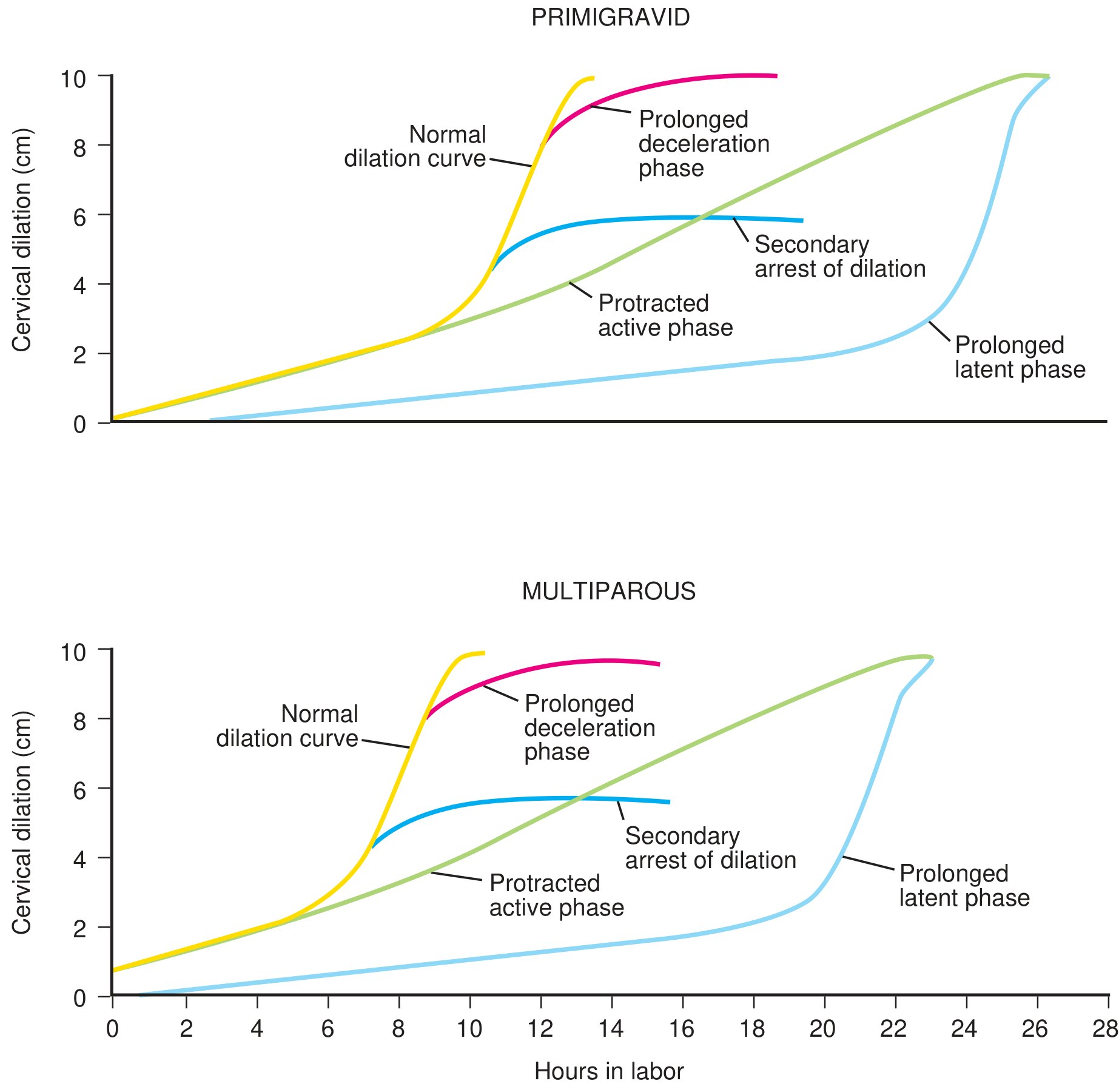
The normal dilation curve (yellow) shows a characteristic sigmoid shape. Abnormal variants include prolonged latent phase, protracted active phase, and secondary arrest of dilation.
4. Cardinal Movements of Labor (Mechanism of Delivery)
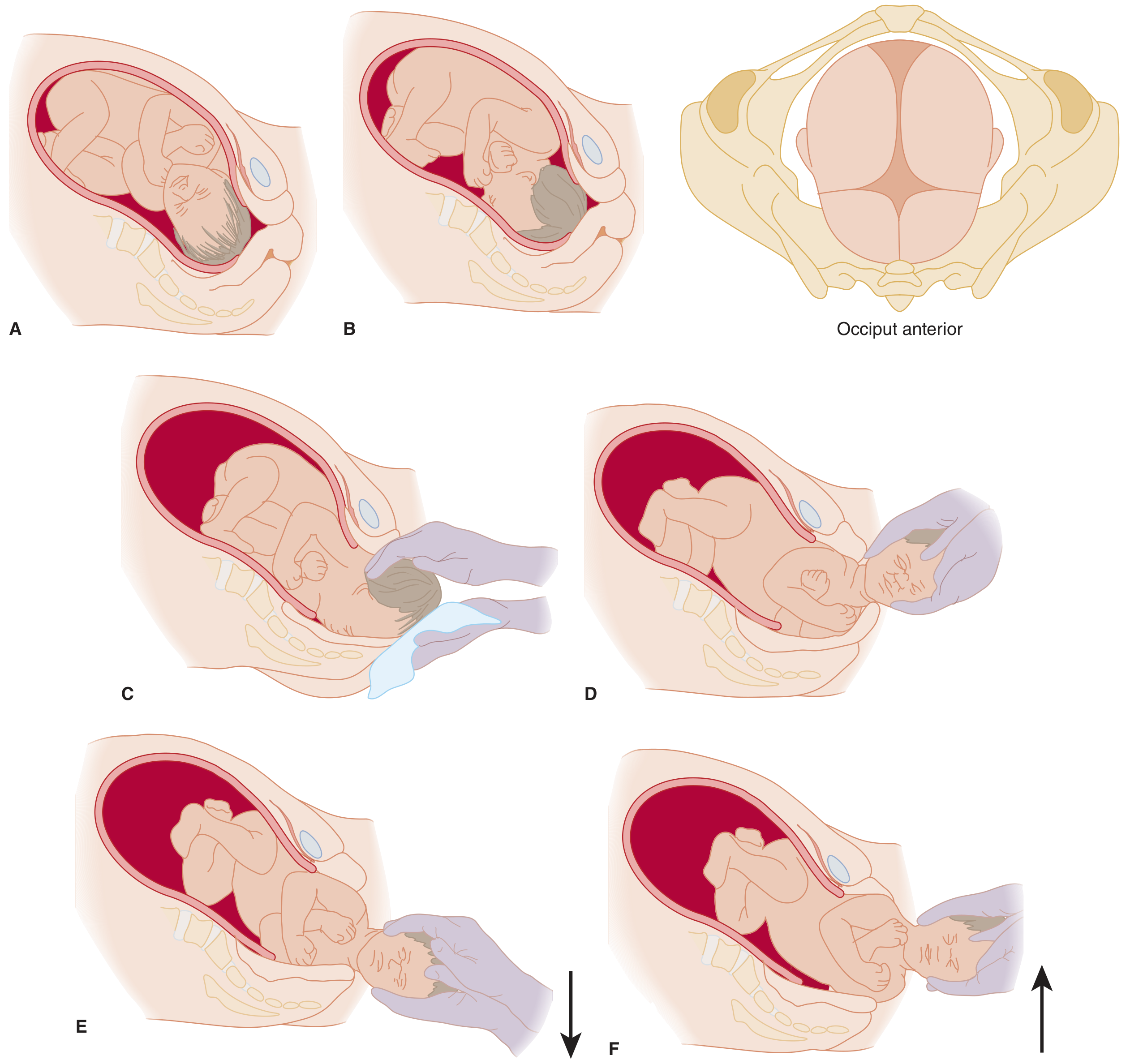
Because the pelvic inlet, midplane, and outlet each have different diameters, the fetal head must rotate at specific points during descent. These six cardinal movements describe vertex (head-first) delivery in the occiput anterior (OA) position:
- Engagement - biparietal diameter passes below the pelvic inlet; presenting part at or below ischial spines (station 0)
- Flexion - chin flexes onto chest, reducing the presenting diameter (suboccipitobregmatic) for easier passage
- Descent - progressive downward movement through the birth canal
- Internal rotation - occiput rotates from transverse to anterior (OA), aligning with the AP diameter of the pelvic outlet
- Extension - as the head reaches the introitus, it extends under the pubic symphysis; the head is delivered by extension
- External rotation (restitution) - after head delivery, the occiput rotates back to face the maternal thigh, reflecting the shoulders aligning in the AP diameter
5. Second Stage of Labor
- Begins at full cervical dilation (10 cm) - patient experiences urge to push
- Average duration: 20 minutes (multiparas) / 50 minutes (nulliparas)
- Alert for CPD if pushing exceeds 2 hours without epidural or 3 hours with epidural in nulliparas - but can continue if FHR is reassuring and descent is progressing
- Fetal heart auscultation every 15 minutes in low-risk, every 10 minutes in higher-risk
Delivery Technique
Head delivery:
- Support the perineum with a sterile towel (posterior hand) and control the fetal head (anterior hand)
- Ask the mother to breathe through contractions (not bear down) to allow slow, controlled expulsion and reduce perineal lacerations
- Head delivers with extension
- After head delivery: suction mouth, pharynx, then nose (if meconium - use DeLee trap via wall suction; do not stimulate breathing until laryngoscopy confirms no meconium below the cords)
- Check for nuchal cord - reduce over the head if possible; if too tight, clamp and cut
Shoulder delivery:
- Anterior shoulder: gentle downward traction + maternal expulsive effort; hyperflexion of maternal hips helps
- Posterior shoulder: gentle upward traction; instruct mother to push gently (excessive effort causes perineal lacerations)
- Remainder of body delivers easily after shoulders
After full delivery:
- Place baby on mother's abdomen on warm towels
- Delay cord clamping: at least 1-3 minutes after birth (increases neonatal iron stores)
- Double-clamp cord 3 cm from umbilicus and cut; obtain cord blood/pH if desired
6. Episiotomy
- Not routine - reserved for: fetal distress requiring expedited delivery, shoulder dystocia, or operative delivery (forceps/vacuum)
- Types:
- Median (midline): easier, less discomfort during recovery, but higher risk of 3rd/4th degree extension
- Mediolateral (45° from midline): lower risk of sphincter/rectal injury, but more painful recovery
- Technique: inject 5-10 mL of 1% lidocaine into posterior fourchette → 2-3 cm scissors incision while protecting fetal head
- Repair episiotomy and lacerations after placenta delivery
Laceration classification:
- 1st degree: skin only
- 2nd degree: skin + perineal muscle
- 3rd degree: involves anal sphincter
- 4th degree: through rectal mucosa
7. Third Stage of Labor (Placental Delivery)
- Placenta usually delivers 10-30 minutes after infant
- Signs of placental separation:
- Lengthening of the umbilical cord
- Sudden gush of blood
- Change in fundal contour (uterus becomes globular and rises)
- Management:
- Allow spontaneous separation - do NOT apply aggressive cord traction (risk: uterine inversion, cord avulsion)
- Gentle cord traction once separation signs appear
- Uterine massage + immediate breastfeeding promote uterine contraction
- Oxytocin (10-40 units in 1 L NS at 250 mL/hr, or 10 units IM) after delivery to prevent atony
8. Fourth Stage (First Hour Postpartum)
The period requiring most vigilance for hemorrhage.
Monitor for:
- Excessive vaginal bleeding
- Boggy, enlarging fundus
- Hypotension
Uterotonic agents for atony:
| Drug | Route | Dose |
|---|---|---|
| Oxytocin | IV/IM | 20-40 units/L IV or 20 units IM |
| Methylergonovine (Methergine) | IM | 0.2 mg q20 min (avoid in hypertension) |
| Carboprost (PGF₂α / Hemabate) | IM | 250 µg q15-20 min, up to 3 doses |
| Misoprostol | Oral/SL/rectal/vaginal | 200-1000 µg (widely used off-label) |
- Uterine massage: most often sufficient to contract the fundus and stop bleeding
- Bimanual massage alongside uterotonics for persistent atony
- Estimated normal blood loss in vaginal delivery: <500 mL
9. Neonatal Assessment - Apgar Score
Calculated at 1 minute and 5 minutes after delivery:
| Sign | 0 | 1 | 2 |
|---|---|---|---|
| Activity (tone) | Absent | Arms/legs flexed | Active movement |
| Pulse | Absent | <100 bpm | >100 bpm |
| Grimace (reflexes) | No response | Grimace | Sneeze/cough/pull away |
| Appearance (color) | Blue/pale | Blue extremities | Pink all over |
| Respiration | Absent | Weak/irregular | Strong cry |
- Score 7-10: normal
- Score <7: needs intervention (positive-pressure ventilation if HR <100 or apneic after 30 seconds)
10. Initial Maternal Admission Assessment
Vital signs: fever, tachycardia, elevated BP
Fetal heart tones: bradycardia or tachycardia
Positioning: avoid prolonged supine (compresses IVC → maternal hypotension → fetal hypoperfusion); use left lateral decubitus
Abdominal exam: fundal height, uterine tenderness, contraction frequency
Perineal exam: check for HSV lesions (contraindication to vaginal delivery)
Fetal heart tones: bradycardia or tachycardia
Positioning: avoid prolonged supine (compresses IVC → maternal hypotension → fetal hypoperfusion); use left lateral decubitus
Abdominal exam: fundal height, uterine tenderness, contraction frequency
Perineal exam: check for HSV lesions (contraindication to vaginal delivery)
Pelvic exam:
- If vaginal bleeding present: bedside ultrasound first to rule out placenta previa before any pelvic exam
- Sterile speculum to assess membrane status, cervical dilation, fetal station and presentation
- If membranes ruptured: no digital exam (even one exam increases infection risk; avoid lubricant as it causes false-positive nitrazine test)
11. Key Complications to Know
| Complication | Management |
|---|---|
| Umbilical cord prolapse | Elevate presenting part manually, keep hand in vagina, Trendelenburg position, emergent C-section; do NOT reduce cord |
| Shoulder dystocia | McRoberts maneuver (hyperflexion of hips), suprapubic pressure, episiotomy if needed |
| Postpartum hemorrhage | Bimanual massage + uterotonics; estimated blood loss >500 mL is pathologic |
| Meconium | DeLee suction, do not stimulate before laryngoscopy, prepare neonatal resuscitation team |
Textbook of Family Medicine, 9e, pp. 502-504 | Tintinalli's Emergency Medicine, 8e, pp. 681-683 | Roberts and Hedges' Clinical Procedures in Emergency Medicine
This is a shared conversation. Sign in to Orris to start your own chat.