
Give impression for the hypoxemia - NCCU BERROYA, JOHN 37/M NCCU3 2109154493 Informant: Mother Admitted Aug 9 due to facial asymmetry, right-sided weakness, loss of consciousness and diaphoresis PULMO PROFILE Non-smoker (+) Bronchia Asthma (Childhood) -No PFTs -On Symbicort (non-compliant) -No recent exacerbation (+) Allergic Rhinitis -Avamys nasal spray (+) Previous sleep studies - unrecalled results (+) COVID, non-critical (2021) STOPBANG 6 (Snoring, Daytime sleepiness, HTN, BMI 59, Neck circumference >40, male) BMI 59.34 (+) Throat mass (?) - sen by ENT advised elective excision (?) Past Medical History: (+) HTN (+) Congestive Heart Failure (+) Severe Hepatic Steatosis (+) Bilateral lower extremity Lymphedema (+) CKD At the ER BAT Activated Cranial MRI done showing intraparenchymal hemorrahge Started on Totilac At NCCU (+) drowsy (+) fluctuating o2 sats - hooked to 5LPM NC ABG done 8/10 ABG 7.43/52/70/34.5/94.9% on 5LPM Compensated metabolic alkalosis with suspected underlying respiratory acidosis with inadequate oxygenation PFR Seen awake Hooked to BIPAP Pinsp 10 PEEP 5 Fio2 60% No increase in work of breathing No desaturations BP 110/80 HR 80s RR 24-26 O2 sat 96% VTEs 340s Ppeak 15 (+) Rhonchi, bilateral Review of diagnostics 8/10 CXR The lungs are hypoaerated rendering bronchovascular crowding. Hazy and streaky densities are seen in both mid to lower lungs. Heart is enlarged. Both hemidiaphragms and costophrenic sulci are intact. The visualized osseous structures are unremarkable. 8/10 Cranial MRI Hyperacute intraparenchymal hemorrhage centered in the LEFT capsuloganglionic region, with surrounding perihematomal edema and mass effects, as detailed Subtle indentation of the LEFT cerebral peduncle, for which beginning uncal herniation is considered. Follow up is recommended. Chronic hemorrhage, RIGHT basal ganglia Chronic lacunar infarct with hemosiderin deposit, pons Microvascular ischemic white matter changes, both cerebral hemispheres (Fazekas 2) No critical stenosis, aneurysm or vascular malformation on time-of-flight angiography 8/9 Hgb 16.4 Hct 51 WBC 11830 N 62 L 23 Plt 346K 8/9 PT 13.3 INR 1.1 PTT 34.6 8/10 Na 143 K 3.5 Crea 1.7 BUN 16 Plans initially >ABG 1hr post BIPAP >Start Salbutamol + Ipratropium neb now then q6 >Budesonide 250mcg neb q12; Latest ABG 8/12 7.39 (7.45) pCO2 50 (48) pO2 89 (77) HCO3 30.3 (33.4) sO2 98 at 50 Compensated respiratory acidosis with adequate O2 for age DFiO2 44.94 PFR 178
-
Obesity Hypoventilation Syndrome (OHS) - most likely dominant etiology
- BMI 59.34 (Class III/Super obesity)
- STOP-BANG score 6 - high risk for Obstructive Sleep Apnea
- Previous sleep studies (results unrecalled)
- Baseline hypercapnia on ABG (pCO2 52 on 8/10, 50 on 8/12) with compensatory metabolic alkalosis (HCO3 34.5 → 30.3), consistent with chronic CO2 retention
- Responds to BIPAP (SpO2 improved to 96-98%)
-
Obstructive Sleep Apnea (OSA) - highly probable
- STOP-BANG 6/8 (Snoring, Daytime sleepiness, HTN, BMI >35, Neck circumference >40cm, Male)
- Prior sleep studies done (results unrecalled, suggesting prior diagnosis)
- Non-compliant with Symbicort - may reflect broader compliance issues
-
Bronchial Asthma / Airway Disease
- Childhood history, on Symbicort (non-compliant)
- Bilateral rhonchi on auscultation
- Responding partially to salbutamol + ipratropium nebulization
- Note: airflow obstruction may be compounding hypoventilation
-
Cardiogenic pulmonary congestion / Congestive Heart Failure
- Known CHF
- CXR showing cardiomegaly, bilateral mid-to-lower hazy and streaky densities, hypoaeration - consistent with pulmonary vascular congestion / interstitial edema
- Bilateral lower extremity lymphedema supports chronic fluid overload
-
Aspiration or Hypostatic pneumonitis - to consider
- Altered level of consciousness (drowsy, ICH with mass effect)
- Bilateral rhonchi, streaky densities on CXR
- Elevated WBC 11,830
-
Upper airway compromise - to consider
- Throat mass (noted, ENT-evaluated, elective excision advised)
- Relevant in the context of OSA/OHS physiology and BIPAP dependence
| Parameter | 8/10 (5LPM NC) | 8/12 (BIPAP 60%) |
|---|---|---|
| pH | 7.43 | 7.39 |
| pCO2 | 52 | 50 |
| pO2 | 70 | 89 |
| HCO3 | 34.5 | 30.3 |
| SpO2 | 94.9% | 98% |
| P/F Ratio | ~116 | ~178 |
- Improvement in oxygenation on BIPAP (P/F 116 → 178)
- Persistent but stable hypercapnia - consistent with chronic hypoventilator state
- P/F ratio of 178 on FiO2 50% - still in Moderate ARDS range by Berlin criteria (though context favors OHS/CHF/hypoventilation over primary ARDS)
- Repeat CXR to monitor pulmonary congestion
- Echocardiography (assess LV function, pulmonary pressures - PHT likely given OHS/OSA)
- Formal polysomnography or overnight oximetry once stable
- Pulmonary function tests (post-acute phase)
- ENT follow-up re: throat mass (airway patency concern)
- Thyroid function tests (rule out hypothyroidism as contributor to OHS/hypoventilation)
can Severe obesity lead reduced functional residual capacity, v/q mismatch hence hypoxemia?
obesity functional residual capacity V/Q mismatch hypoxemia
Severe Obesity → Reduced FRC → V/Q Mismatch → Hypoxemia
Step 1: Fat Loading Reduces Chest Wall and Lung Compliance
Step 2: Reduced FRC - Primarily via Loss of ERV
"Reduction in FRC is primarily a result of reduced expiratory reserve volume (ERV)... ERV is the most sensitive indicator of the effect of obesity on pulmonary function."
- Barash, Cullen, and Stoelting's Clinical Anesthesia, 9e
Step 3: FRC Falls Below Closing Capacity (CC) - The Critical Threshold
- FRC decreases (from fat loading)
- CC is unchanged (RV and CC are not affected by obesity)
- Therefore FRC approaches or falls below CC during normal tidal ventilation
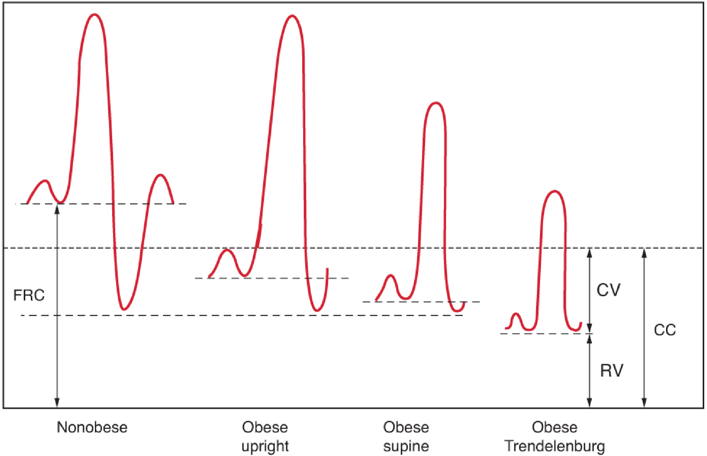
Step 4: Small Airway Closure → V/Q Mismatch and Shunt
| Mechanism | Effect |
|---|---|
| Low V/Q units | Collapsed/poorly ventilated alveoli continue to receive blood flow - blood passes through without adequate O2 loading |
| Intrapulmonary right-to-left shunt | Fully collapsed alveoli (atelectasis) receive perfusion with zero ventilation - true shunt |
| Expiratory flow limitation | Small airway closure causes air trapping and uneven distribution of ventilation |
"Reduced FRC (due to decreased ERV) can result in lung volumes below closing capacity in the course of normal tidal ventilation, leading to small airway closure, ventilation-perfusion mismatch, right-to-left shunting, and arterial hypoxemia."
- Barash, Cullen, and Stoelting's Clinical Anesthesia, 9e
- Reduced FRC, ERV
- Small airway closure
- Ventilation-perfusion inequality
- Expiratory flow limitation
Step 5: Worsened by Recumbency and Sleep
- Supine - abdominal contents further elevate the diaphragm, further reducing FRC
- Asleep - behavioral input to ventilation is lost; chemosensitivity to O2 and CO2 is blunted; ventilatory set point for PaCO2 rises 2-3 mmHg
- OSA superimposed - cyclic complete upper airway obstruction causes repetitive desaturations without recovery to baseline between apneas
"Patients desaturate because of (1) increased V/Q mismatch with increased perfusion to areas of decreased ventilation... and (5) alterations in respiratory mechanics, including reduced functional residual capacity and restricted chest wall expansion."
- Murray & Nadel's Textbook of Respiratory Medicine
Summary: The Complete Chain in This Patient (BERROYA, BMI 59.34)
Severe Obesity (BMI 59)
        ↓
↑ Fat load on thorax/abdomen
        ↓
↓ Chest wall & lung compliance
        ↓
↓ ERV → ↓ FRC (FRC approaches/falls below CC)
        ↓
Small airway closure during normal tidal breathing
        ↓
Low V/Q units + intrapulmonary shunt (dependent lung zones)
        ↓
Arterial hypoxemia (pO2 70 on 5LPM NC)
        ↓
Further worsened by:
- Supine position (ICU)
- Altered consciousness (ICH - ↓ respiratory drive)
- CHF/pulmonary congestion (↑ CC, ↓ compliance further)
- OSA/OHS (cyclic and chronic hypoventilation)