Potassium normal ranges

Normal serum potassium is 3.5 - 5.0 mEq/L (or mmol/L, same numeric value) in adults. Values below 3.5 are hypokalemia; values above 5.0 are hyperkalemia.
Searching sources...
Reading File
Finding Sources
Searching the Web

serum potassium normal range reference values clinical

Here is a complete reference for potassium normal ranges:

Serum Potassium - Normal Reference Ranges

Adults

MeasurementNormal Range
Serum potassium3.5 - 5.0 mEq/L (most labs)
Some lab references3.5 - 5.2 mEq/L
mEq/L = mmol/L for potassium (they are numerically identical)

By Age Group (RCPA Reference Ranges)

AgeNormal Range
Newborn (0 - <1 week)3.8 - 6.5 mmol/L
Infant (1 week - <26 weeks)4.2 - 6.7 mmol/L
Infant/toddler (26 weeks - <2 years)3.9 - 5.6 mmol/L
Child/adolescent (2 - <18 years)3.6 - 5.3 mmol/L
Adult (18+)3.5 - 5.2 mmol/L
Note: Infants and neonates normally have higher potassium than adults.

Clinical Interpretation

ValueClassification
< 3.5 mEq/LHypokalemia
3.5 - 5.0 mEq/LNormal
> 5.0 mEq/LHyperkalemia
Clinically significant thresholds:
  • Severe hypokalemia: < 2.5 mEq/L (cardiac arrhythmia risk)
  • Mild hypokalemia: 3.0 - 3.5 mEq/L
  • Mild hyperkalemia: 5.0 - 5.5 mEq/L
  • Severe hyperkalemia: > 6.5 mEq/L (life-threatening, immediate ECG changes)

Urine Potassium (Spot Sample)

ValueInterpretation
< 20 mEq/LSuggests poor intake, GI loss, or intracellular shift
> 40 mEq/LSuggests renal potassium wasting

Key Points

  • Serum vs plasma: Serum values run ~0.1-0.5 mEq/L higher than plasma due to release from platelets during clotting - this is a known pre-analytic artifact.
  • Pseudohyperkalemia (falsely elevated) can occur with hemolysis, prolonged tourniquet use, or extreme leukocytosis/thrombocytosis.
  • Hypertension patients: Recent evidence suggests an optimal range of 4.1 - 4.7 mmol/L is associated with lowest mortality, narrower than the standard reference range.
  • The normal range may vary slightly between laboratories - always check your lab's specific reference interval.
Sources: Goldman-Cecil Medicine; Schwartz's Principles of Surgery; RCPA Manual; Medscape Reference

Tell me about the diseases of the stomach and duodenum step by step and simplify the topic in surgery.Explain topic from basic to advanced in simple language.Use real life examples and ask me questions to test my understanding.You can use Manipal Manual of Surgery as your reference and make sure you cover everything including exam pearls and high yield stuff

Finding Sources
Diseases of the stomach and duodenum is a big surgical topic. Let me map it out first, then we'll go deep on whichever part you want.

Topic Breakdown

  1. Anatomy & Physiology Basics - stomach regions, blood supply, nerve supply (vagus), gastric secretion (HCl, pepsin, gastrin, somatostatin), duodenal anatomy - foundation for everything else
  2. Peptic Ulcer Disease (PUD) - gastric vs duodenal ulcer, H. pylori, NSAID-induced, pathophysiology, clinical features, investigations (endoscopy, urea breath test)
  3. Complications of Peptic Ulcer - perforation, hemorrhage (bleeding ulcer), gastric outlet obstruction (pyloric stenosis), malignant transformation
  4. Surgery for Peptic Ulcer Disease - vagotomy (truncal, selective, highly selective), gastrectomy types (Billroth I, Billroth II, Roux-en-Y), indications for surgery today vs historically
  5. Gastric Carcinoma - risk factors, early vs advanced gastric cancer, spread patterns (Virchow's node, Krukenberg tumor, Sister Mary Joseph nodule), staging, surgical management (subtotal vs total gastrectomy, lymphadenectomy)
  6. Gastric Lymphoma & GIST - MALT lymphoma, gastrointestinal stromal tumors, management differences from adenocarcinoma
  7. Zollinger-Ellison Syndrome - gastrinoma, MEN-1 association, diagnosis and management
  8. Congenital & Pediatric Conditions - congenital hypertrophic pyloric stenosis (CHPS), duodenal atresia
  9. Post-gastrectomy Syndromes - dumping syndrome (early/late), afferent loop syndrome, blind loop syndrome, bile reflux gastritis, nutritional deficiencies after gastrectomy
  10. Miscellaneous - gastric volvulus, Mallory-Weiss syndrome, Boerhaave syndrome, bezoars, duodenal diverticula

Quick Overview

The stomach and duodenum sit at the crossroads of acid, enzymes, and mechanical digestion, so most surgical disease here falls into three buckets: ulceration (acid-peptic disease and its complications), neoplasia (cancer, lymphoma, GIST), and mechanical/congenital problems (obstruction, atresia, volvulus).
Historically, peptic ulcer surgery (vagotomies, gastrectomies) was extremely common, but since the discovery that H. pylori and NSAIDs cause most ulcers, medical therapy (PPIs + antibiotics) has replaced surgery for uncomplicated PUD. Surgery today is mainly reserved for complications - a perforated ulcer needing emergency repair, uncontrolled bleeding, or obstruction - and for cancer resection. This shift is a favorite exam theme (Manipal loves asking "why has elective ulcer surgery declined?").
Gastric cancer surgery revolves around knowing anatomical spread (which lymph node stations, which distant sites) and choosing between subtotal and total gastrectomy based on tumor location. Post-gastrectomy syndromes are a classic viva topic because they test understanding of the altered anatomy created by each surgical technique.
Which topic do you want to start with?
Asking for Preferences

Which topic should we go deep into first?

Normal serum potassium is 3.5 - 5.0 mEq/L (or mmol/L, same numeric value) in adults. Values below 3.5 are hypokalemia; values above 5.0 are hyperkalemia.
**Searching sources...**Here is a complete reference for potassium normal ranges:

Serum Potassium - Normal Reference Ranges

Adults

MeasurementNormal Range
Serum potassium3.5 - 5.0 mEq/L (most labs)
Some lab references3.5 - 5.2 mEq/L
mEq/L = mmol/L for potassium (they are numerically identical)

By Age Group (RCPA Reference Ranges)

AgeNormal Range
Newborn (0 - <1 week)3.8 - 6.5 mmol/L
Infant (1 week - <26 weeks)4.2 - 6.7 mmol/L
Infant/toddler (26 weeks - <2 years)3.9 - 5.6 mmol/L
Child/adolescent (2 - <18 years)3.6 - 5.3 mmol/L
Adult (18+)3.5 - 5.2 mmol/L
Note: Infants and neonates normally have higher potassium than adults.

Clinical Interpretation

ValueClassification
< 3.5 mEq/LHypokalemia
3.5 - 5.0 mEq/LNormal
> 5.0 mEq/LHyperkalemia
Clinically significant thresholds:
  • Severe hypokalemia: < 2.5 mEq/L (cardiac arrhythmia risk)
  • Mild hypokalemia: 3.0 - 3.5 mEq/L
  • Mild hyperkalemia: 5.0 - 5.5 mEq/L
  • Severe hyperkalemia: > 6.5 mEq/L (life-threatening, immediate ECG changes)

Urine Potassium (Spot Sample)

ValueInterpretation
< 20 mEq/LSuggests poor intake, GI loss, or intracellular shift
> 40 mEq/LSuggests renal potassium wasting

Key Points

  • Serum vs plasma: Serum values run ~0.1-0.5 mEq/L higher than plasma due to release from platelets during clotting - this is a known pre-analytic artifact.
  • Pseudohyperkalemia (falsely elevated) can occur with hemolysis, prolonged tourniquet use, or extreme leukocytosis/thrombocytosis.
  • Hypertension patients: Recent evidence suggests an optimal range of 4.1 - 4.7 mmol/L is associated with lowest mortality, narrower than the standard reference range.
  • The normal range may vary slightly between laboratories - always check your lab's specific reference interval.
Sources: Goldman-Cecil Medicine; Schwartz's Principles of Surgery; RCPA Manual; Medscape ReferenceDiseases of the stomach and duodenum is a big surgical topic. Let me map it out first, then we'll go deep on whichever part you want.

Topic Breakdown

  1. Anatomy & Physiology Basics - stomach regions, blood supply, nerve supply (vagus), gastric secretion (HCl, pepsin, gastrin, somatostatin), duodenal anatomy - foundation for everything else
  2. Peptic Ulcer Disease (PUD) - gastric vs duodenal ulcer, H. pylori, NSAID-induced, pathophysiology, clinical features, investigations (endoscopy, urea breath test)
  3. Complications of Peptic Ulcer - perforation, hemorrhage (bleeding ulcer), gastric outlet obstruction (pyloric stenosis), malignant transformation
  4. Surgery for Peptic Ulcer Disease - vagotomy (truncal, selective, highly selective), gastrectomy types (Billroth I, Billroth II, Roux-en-Y), indications for surgery today vs historically
  5. Gastric Carcinoma - risk factors, early vs advanced gastric cancer, spread patterns (Virchow's node, Krukenberg tumor, Sister Mary Joseph nodule), staging, surgical management (subtotal vs total gastrectomy, lymphadenectomy)
  6. Gastric Lymphoma & GIST - MALT lymphoma, gastrointestinal stromal tumors, management differences from adenocarcinoma
  7. Zollinger-Ellison Syndrome - gastrinoma, MEN-1 association, diagnosis and management
  8. Congenital & Pediatric Conditions - congenital hypertrophic pyloric stenosis (CHPS), duodenal atresia
  9. Post-gastrectomy Syndromes - dumping syndrome (early/late), afferent loop syndrome, blind loop syndrome, bile reflux gastritis, nutritional deficiencies after gastrectomy
  10. Miscellaneous - gastric volvulus, Mallory-Weiss syndrome, Boerhaave syndrome, bezoars, duodenal diverticula

Quick Overview

The stomach and duodenum sit at the crossroads of acid, enzymes, and mechanical digestion, so most surgical disease here falls into three buckets: ulceration (acid-peptic disease and its complications), neoplasia (cancer, lymphoma, GIST), and mechanical/congenital problems (obstruction, atresia, volvulus).
Historically, peptic ulcer surgery (vagotomies, gastrectomies) was extremely common, but since the discovery that H. pylori and NSAIDs cause most ulcers, medical therapy (PPIs + antibiotics) has replaced surgery for uncomplicated PUD. Surgery today is mainly reserved for complications - a perforated ulcer needing emergency repair, uncontrolled bleeding, or obstruction - and for cancer resection. This shift is a favorite exam theme (Manipal loves asking "why has elective ulcer surgery declined?").
Gastric cancer surgery revolves around knowing anatomical spread (which lymph node stations, which distant sites) and choosing between subtotal and total gastrectomy based on tumor location. Post-gastrectomy syndromes are a classic viva topic because they test understanding of the altered anatomy created by each surgical technique.
Which topic do you want to start with?
Finding Sources
Finding Sources
Finding Sources
Reading File
Finding Sources
Finding Sources

Topic 1: Anatomy & Physiology of the Stomach and Duodenum (The Foundation)

Before we can understand diseases, we need to understand the normal machine that breaks down. Think of this like learning how a car engine works before learning why it breaks down. Let's build this step by step.

Step 1: The Stomach - Basic Layout

Picture the stomach as a J-shaped bag sitting in your upper left abdomen, just below your ribs. It has:
  • Fundus - the dome at the top (where swallowed air collects - that's why you burp!)
  • Body - the main chamber where food mixes
  • Antrum - the lower narrow part, acts like a "grinder" before pushing food out
  • Pylorus - the exit gate (a tight ring of muscle) that controls what leaves into the duodenum
Real-life example: Think of the stomach like a washing machine with a locked door. Food gets churned and mixed with acid (the "wash cycle"), and the pylorus is the locked door that only opens once the food (clothes) is broken down into small enough particles (liquid, <2mm) to pass through.

Step 2: Blood Supply (This is HIGH YIELD for exams)

Surgeons care intensely about blood supply because bleeding ulcers and surgical resections depend on it.
  • Lesser curve (the short, inner curve): supplied by left gastric artery (from the coeliac axis) + right gastric artery (from hepatic artery)
  • Greater curve (the long, outer curve): supplied by right gastroepiploic artery + left gastroepiploic artery (from splenic artery)
  • Fundus: supplied by short gastric arteries (vasa brevia) from the splenic artery
  • Gastroduodenal artery runs directly behind the first part of the duodenum
Exam Pearl: A posterior duodenal ulcer erodes into the gastroduodenal artery because that artery runs right behind the first part of the duodenum. This is THE classic cause of massive, life-threatening upper GI bleeding in surgery exams. Anterior ulcers, by contrast, tend to perforate (because there's nothing solid in front to erode into - just the peritoneal cavity).
Think of it like this: Back wall ulcer = bleeding (hits a artery). Front wall ulcer = perforation (hits open space).

Step 3: The Duodenum - Four Parts

The duodenum is C-shaped, curling around the head of the pancreas. It has 4 parts:
  1. First (superior) part - ~5cm, where most duodenal ulcers occur; behind it runs the gastroduodenal artery (danger zone!)
  2. Second (descending) part - contains the ampulla of Vater (where bile duct + pancreatic duct empty in)
  3. Third (horizontal) part - crosses in front of the aorta and vertebral column; can get compressed (SMA syndrome)
  4. Fourth (ascending) part - joins the jejunum at the duodenojejunal flexure (ligament of Treitz) - this landmark is important because it's how surgeons/radiologists distinguish upper GI bleed from lower GI bleed
Real-life example: Think of the duodenum like a roundabout (traffic circle) on a highway - it's the junction point where the stomach's output, the liver's bile, and the pancreas's enzymes all merge before heading down the small intestine highway.

Step 4: Gastric Acid Secretion (The Physiology Behind Peptic Ulcers)

This is the single most important physiology concept for understanding Topic 2 (Peptic Ulcer Disease), so let's nail it now.
The 3 stimulators of acid secretion, acting on the parietal cell:
  1. Gastrin (from G cells in the antrum) - released when food stretches the stomach or protein is present
  2. Histamine (from ECL cells - enterochromaffin-like cells) - this is why H2-blockers (ranitidine, famotidine) work
  3. Acetylcholine (from vagus nerve) - this is why vagotomy (cutting the vagus nerve) reduces acid
Simple analogy: Think of acid secretion like a factory with 3 supervisors telling workers (parietal cells) to make more product (HCl acid):
  • Supervisor 1 = Gastrin (hormone, travels in blood)
  • Supervisor 2 = Histamine (local chemical messenger)
  • Supervisor 3 = Vagus nerve (direct nerve wire)
All 3 supervisors ultimately push the parietal cell to pump out HCl. This is why PPIs (proton pump inhibitors) are the most effective drug class - they block the final common pathway (the proton pump itself), rather than blocking just one supervisor.
Exam Pearl: This is exactly why truncal vagotomy (an old surgical treatment for ulcers) reduced acid but also caused side effects - the vagus nerve doesn't just talk to the stomach, it also controls gastric emptying and gallbladder function, so cutting it causes gastric stasis and diarrhea (we'll cover this in the surgery topic).

Quick Recap (before you answer questions)

StructureKey Point
FundusAir collection, burping
AntrumGrinding, houses G cells (gastrin)
PylorusExit gate
First part of duodenumUlcer hotspot; gastroduodenal artery behind it
Ampulla of VaterBile + pancreatic duct entry (2nd part of duodenum)
Acid stimulatorsGastrin, Histamine, Acetylcholine → all act on parietal cell

Test Your Understanding - Answer These:

  1. A patient has a posterior duodenal ulcer that suddenly causes massive vomiting of blood. Which artery is most likely eroded, and why does the location (posterior vs anterior) matter?
  2. If you give a patient an H2-blocker like ranitidine, which of the 3 "supervisors" of acid secretion are you blocking?
  3. A surgeon cuts the vagus nerve trunk to reduce stomach acid in an old-style ulcer operation. Besides reducing acid, name one side effect you'd expect, based on what else the vagus nerve does.
Try answering these three, and I'll check your answers and then we'll move on to Topic 2: Peptic Ulcer Disease (causes, H. pylori, NSAIDs, clinical features) - which builds directly on what we just learned.

Phone number one, uh, you would... Which arch you would be eroded. It would be... The gastroduodenal artery. I don't know why the location would matter. Uh, for number two... You are blocking all three of the supervises. You are blocking the, the proton pump pathway. Number three. You would have gastric stasis and diarrhea.

Finding Sources
Finding Sources
Finding Sources

Feedback on Your Answers

Q1 - Correct, but let's fix the "why": Yes, the gastroduodenal artery. Location matters like this: the artery runs directly behind the first part of the duodenum. A posterior wall ulcer eats backward and erodes straight into that artery, causing massive bleeding. An anterior wall ulcer, on the other hand, has nothing solid behind it (just the open peritoneal cavity), so it tends to perforate instead. Simple rule to remember: "Posterior = Bleeds. Anterior = Bursts."
Q2 - Partially incorrect, important correction: Ranitidine (an H2-blocker) blocks only the histamine pathway (Supervisor 2) - it doesn't touch gastrin or acetylcholine directly. You were thinking of the PPI (proton pump inhibitor), which blocks the final common pathway (the proton pump itself) - that's why PPIs work regardless of which of the 3 supervisors is shouting, and why they're more powerful than H2-blockers. Good instinct connecting to the pump, just the wrong drug class.
Q3 - Correct. Vagotomy removes the nerve signal for gastric emptying too, causing gastric stasis (delayed emptying), and it disrupts normal bowel/biliary motility, contributing to post-vagotomy diarrhea. Solid answer.

Topic 2: Peptic Ulcer Disease (PUD)

Now that you know how acid is made, let's see what happens when the balance between acid/pepsin (attack) and mucosal defense (protection) breaks down.

Step 1: The Basic Concept - A Broken Balance

Think of the stomach lining like a castle wall protecting the tissue from acid inside. Two things guard the wall:
  • Mucus-bicarbonate layer (a protective coat)
  • Good blood flow (brings nutrients, removes acid that leaks through)
An ulcer happens when attack forces (acid, pepsin, H. pylori, NSAIDs) overwhelm defense forces, punching a hole through the wall.

Step 2: The Two Main Culprits

1. Helicobacter pylori (H. pylori)
  • A spiral bacterium that burrows into the mucus layer of the antrum
  • It weakens the mucus-bicarbonate barrier and triggers chronic inflammation (gastritis)
  • Found in the vast majority of duodenal ulcers and most gastric ulcers
  • Real-life example: Think of H. pylori like termites in a wooden fence - they don't attack all at once, but they quietly weaken the structure until a hole forms with normal wear and tear (acid).
2. NSAIDs (Aspirin, Ibuprofen, Diclofenac)
  • Mechanism: NSAIDs block the COX-1 enzyme, which normally makes prostaglandins that protect the stomach lining (they boost mucus, bicarbonate, and blood flow)
  • No COX-1 → no protective prostaglandins → the castle wall gets thin and weak, even without any bacteria involved
  • Real-life example: An elderly patient taking daily ibuprofen for knee arthritis develops a stomach ulcer with no H. pylori found - this is classic NSAID-induced injury, direct chemical damage to the defense system, not an infection.
Exam Pearl: COX-2 selective NSAIDs (like celecoxib) cause fewer ulcers because COX-2 is mostly involved in inflammation/pain, not stomach protection - but they raise cardiovascular risk instead. This trade-off is a favorite viva question.

Step 3: Gastric Ulcer vs Duodenal Ulcer - The Classic Comparison Table

FeatureDuodenal Ulcer (DU)Gastric Ulcer (GU)
Acid levelsNormal or increasedNormal or low
AgeYounger (30-50)Older (>50)
Pain timingRelieved by food ("food helps")Worsened by food ("food hurts")
Malignancy riskEssentially noneMust always rule out cancer
H. pylori association~90-95%~70-80%
Location risk1st part of duodenumLesser curvature, antrum
Real-life example to remember pain timing: A duodenal ulcer patient often wakes up at 2-3 AM with pain (acid builds up overnight with nothing to buffer it) and feels better after eating breakfast. A gastric ulcer patient feels fine before eating but develops pain soon after a meal, because food actually stimulates more acid/gastrin release, right where the ulcer is.
Critical Exam Pearl: Every gastric ulcer must be biopsied (usually 6-8 biopsies from the ulcer edge) to rule out gastric cancer, because a malignant ulcer can look identical to a benign one on endoscopy. Duodenal ulcers are essentially never biopsied for cancer because duodenal cancer is rare.

Step 4: Clinical Features

  • Epigastric pain - burning, gnawing
  • Duodenal ulcer: pain-food-relief pattern, nocturnal pain
  • Gastric ulcer: pain-food-pain pattern, may have weight loss (fear of eating)
  • Nausea, bloating, early satiety in some patients

Step 5: Investigations (High Yield)

  1. Upper GI endoscopy (OGD) - gold standard, allows direct visualization + biopsy
  2. H. pylori testing:
    • Urea breath test - non-invasive, good for confirming eradication after treatment
    • Rapid urease test (CLO test) - done on biopsy sample during endoscopy
    • Stool antigen test
    • Serology - only tells you about past exposure, not active infection (can't confirm cure)
Exam Pearl: Don't use serology to check if H. pylori treatment worked - antibodies stay positive for a long time even after the bacteria is gone. Use urea breath test or stool antigen instead.

Step 6: Basic Treatment Concept (Medical, Not Surgical - Surgery Topic Comes Later)

  • Triple therapy: PPI + Amoxicillin + Clarithromycin (7-14 days) - eradicates H. pylori
  • Stop the NSAID if that's the cause, or add a PPI cover if it can't be stopped
  • Surgery is now reserved almost exclusively for complications (which we'll cover next)

Quick Recap Table

ConceptKey Point
H. pylori mechanismWeakens mucus barrier, chronic gastritis
NSAID mechanismBlocks COX-1 → no protective prostaglandins
DU pain patternFood relieves, nocturnal pain
GU pain patternFood worsens, must biopsy for cancer
Best non-invasive H. pylori cure testUrea breath test or stool antigen (NOT serology)

Test Your Understanding:

  1. A 65-year-old woman on daily aspirin for her heart develops a stomach ulcer. Endoscopy shows no H. pylori. Explain in your own words why she still got an ulcer.
  2. A 35-year-old man says his stomach pain wakes him up at night but gets better after breakfast. Is this more likely a gastric ulcer or duodenal ulcer, and why?
  3. Why must every gastric ulcer be biopsied, but a duodenal ulcer usually isn't?
Give these a shot, and once you answer, we'll move to Topic 3: Complications of Peptic Ulcer Disease - perforation, bleeding, and gastric outlet obstruction, which is where surgery really comes into play.

Uh, she got an ulcer because of chronic NSAID use. And that damaged, uh, the stomach lining because... NSAID is a COX-1... COX-1 inhibitor. So if there is no COX-1... And COX... Sorry. And if, and COX-1 makes prostaglandins... If there's no COX-1... Um... I can't remember what else I can say about this. Number two. This is a duodenal ulcer because... It is a build-up of acid. After, uh, uh, a few hours after the meal, and that causes the pain. Uh, number three... Uh... Gastric ulcer can also present as a gastric cancer. And they have a higher risk of malignancy. That is why gastric, uh, ulcer must be biopsied.

Finding Sources
Finding Sources
Finding Sources

Feedback on Your Answers

Q1 - Correct, let's complete the thought: Exactly right up to "if there's no COX-1..." - here's the missing piece: COX-1 normally makes prostaglandins that protect the stomach lining by boosting mucus production, bicarbonate secretion, and blood flow to the mucosa. Block COX-1 with aspirin → no prostaglandins → the protective "castle wall" gets thin → acid burns straight through, even without any bacteria involved. Good reasoning, just needed that final link.
Q2 - Correct. Duodenal ulcer: acid builds up overnight with nothing to buffer it, causing nocturnal pain, and food (which buffers acid and also inhibits some acid release via mechanisms) relieves it.
Q3 - Correct. Gastric ulcers can look benign on endoscopy but actually be malignant, so biopsy is mandatory to rule out cancer. Duodenal ulcers essentially never turn out to be cancer, so it's skipped there.
You're getting the core logic. Let's move to where surgery really becomes relevant.

Topic 3: Complications of Peptic Ulcer Disease

This is the topic surgeons live in, because medical therapy has almost eliminated elective ulcer surgery - but complications still land on the operating table. There are 3 major complications. Think of them as "the 3 ways an ulcer can turn into an emergency."

Complication 1: Perforation

What happens: The ulcer eats all the way through the wall, and stomach/duodenal contents (acid, food, bacteria) spill into the peritoneal cavity, causing sudden chemical peritonitis.
Real-life example: Imagine a water pipe bursting inside your house wall - suddenly water (acid) is spraying everywhere it shouldn't be, causing damage far from the original leak site. That's exactly what happens when gastric/duodenal contents hit the peritoneum - it causes a diffuse, severe chemical burn of the abdominal lining.
Clinical features (classic triad-like presentation):
  • Sudden, severe "knife-like" epigastric pain
  • Pain quickly becomes generalized across the whole abdomen
  • Board-like rigidity of the abdominal wall (involuntary guarding) - a classic exam buzzword
  • Patient lies still (movement worsens pain) - unlike colicky pain where patients writhe around
Exam Pearl: The classic teaching says "sudden onset, board-like rigidity" but real-world/Bailey & Love explicitly notes this classic dramatic presentation is now seen less often, especially in elderly patients on steroids or NSAIDs, who may have a much subtler presentation. This is a common trick in exams - don't assume every perforation looks textbook.
Investigation:
  • Erect chest X-ray - look for free air under the diaphragm (seen in just over half of cases only - so a normal X-ray does NOT rule out perforation!)
  • CT scan - more accurate, now the go-to investigation
Management concept: Emergency surgery - classically Graham's omental patch repair (a piece of omentum is used to plug/patch the perforation), plus peritoneal lavage. We'll cover surgical technique details in the Surgery topic.

Complication 2: Hemorrhage (Bleeding Ulcer)

What happens: The ulcer erodes into a blood vessel. Remember Topic 1 - posterior duodenal ulcers erode into the gastroduodenal artery, causing the most dangerous bleeds.
Clinical features:
  • Hematemesis (vomiting blood - fresh red or "coffee-ground")
  • Melena (black, tarry, foul-smelling stools from digested blood)
  • Signs of hypovolemic shock if severe (tachycardia, hypotension)
High-Yield Concept - The Forrest Classification: This is THE tool used at endoscopy to predict rebleeding risk and decide who needs endoscopic therapy.
Forrest ClassEndoscopic FindingRebleed RiskNeeds Endoscopic Therapy?
IAActive spurting vesselVery highYes
IBActive oozingHighYes
IIAVisible non-bleeding vesselHighYes
IIBAdherent clotModerateConsider
IICFlat pigmented spotLowNo
IIIClean ulcer baseVery lowNo
Real-life example: Think of the Forrest classification like a fire department triage system - a spurting vessel (Class IA) is an active fire that needs immediate action, while a clean ulcer base (Class III) is just old ash with no risk of reigniting.
Management concept: Resuscitation first (IV fluids, blood transfusion if needed) → urgent endoscopy for diagnosis + treatment (injection of adrenaline, thermal coagulation, or clips) → surgery only if endoscopic therapy fails or rebleeding recurs.

Complication 3: Gastric Outlet Obstruction (GOO)

What happens: Chronic scarring and edema from a duodenal or pyloric channel ulcer narrows the outlet, so the stomach can't empty properly. (Note: in modern practice, GOO is now seen more often from malignancy - gastric or periampullary cancer - than from benign peptic scarring, since ulcers are treated early with PPIs.)
Clinical features:
  • Early satiety, abdominal fullness
  • Nausea and vomiting - classically of previously ingested, undigested food eaten hours or even a day earlier
  • Weight loss over time
  • Succussion splash - a sloshing sound heard on shaking the patient's abdomen, due to retained fluid in a distended stomach
Real-life example: Think of the pylorus like a clogged drain in a sink - water (food) keeps backing up and overflowing (vomiting) because it can't get through the narrowed exit. Over time, the "sink" (stomach) stretches out from constantly holding backed-up contents.
Metabolic Exam Pearl: Chronic vomiting from GOO classically causes a hypochloremic, hypokalemic metabolic alkalosis (loss of HCl and K+ in vomitus, with compensatory kidney changes worsening the alkalosis further - "paradoxical aciduria"). This is a very frequently tested lab pattern.
Management concept: Correct the fluid/electrolyte imbalance first, decompress the stomach (NG tube), then treat the cause - endoscopic balloon dilation for benign strictures, or surgery (gastrojejunostomy/pyloroplasty) for refractory benign disease or palliation of malignant obstruction.

Quick Recap Table

ComplicationKey BuzzwordDanger
PerforationBoard-like rigidity, free air under diaphragmChemical peritonitis
HemorrhagePosterior DU → gastroduodenal arteryHypovolemic shock
GOOSuccussion splash, hypochloremic hypokalemic alkalosisDehydration, malnutrition

Test Your Understanding:

  1. A patient with a known history of ulcers presents with sudden severe abdominal pain and a rigid abdomen. His chest X-ray shows no free air under the diaphragm. Can you rule out perforation? Why or why not?
  2. On endoscopy, a bleeding ulcer shows a visible vessel that is not actively bleeding (Forrest IIA). Should this patient get endoscopic therapy, and why does this matter clinically?
  3. A patient with chronic vomiting from gastric outlet obstruction is found to have low potassium and low chloride with a metabolic alkalosis. Explain in simple terms why vomiting causes this specific electrolyte pattern.
Answer these, and we'll move to Topic 4: Surgery for Peptic Ulcer Disease (vagotomy types, gastrectomy reconstructions) next.

Question one. I cannot rule out perforation because, mm-mm. 50% of the people will not... ... have free air under the diaphragm, so I would like to do a CT. Uh, to see... To make it more accurate, to see what's, uh, what's, what's going on. Number two... Yes, this person should be getting, uh, an endoscopic therapy. I do not know why it matters clinically. But we want to make sure the patient is hemodynamically stable. Number three. Uh, vomiting will cause all the gastric contents to be... vomited out. Um, there'll be loss of electrolytes. And that, that is the only thing I know.

Finding Sources
Finding Sources
Finding Sources

Feedback on Your Answers

Q1 - Correct. Free air is absent in nearly half of perforations, so a normal chest X-ray never rules it out. CT is now the more accurate, go-to investigation for exactly that reason.
Q2 - Correct action, let's complete the "why": Yes, Forrest IIA (visible non-bleeding vessel) needs endoscopic therapy. The clinical reason isn't primarily about hemodynamic stability at that exact moment - it's about preventing a future catastrophic rebleed. A visible vessel is like an unexploded landmine sitting in the ulcer base; even though it isn't actively bleeding right now, it has a high probability of bleeding again, and a second bleed can be fatal. Treating it now (clipping, injection, thermal therapy) defuses that landmine before it goes off.
Q3 - Partially correct, let's fill the gap (this is a favorite exam trick): You're right that gastric contents get lost. Specifically: HCl (hydrogen and chloride ions) are lost in vomit, directly causing alkalosis (less H+ in the body) and hypochloremia. Here's the twist that examiners love: the body also becomes volume-depleted, so the kidneys activate aldosterone to hang onto sodium and water - but aldosterone forces the kidney to dump potassium and hydrogen ions into urine to do this. So the kidney, trying to fix volume depletion, actually worsens the alkalosis and causes hypokalemia. This is called "paradoxical aciduria" - the urine is acidic despite the blood being alkalotic. It's one of the most tested acid-base scenarios in surgery exams.

Topic 4: Surgery for Peptic Ulcer Disease

Now we get to the operating table. First, the most important concept to understand before memorizing procedures:

The Big Picture Shift (Exam Favorite Question)

Before H. pylori was discovered (1980s) and before PPIs existed, elective surgery for uncomplicated ulcers was common. Today, medical therapy is so effective that elective ulcer surgery has almost disappeared. Surgery is now used almost exclusively for complications - perforation, uncontrolled bleeding, or obstruction - which is exactly what we covered in Topic 3.
Real-life analogy: Think of it like treating high blood pressure. Decades ago, before good drugs existed, doctors sometimes did surgery on nerves to lower blood pressure. Now that we have effective pills, that surgery is essentially obsolete - reserved only for rare situations medicine can't fix. Ulcer surgery followed the exact same path once PPIs and H. pylori eradication arrived.

Step 1: Vagotomy - The Old-School Acid-Reduction Operations

Remember Topic 1 - acetylcholine (via the vagus nerve) is one of the 3 "supervisors" telling parietal cells to make acid. Vagotomy cuts that wire. There are 3 types, and the differences matter a lot for exams:
TypeWhat's CutAcid ReductionProblemNeeds Drainage Procedure?
Truncal vagotomyBoth main vagal trunks at the esophagusMost completeDenervates the whole stomach, liver, gallbladder, intestines too → gastric stasis, diarrhea, gallstonesYes - because it paralyzes the antropyloric pump
Selective vagotomyOnly the nerve branches to stomach (spares liver/biliary/intestinal branches)GoodRarely used nowYes
Highly selective vagotomy (HSV) (also called parietal cell vagotomy)Only the nerves to the acid-secreting body/fundus, sparing the nerve to the antrum/pylorus (nerve of Latarjet)Good, more targetedLowest complication rate of all vagotomy typesNo - pylorus still functions normally
Why drainage procedures matter: Truncal and selective vagotomy paralyze the pylorus's ability to relax and open, so surgeons must add a drainage procedure - most commonly a Heineke-Mikulicz pyloroplasty (a longitudinal cut across the pylorus, sewn transversely to widen it) - otherwise the stomach can't empty. Highly selective vagotomy specifically spares this nerve, so no drainage procedure is needed. This is a classic exam distinction.
Exam Pearl (recurrence vs complication rates): HSV has a higher ulcer recurrence rate than truncal vagotomy + antrectomy, but a much lower complication/side-effect rate. This trade-off (fewer side effects vs slightly more recurrence) is a favorite viva question - "why would you choose HSV despite higher recurrence?" Answer: because the side effects of the more aggressive operations (dumping, diarrhea, bile reflux) are often worse for quality of life than a treatable recurrence.

Step 2: Gastrectomy-Based Procedures - Removing Part of the Stomach

These remove the acid/gastrin-producing tissue directly, then reconstruct how the remaining stomach connects to the intestine.
Billroth I (Gastroduodenostomy)
  • Remove the distal stomach (antrum) → connect the remaining stomach directly to the duodenum
  • Real-life analogy: Think of it like removing a section of pipe and directly rejoining the two remaining ends - simple, preserves the normal route (bile and pancreatic juice still meet food at the usual point)
  • More "physiological" - food still passes through the duodenum
Billroth II (Gastrojejunostomy / Polya)
  • Remove the distal stomach → close off the duodenal stump → connect the remaining stomach directly to a loop of jejunum, bypassing the duodenum
  • Real-life analogy: Instead of rejoining the original pipe, you cap off the old exit and build a brand-new pipe connection further downstream
  • Creates an afferent loop (duodenum + proximal jejunum, carrying bile/pancreatic juice) and an efferent loop (carries food onward) - this altered anatomy is exactly why afferent loop syndrome exists (we'll cover this in Post-gastrectomy Syndromes)
Roux-en-Y Gastrojejunostomy
  • A more modern reconstruction: the jejunum is divided, one limb is connected to the stomach, and bile/pancreatic secretions are diverted lower down through a separate limb
  • Advantage: Significantly reduces bile reflux gastritis because bile no longer washes back up into the stomach
  • Increasingly preferred, especially after total gastrectomy for cancer
Exam Pearl: Billroth II patients are notoriously difficult for ERCP (endoscopic bile duct procedures) later in life, because the altered anatomy (bypassed duodenum, blind afferent loop) makes it hard to reach the ampulla of Vater endoscopically. This comes up in GI/surgery integration questions.

Step 3: Putting It Together - Which Operation for Which Complication?

Complication (from Topic 3)Typical Surgical Approach
PerforationGraham's omental patch (simple closure + omentum patch), rarely definitive acid-reducing surgery in the emergency setting
Uncontrolled bleedingEndoscopic control first; if failed, surgical under-running of the bleeding vessel ± acid-reducing procedure
Gastric outlet obstruction (benign)Gastrojejunostomy or pyloroplasty to bypass/widen the narrowed segment

Quick Recap Table

ConceptKey Point
Why elective ulcer surgery declinedPPIs + H. pylori eradication treat the cause medically
HSVNo drainage needed, fewer side effects, slightly higher recurrence
Truncal/selective vagotomyNeeds drainage procedure (pyloroplasty)
Billroth IStomach → duodenum directly
Billroth IIStomach → jejunum, duodenum bypassed (afferent/efferent loops created)
Roux-en-YDiverts bile away from stomach, reduces bile reflux

Test Your Understanding:

  1. Why does truncal vagotomy require an additional drainage procedure like pyloroplasty, but highly selective vagotomy does not?
  2. A patient who had a Billroth II gastrectomy years ago now needs an ERCP for a gallstone in the bile duct. Why might this procedure be technically more difficult in this patient compared to someone with normal anatomy?
  3. Why has elective surgery for uncomplicated peptic ulcer disease become rare today, and in what situations is surgery still needed?
Answer these, and next we'll move to Topic 5: Gastric Carcinoma - a big exam topic covering risk factors, spread patterns (Virchow's node, Krukenberg tumor), and surgical management.

Move on to the next topic

Finding Sources
Finding Sources
Finding Sources
Searching Images

gastric carcinoma linitis plastica endoscopic image

This composite medical image provides a multimodal view of gastric pathology, specifically linitis plastica-like findings associated with metastatic lobular breast carcinoma. Panel (a) is an upper gastrointestinal endoscopy showing the gastric lumen. The mucosal rugae are notably edematous, thickened, and lack normal distensibility, presenting a reddish-pink, glistening surface. Panel (b) is an axial contrast-enhanced CT scan of the upper abdomen. It demonstrates significant, diffuse, and circumferential thickening of the gastric wall (linitis plastica appearance) with prominent contrast enhancement. The stomach wall appears hyper-attenuating compared to typical gastric tissue. These visual findings are clinically significant for identifying infiltrative gastric lesions (Borrmann type 4), which in this case represent secondary involvement of the stomach by metastatic disease. The image is useful for medical education regarding the diagnostic correlation between endoscopic mucosal changes and cross-sectional radiological findings in oncological gastrointestinal pathology.

This composite medical image provides a multimodal view of gastric pathology, specifically linitis plastica-like findings associated with metastatic lobular breast carcinoma. Panel (a) is an upper gastrointestinal endoscopy showing the gastric lumen. The mucosal rugae are notably edematous, thickened, and lack normal distensibility, presenting a reddish-pink, glistening surface. Panel (b) is an axial contrast-enhanced CT scan of the upper abdomen. It demonstrates significant, diffuse, and circumferential thickening of the gastric wall (linitis plastica appearance) with prominent contrast enhancement. The stomach wall appears hyper-attenuating compared to typical gastric tissue. These visual findings are clinically significant for identifying infiltrative gastric lesions (Borrmann type 4), which in this case represent secondary involvement of the stomach by metastatic disease. The image is useful for medical education regarding the diagnostic correlation between endoscopic mucosal changes and cross-sectional radiological findings in oncological gastrointestinal pathology.

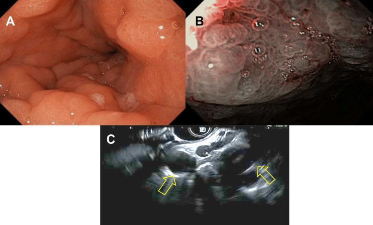
This composite clinical figure displays diagnostic images from an upper gastrointestinal evaluation. Panel A is a standard white-light endoscopic view of the gastric antrum showing longitudinal folds with several white spots, indicating an inflamed gastric mucosa. Panel B provides a high-contrast view using Narrow-Band Imaging (NBI), which highlights a distorted, nodular mucosal surface with irregular vascular patterns and darker coloration, suggestive of infiltrative pathology like linitis plastica. Panel C presents an endoscopic ultrasound (EUS) image where yellow arrows indicate multiple enlarged, hypoechoic lymph nodes. Collectively, these images illustrate a diagnostic workup for gastric wall thickening and lymphadenopathy, clinically relevant for identifying metastatic disease such as lobular breast carcinoma or primary gastric malignancy. The target audience includes gastroenterologists and oncology residents focusing on endoscopic diagnostic techniques.

This composite clinical figure displays diagnostic images from an upper gastrointestinal evaluation. Panel A is a standard white-light endoscopic view of the gastric antrum showing longitudinal folds with several white spots, indicating an inflamed gastric mucosa. Panel B provides a high-contrast view using Narrow-Band Imaging (NBI), which highlights a distorted, nodular mucosal surface with irregular vascular patterns and darker coloration, suggestive of infiltrative pathology like linitis plastica. Panel C presents an endoscopic ultrasound (EUS) image where yellow arrows indicate multiple enlarged, hypoechoic lymph nodes. Collectively, these images illustrate a diagnostic workup for gastric wall thickening and lymphadenopathy, clinically relevant for identifying metastatic disease such as lobular breast carcinoma or primary gastric malignancy. The target audience includes gastroenterologists and oncology residents focusing on endoscopic diagnostic techniques.

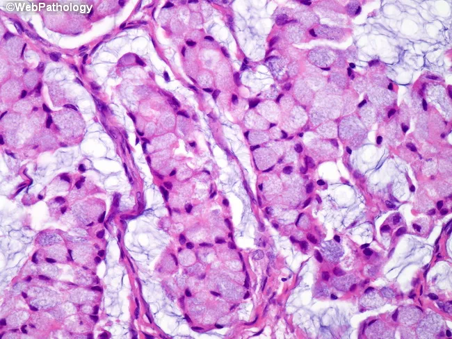
This histopathology image depicts an ovarian metastasis consistent with a Krukenberg tumor. The specimen is an ovarian tissue section prepared for light microscopy and stained with Hematoxylin and Eosin. The primary neoplasm is diffuse-type gastric adenocarcinoma presenting as linitis plastica; however, this image centers on metastatic ovarian involvement. Morphology shows nests and clusters of pleomorphic signet-ring cells with intracellular mucin vacuoles that displace the nucleus to the periphery, producing the characteristic signet-ring appearance. Cells are embedded in a fibromyxoid stromal background with infiltrative spread, occasional acinar-like architecture, and variable cytoplasmic eosinophilia. The mucin-rich cytoplasm yields a pale to empty-appearing cell, contributing to overall discohesive, signet-ring phenotype. The lesion commonly involves both ovaries, though laterality cannot be determined from a single slide. These features reflect metastatic spread from a primary diffuse gastric carcinoma (linitis plastica). Clinically, recognition of signet-ring cell adenocarcinoma in the ovary carries high diagnostic significance: it implies a gastric origin, guides endoscopic evaluation, staging, and systemic therapy, and correlates with poorer prognosis. Differential diagnoses include primary ovarian signet-ring cell/metastatic tumors from breast or colorectal primaries; however, bilateral ovarian involvement with mucin production and the histo-pattern described supports Krukenberg metastasis from gastric cancer.

This histopathology image depicts an ovarian metastasis consistent with a Krukenberg tumor. The specimen is an ovarian tissue section prepared for light microscopy and stained with Hematoxylin and Eosin. The primary neoplasm is diffuse-type gastric adenocarcinoma presenting as linitis plastica; however, this image centers on metastatic ovarian involvement. Morphology shows nests and clusters of pleomorphic signet-ring cells with intracellular mucin vacuoles that displace the nucleus to the periphery, producing the characteristic signet-ring appearance. Cells are embedded in a fibromyxoid stromal background with infiltrative spread, occasional acinar-like architecture, and variable cytoplasmic eosinophilia. The mucin-rich cytoplasm yields a pale to empty-appearing cell, contributing to overall discohesive, signet-ring phenotype. The lesion commonly involves both ovaries, though laterality cannot be determined from a single slide. These features reflect metastatic spread from a primary diffuse gastric carcinoma (linitis plastica). Clinically, recognition of signet-ring cell adenocarcinoma in the ovary carries high diagnostic significance: it implies a gastric origin, guides endoscopic evaluation, staging, and systemic therapy, and correlates with poorer prognosis. Differential diagnoses include primary ovarian signet-ring cell/metastatic tumors from breast or colorectal primaries; however, bilateral ovarian involvement with mucin production and the histo-pattern described supports Krukenberg metastasis from gastric cancer.

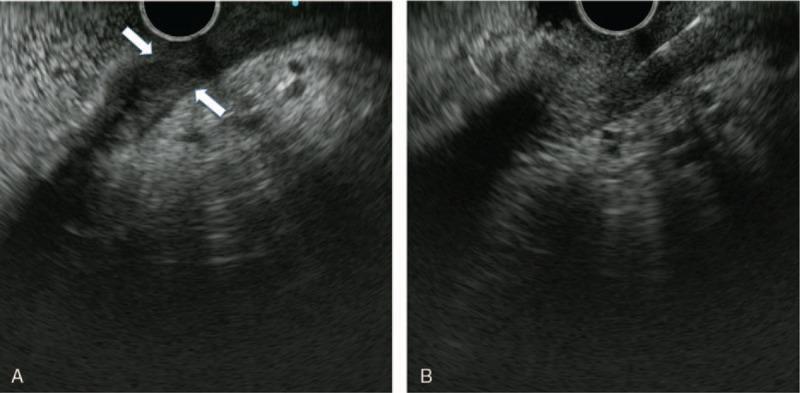
This diagnostic image consists of two panels (A and B) showing endoscopic ultrasound (EUS) of the gastric wall. Panel A demonstrates significant, diffuse gastric wall thickening (measured up to 9.3 mm) primarily located at the greater curvature. The typical five-layer sonographic architecture of the gastric wall is notably lost or 'unclear,' replaced by a relatively homogeneous, slightly low-level (hypoechoic) echo pattern throughout all layers. Panel B illustrates an endoscopic ultrasound-guided fine needle aspiration (EUS-FNA) procedure. A linear echogenic needle is visible penetrating the thickened gastric wall in a parallel orientation to the wall surface, directed toward the lesion or perigastric fluid. These findings are clinically significant for the evaluation of infiltrative gastric processes, such as linitis plastica or signet ring cell carcinoma, where normal wall stratification is often disrupted by malignant infiltration.

This diagnostic image consists of two panels (A and B) showing endoscopic ultrasound (EUS) of the gastric wall. Panel A demonstrates significant, diffuse gastric wall thickening (measured up to 9.3 mm) primarily located at the greater curvature. The typical five-layer sonographic architecture of the gastric wall is notably lost or 'unclear,' replaced by a relatively homogeneous, slightly low-level (hypoechoic) echo pattern throughout all layers. Panel B illustrates an endoscopic ultrasound-guided fine needle aspiration (EUS-FNA) procedure. A linear echogenic needle is visible penetrating the thickened gastric wall in a parallel orientation to the wall surface, directed toward the lesion or perigastric fluid. These findings are clinically significant for the evaluation of infiltrative gastric processes, such as linitis plastica or signet ring cell carcinoma, where normal wall stratification is often disrupted by malignant infiltration.

Topic 5: Gastric Carcinoma

This is one of the highest-yield oncology-surgery topics because examiners love testing the classic "eponymous signs" of spread. Let's build it step by step.

Step 1: Risk Factors - Think "Chronic Irritation Over Time"

Gastric cancer develops from years of chronic mucosal injury. Key risk factors:
  • H. pylori infection - the single biggest risk factor (causes chronic gastritis → atrophic gastritis → intestinal metaplasia → dysplasia → cancer, a well-known stepwise progression called the Correa cascade)
  • Diet - smoked, salted, pickled foods (high nitrates/nitrites), low fruit/vegetable intake
  • Smoking
  • Blood group A (classic exam factoid, mechanism not fully understood)
  • Pernicious anemia / atrophic gastritis - loss of acid production, bacterial overgrowth, nitrosamine formation
  • Family history / hereditary diffuse gastric cancer (CDH1 gene mutation)
  • Previous partial gastrectomy (bile reflux onto the remnant stomach mucosa over decades - remember this connects to Topic 4!)
Real-life example: Think of the stomach lining like soil that's been repeatedly exposed to acid rain (H. pylori, salt, smoke) for decades. Eventually the soil quality degrades (atrophic gastritis), and abnormal "weeds" (dysplasia) start growing, which can eventually turn into cancer.

Step 2: Two Main Types (Worth Knowing)

  • Intestinal type - well-differentiated, forms a gland-like structure, associated with H. pylori/atrophic gastritis, more common in older men, seen in high-incidence areas
  • Diffuse type - poorly differentiated, individual cells infiltrate the wall (classic signet-ring cells - mucin pushes the nucleus to one side, making the cell look like a ring), spreads along the wall causing linitis plastica ("leather bottle stomach" - a rigid, thickened, non-distensible stomach), worse prognosis, seen in younger patients, linked to CDH1 mutations
Real-life example: Intestinal type is like a single tumor growing as a lump (like a golf ball), while diffuse type is like ink soaking through paper - it infiltrates diffusely through the wall rather than forming one discrete mass, which is why linitis plastica stomachs feel rigid and leathery on imaging, not lumpy.

Step 3: The Classic "Eponymous Signs" of Spread (VERY High Yield)

Gastric cancer has a legendary list of named signs marking distant spread - these are asked constantly in exams:
SignSiteWhat It Means
Virchow's nodeLeft supraclavicular lymph nodeCancer has spread via thoracic duct to distant nodes
Sister Mary Joseph nodulePeriumbilical noduleMetastasis to the umbilicus (via ligaments/peritoneal spread)
Krukenberg tumorOvary (often bilateral)"Drop metastases" - cells seed onto the ovaries via peritoneal fluid; histology shows classic signet-ring cells
Blumer's shelfPouch of Douglas (felt on rectal exam)Peritoneal deposits gravitate to the pelvis
Irish nodeLeft axillary nodeAnother distant nodal spread site
Here's the histology of a Krukenberg tumor showing the classic signet-ring cells:
Krukenberg tumor histology showing signet-ring cells
Real-life example to remember these: Imagine gastric cancer cells as seeds blown by the wind (lymphatic/peritoneal fluid). Some seeds travel up through lymphatic channels all the way to the neck (Virchow's node), some float down with gravity in peritoneal fluid to the pelvis (Blumer's shelf) or land on the ovaries (Krukenberg), and some travel along the ligament to the belly button (Sister Mary Joseph). All of these findings mean the disease is already metastatic - unfortunately, by the time these signs appear, curative surgery is usually no longer possible.
Exam Pearl: All of these eponymous signs indicate incurable, stage IV disease. Finding any one of them on physical exam should immediately shift the conversation from "how do we resect this" to "how do we palliate this."

Step 4: Surgical Management - Location Decides the Operation

  • Distal/antral tumorsSubtotal (partial) gastrectomy with adequate margins, usually reconstructed as Billroth II or Roux-en-Y
  • Proximal tumors (body/cardia) or diffuse/linitis plasticaTotal gastrectomy, reconstructed with Roux-en-Y esophagojejunostomy
  • Lymphadenectomy - removing regional lymph nodes (D1 vs D2 dissection is a major topic in Eastern vs Western surgical practice, with D2 favored in Japan/Korea and increasingly worldwide)
Exam Pearl: Despite doing a more extensive resection, there is very little functional difference in quality of life between total and subtotal gastrectomy patients long-term - both need careful nutritional follow-up (B12 injections for life after total gastrectomy, since intrinsic factor is gone).

Step 5: Early vs Advanced Gastric Cancer

  • Early gastric cancer (EGC) - confined to mucosa/submucosa, regardless of lymph node status. Very good prognosis if caught here (common in Japan due to screening programs). Can sometimes be treated with endoscopic mucosal resection (EMR) or endoscopic submucosal dissection (ESD) instead of surgery.
  • Advanced gastric cancer - invades muscularis propria or beyond. Needs formal gastrectomy + lymphadenectomy ± chemotherapy.

Quick Recap Table

ConceptKey Point
Biggest risk factorH. pylori (Correa cascade)
Intestinal typeGland-forming, older patients, H. pylori-linked
Diffuse typeSignet-ring cells, linitis plastica, younger patients, CDH1
Virchow's/SMJ/Krukenberg/Blumer'sAll signal incurable stage IV disease
Distal tumorSubtotal gastrectomy
Proximal/diffuse tumorTotal gastrectomy + Roux-en-Y

Test Your Understanding:

  1. A 60-year-old man with known gastric cancer is found on physical exam to have a hard, enlarged lymph node above his left collarbone. What is this called, and what does it mean for his treatment plan?
  2. Why does diffuse-type gastric cancer with signet-ring cells tend to cause a rigid, non-distensible "leather bottle" stomach, while intestinal-type cancer typically forms a discrete mass?
  3. A patient presents with a new ovarian mass. Biopsy shows signet-ring cells. What primary cancer should you suspect, and what is this metastatic pattern called?
Answer when ready, and we'll move to Topic 6: Gastric Lymphoma & GIST next - a shorter topic that contrasts nicely with adenocarcinoma.

Tell me about colorectal polyps and colorectal cancer step by step and simplify the topic in surgery.Explain topic from basic to advanced in simple language.Use real life examples and ask me questions to test my understanding.You can use Manipal Manual of Surgery as your reference and make sure you cover everything including exam pearls and high yield stuff tested in surgery exams.You man include stuff from Browse’s textbook because that is where my examiners choose their questions from

Finding Sources
Colorectal polyps and cancer is another big surgical topic. Here's the map before we go deep on any one part.

Topic Breakdown

  1. Colon & Rectum Basics - anatomy (ascending/transverse/descending/sigmoid/rectum), blood supply (SMA vs IMA territory), lymphatic drainage - the foundation
  2. Colorectal Polyps - types (adenomatous, hyperplastic, hamartomatous, inflammatory), adenoma-carcinoma sequence, malignant potential factors (size, villous type, dysplasia)
  3. Polyposis Syndromes - FAP, Lynch syndrome (HNPCC), Peutz-Jeghers, juvenile polyposis, MUTYH-associated polyposis - genetics and cancer risk
  4. Colorectal Carcinoma - Etiology & Pathology - risk factors, molecular pathways (APC, KRAS, p53), left-sided vs right-sided cancer differences
  5. Colorectal Carcinoma - Clinical Presentation & Screening - symptoms by location, screening guidelines (colonoscopy, FIT test), staging (TNM, Dukes)
  6. Investigations & Staging - colonoscopy, CT, CEA, MRI rectum, staging systems
  7. Surgical Management - right hemicolectomy, left hemicolectomy, sigmoid colectomy, anterior resection, abdominoperineal resection (APR), total mesorectal excision (TME)
  8. Rectal Cancer Specifics - neoadjuvant chemoradiation, sphincter-preserving surgery vs APR, TME principles
  9. Complications & Emergency Presentations - obstruction, perforation, management of acute large bowel obstruction
  10. Colorectal Liver Metastases - resectability criteria, staged resections, chemotherapy

Quick Overview

Colorectal disease in surgery splits into two connected stories: polyps (the precursor lesions) and cancer (the endpoint). The key unifying concept is the adenoma-carcinoma sequence - most colorectal cancers arise from adenomatous polyps over years, through a stepwise accumulation of mutations (APC, KRAS, p53). This is why colonoscopic screening and polypectomy actually prevent cancer, not just detect it early.
Location matters enormously in this topic. Right-sided cancers tend to present with anemia and occult bleeding (wide lumen, liquid stool, tumors grow large before obstructing), while left-sided cancers present with obstruction and altered bowel habit (narrow lumen, solid stool). Surgical management follows the blood supply and lymphatic drainage of each segment, so knowing the anatomy in Topic 1 makes every operation in Topic 7 logical rather than something to memorize by rote.
Rectal cancer is treated as its own subspecialty within this topic because of two things unique to the rectum: no serosa (different spread pattern) and proximity to the sphincter (raising the constant question "can we save the sphincter or does the patient need a permanent stoma"). This is where TME, neoadjuvant therapy, and the APR vs anterior resection decision become major exam themes, especially from Bailey & Love and Browse's.
Which topic should we start with?
Asking for Preferences

Which topic should we go deep into first?

Normal serum potassium is 3.5 - 5.0 mEq/L (or mmol/L, same numeric value) in adults. Values below 3.5 are hypokalemia; values above 5.0 are hyperkalemia.
**Searching sources...**Here is a complete reference for potassium normal ranges:

Serum Potassium - Normal Reference Ranges

Adults

MeasurementNormal Range
Serum potassium3.5 - 5.0 mEq/L (most labs)
Some lab references3.5 - 5.2 mEq/L
mEq/L = mmol/L for potassium (they are numerically identical)

By Age Group (RCPA Reference Ranges)

AgeNormal Range
Newborn (0 - <1 week)3.8 - 6.5 mmol/L
Infant (1 week - <26 weeks)4.2 - 6.7 mmol/L
Infant/toddler (26 weeks - <2 years)3.9 - 5.6 mmol/L
Child/adolescent (2 - <18 years)3.6 - 5.3 mmol/L
Adult (18+)3.5 - 5.2 mmol/L
Note: Infants and neonates normally have higher potassium than adults.

Clinical Interpretation

ValueClassification
< 3.5 mEq/LHypokalemia
3.5 - 5.0 mEq/LNormal
> 5.0 mEq/LHyperkalemia
Clinically significant thresholds:
  • Severe hypokalemia: < 2.5 mEq/L (cardiac arrhythmia risk)
  • Mild hypokalemia: 3.0 - 3.5 mEq/L
  • Mild hyperkalemia: 5.0 - 5.5 mEq/L
  • Severe hyperkalemia: > 6.5 mEq/L (life-threatening, immediate ECG changes)

Urine Potassium (Spot Sample)

ValueInterpretation
< 20 mEq/LSuggests poor intake, GI loss, or intracellular shift
> 40 mEq/LSuggests renal potassium wasting

Key Points

  • Serum vs plasma: Serum values run ~0.1-0.5 mEq/L higher than plasma due to release from platelets during clotting - this is a known pre-analytic artifact.
  • Pseudohyperkalemia (falsely elevated) can occur with hemolysis, prolonged tourniquet use, or extreme leukocytosis/thrombocytosis.
  • Hypertension patients: Recent evidence suggests an optimal range of 4.1 - 4.7 mmol/L is associated with lowest mortality, narrower than the standard reference range.
  • The normal range may vary slightly between laboratories - always check your lab's specific reference interval.
Sources: Goldman-Cecil Medicine; Schwartz's Principles of Surgery; RCPA Manual; Medscape ReferenceDiseases of the stomach and duodenum is a big surgical topic. Let me map it out first, then we'll go deep on whichever part you want.

Topic Breakdown

  1. Anatomy & Physiology Basics - stomach regions, blood supply, nerve supply (vagus), gastric secretion (HCl, pepsin, gastrin, somatostatin), duodenal anatomy - foundation for everything else
  2. Peptic Ulcer Disease (PUD) - gastric vs duodenal ulcer, H. pylori, NSAID-induced, pathophysiology, clinical features, investigations (endoscopy, urea breath test)
  3. Complications of Peptic Ulcer - perforation, hemorrhage (bleeding ulcer), gastric outlet obstruction (pyloric stenosis), malignant transformation
  4. Surgery for Peptic Ulcer Disease - vagotomy (truncal, selective, highly selective), gastrectomy types (Billroth I, Billroth II, Roux-en-Y), indications for surgery today vs historically
  5. Gastric Carcinoma - risk factors, early vs advanced gastric cancer, spread patterns (Virchow's node, Krukenberg tumor, Sister Mary Joseph nodule), staging, surgical management (subtotal vs total gastrectomy, lymphadenectomy)
  6. Gastric Lymphoma & GIST - MALT lymphoma, gastrointestinal stromal tumors, management differences from adenocarcinoma
  7. Zollinger-Ellison Syndrome - gastrinoma, MEN-1 association, diagnosis and management
  8. Congenital & Pediatric Conditions - congenital hypertrophic pyloric stenosis (CHPS), duodenal atresia
  9. Post-gastrectomy Syndromes - dumping syndrome (early/late), afferent loop syndrome, blind loop syndrome, bile reflux gastritis, nutritional deficiencies after gastrectomy
  10. Miscellaneous - gastric volvulus, Mallory-Weiss syndrome, Boerhaave syndrome, bezoars, duodenal diverticula

Quick Overview

The stomach and duodenum sit at the crossroads of acid, enzymes, and mechanical digestion, so most surgical disease here falls into three buckets: ulceration (acid-peptic disease and its complications), neoplasia (cancer, lymphoma, GIST), and mechanical/congenital problems (obstruction, atresia, volvulus).
Historically, peptic ulcer surgery (vagotomies, gastrectomies) was extremely common, but since the discovery that H. pylori and NSAIDs cause most ulcers, medical therapy (PPIs + antibiotics) has replaced surgery for uncomplicated PUD. Surgery today is mainly reserved for complications - a perforated ulcer needing emergency repair, uncontrolled bleeding, or obstruction - and for cancer resection. This shift is a favorite exam theme (Manipal loves asking "why has elective ulcer surgery declined?").
Gastric cancer surgery revolves around knowing anatomical spread (which lymph node stations, which distant sites) and choosing between subtotal and total gastrectomy based on tumor location. Post-gastrectomy syndromes are a classic viva topic because they test understanding of the altered anatomy created by each surgical technique.
Which topic do you want to start with?# Topic 1: Anatomy & Physiology of the Stomach and Duodenum (The Foundation)
Before we can understand diseases, we need to understand the normal machine that breaks down. Think of this like learning how a car engine works before learning why it breaks down. Let's build this step by step.

Step 1: The Stomach - Basic Layout

Picture the stomach as a J-shaped bag sitting in your upper left abdomen, just below your ribs. It has:
  • Fundus - the dome at the top (where swallowed air collects - that's why you burp!)
  • Body - the main chamber where food mixes
  • Antrum - the lower narrow part, acts like a "grinder" before pushing food out
  • Pylorus - the exit gate (a tight ring of muscle) that controls what leaves into the duodenum
Real-life example: Think of the stomach like a washing machine with a locked door. Food gets churned and mixed with acid (the "wash cycle"), and the pylorus is the locked door that only opens once the food (clothes) is broken down into small enough particles (liquid, <2mm) to pass through.

Step 2: Blood Supply (This is HIGH YIELD for exams)

Surgeons care intensely about blood supply because bleeding ulcers and surgical resections depend on it.
  • Lesser curve (the short, inner curve): supplied by left gastric artery (from the coeliac axis) + right gastric artery (from hepatic artery)
  • Greater curve (the long, outer curve): supplied by right gastroepiploic artery + left gastroepiploic artery (from splenic artery)
  • Fundus: supplied by short gastric arteries (vasa brevia) from the splenic artery
  • Gastroduodenal artery runs directly behind the first part of the duodenum
Exam Pearl: A posterior duodenal ulcer erodes into the gastroduodenal artery because that artery runs right behind the first part of the duodenum. This is THE classic cause of massive, life-threatening upper GI bleeding in surgery exams. Anterior ulcers, by contrast, tend to perforate (because there's nothing solid in front to erode into - just the peritoneal cavity).
Think of it like this: Back wall ulcer = bleeding (hits a artery). Front wall ulcer = perforation (hits open space).

Step 3: The Duodenum - Four Parts

The duodenum is C-shaped, curling around the head of the pancreas. It has 4 parts:
  1. First (superior) part - ~5cm, where most duodenal ulcers occur; behind it runs the gastroduodenal artery (danger zone!)
  2. Second (descending) part - contains the ampulla of Vater (where bile duct + pancreatic duct empty in)
  3. Third (horizontal) part - crosses in front of the aorta and vertebral column; can get compressed (SMA syndrome)
  4. Fourth (ascending) part - joins the jejunum at the duodenojejunal flexure (ligament of Treitz) - this landmark is important because it's how surgeons/radiologists distinguish upper GI bleed from lower GI bleed
Real-life example: Think of the duodenum like a roundabout (traffic circle) on a highway - it's the junction point where the stomach's output, the liver's bile, and the pancreas's enzymes all merge before heading down the small intestine highway.

Step 4: Gastric Acid Secretion (The Physiology Behind Peptic Ulcers)

This is the single most important physiology concept for understanding Topic 2 (Peptic Ulcer Disease), so let's nail it now.
The 3 stimulators of acid secretion, acting on the parietal cell:
  1. Gastrin (from G cells in the antrum) - released when food stretches the stomach or protein is present
  2. Histamine (from ECL cells - enterochromaffin-like cells) - this is why H2-blockers (ranitidine, famotidine) work
  3. Acetylcholine (from vagus nerve) - this is why vagotomy (cutting the vagus nerve) reduces acid
Simple analogy: Think of acid secretion like a factory with 3 supervisors telling workers (parietal cells) to make more product (HCl acid):
  • Supervisor 1 = Gastrin (hormone, travels in blood)
  • Supervisor 2 = Histamine (local chemical messenger)
  • Supervisor 3 = Vagus nerve (direct nerve wire)
All 3 supervisors ultimately push the parietal cell to pump out HCl. This is why PPIs (proton pump inhibitors) are the most effective drug class - they block the final common pathway (the proton pump itself), rather than blocking just one supervisor.
Exam Pearl: This is exactly why truncal vagotomy (an old surgical treatment for ulcers) reduced acid but also caused side effects - the vagus nerve doesn't just talk to the stomach, it also controls gastric emptying and gallbladder function, so cutting it causes gastric stasis and diarrhea (we'll cover this in the surgery topic).

Quick Recap (before you answer questions)

StructureKey Point
FundusAir collection, burping
AntrumGrinding, houses G cells (gastrin)
PylorusExit gate
First part of duodenumUlcer hotspot; gastroduodenal artery behind it
Ampulla of VaterBile + pancreatic duct entry (2nd part of duodenum)
Acid stimulatorsGastrin, Histamine, Acetylcholine → all act on parietal cell

Test Your Understanding - Answer These:

  1. A patient has a posterior duodenal ulcer that suddenly causes massive vomiting of blood. Which artery is most likely eroded, and why does the location (posterior vs anterior) matter?
  2. If you give a patient an H2-blocker like ranitidine, which of the 3 "supervisors" of acid secretion are you blocking?
  3. A surgeon cuts the vagus nerve trunk to reduce stomach acid in an old-style ulcer operation. Besides reducing acid, name one side effect you'd expect, based on what else the vagus nerve does.
Try answering these three, and I'll check your answers and then we'll move on to Topic 2: Peptic Ulcer Disease (causes, H. pylori, NSAIDs, clinical features) - which builds directly on what we just learned.## Feedback on Your Answers
Q1 - Correct, but let's fix the "why": Yes, the gastroduodenal artery. Location matters like this: the artery runs directly behind the first part of the duodenum. A posterior wall ulcer eats backward and erodes straight into that artery, causing massive bleeding. An anterior wall ulcer, on the other hand, has nothing solid behind it (just the open peritoneal cavity), so it tends to perforate instead. Simple rule to remember: "Posterior = Bleeds. Anterior = Bursts."
Q2 - Partially incorrect, important correction: Ranitidine (an H2-blocker) blocks only the histamine pathway (Supervisor 2) - it doesn't touch gastrin or acetylcholine directly. You were thinking of the PPI (proton pump inhibitor), which blocks the final common pathway (the proton pump itself) - that's why PPIs work regardless of which of the 3 supervisors is shouting, and why they're more powerful than H2-blockers. Good instinct connecting to the pump, just the wrong drug class.
Q3 - Correct. Vagotomy removes the nerve signal for gastric emptying too, causing gastric stasis (delayed emptying), and it disrupts normal bowel/biliary motility, contributing to post-vagotomy diarrhea. Solid answer.

Topic 2: Peptic Ulcer Disease (PUD)

Now that you know how acid is made, let's see what happens when the balance between acid/pepsin (attack) and mucosal defense (protection) breaks down.

Step 1: The Basic Concept - A Broken Balance

Think of the stomach lining like a castle wall protecting the tissue from acid inside. Two things guard the wall:
  • Mucus-bicarbonate layer (a protective coat)
  • Good blood flow (brings nutrients, removes acid that leaks through)
An ulcer happens when attack forces (acid, pepsin, H. pylori, NSAIDs) overwhelm defense forces, punching a hole through the wall.

Step 2: The Two Main Culprits

1. Helicobacter pylori (H. pylori)
  • A spiral bacterium that burrows into the mucus layer of the antrum
  • It weakens the mucus-bicarbonate barrier and triggers chronic inflammation (gastritis)
  • Found in the vast majority of duodenal ulcers and most gastric ulcers
  • Real-life example: Think of H. pylori like termites in a wooden fence - they don't attack all at once, but they quietly weaken the structure until a hole forms with normal wear and tear (acid).
2. NSAIDs (Aspirin, Ibuprofen, Diclofenac)
  • Mechanism: NSAIDs block the COX-1 enzyme, which normally makes prostaglandins that protect the stomach lining (they boost mucus, bicarbonate, and blood flow)
  • No COX-1 → no protective prostaglandins → the castle wall gets thin and weak, even without any bacteria involved
  • Real-life example: An elderly patient taking daily ibuprofen for knee arthritis develops a stomach ulcer with no H. pylori found - this is classic NSAID-induced injury, direct chemical damage to the defense system, not an infection.
Exam Pearl: COX-2 selective NSAIDs (like celecoxib) cause fewer ulcers because COX-2 is mostly involved in inflammation/pain, not stomach protection - but they raise cardiovascular risk instead. This trade-off is a favorite viva question.

Step 3: Gastric Ulcer vs Duodenal Ulcer - The Classic Comparison Table

FeatureDuodenal Ulcer (DU)Gastric Ulcer (GU)
Acid levelsNormal or increasedNormal or low
AgeYounger (30-50)Older (>50)
Pain timingRelieved by food ("food helps")Worsened by food ("food hurts")
Malignancy riskEssentially noneMust always rule out cancer
H. pylori association~90-95%~70-80%
Location risk1st part of duodenumLesser curvature, antrum
Real-life example to remember pain timing: A duodenal ulcer patient often wakes up at 2-3 AM with pain (acid builds up overnight with nothing to buffer it) and feels better after eating breakfast. A gastric ulcer patient feels fine before eating but develops pain soon after a meal, because food actually stimulates more acid/gastrin release, right where the ulcer is.
Critical Exam Pearl: Every gastric ulcer must be biopsied (usually 6-8 biopsies from the ulcer edge) to rule out gastric cancer, because a malignant ulcer can look identical to a benign one on endoscopy. Duodenal ulcers are essentially never biopsied for cancer because duodenal cancer is rare.

Step 4: Clinical Features

  • Epigastric pain - burning, gnawing
  • Duodenal ulcer: pain-food-relief pattern, nocturnal pain
  • Gastric ulcer: pain-food-pain pattern, may have weight loss (fear of eating)
  • Nausea, bloating, early satiety in some patients

Step 5: Investigations (High Yield)

  1. Upper GI endoscopy (OGD) - gold standard, allows direct visualization + biopsy
  2. H. pylori testing:
    • Urea breath test - non-invasive, good for confirming eradication after treatment
    • Rapid urease test (CLO test) - done on biopsy sample during endoscopy
    • Stool antigen test
    • Serology - only tells you about past exposure, not active infection (can't confirm cure)
Exam Pearl: Don't use serology to check if H. pylori treatment worked - antibodies stay positive for a long time even after the bacteria is gone. Use urea breath test or stool antigen instead.

Step 6: Basic Treatment Concept (Medical, Not Surgical - Surgery Topic Comes Later)

  • Triple therapy: PPI + Amoxicillin + Clarithromycin (7-14 days) - eradicates H. pylori
  • Stop the NSAID if that's the cause, or add a PPI cover if it can't be stopped
  • Surgery is now reserved almost exclusively for complications (which we'll cover next)

Quick Recap Table

ConceptKey Point
H. pylori mechanismWeakens mucus barrier, chronic gastritis
NSAID mechanismBlocks COX-1 → no protective prostaglandins
DU pain patternFood relieves, nocturnal pain
GU pain patternFood worsens, must biopsy for cancer
Best non-invasive H. pylori cure testUrea breath test or stool antigen (NOT serology)

Test Your Understanding:

  1. A 65-year-old woman on daily aspirin for her heart develops a stomach ulcer. Endoscopy shows no H. pylori. Explain in your own words why she still got an ulcer.
  2. A 35-year-old man says his stomach pain wakes him up at night but gets better after breakfast. Is this more likely a gastric ulcer or duodenal ulcer, and why?
  3. Why must every gastric ulcer be biopsied, but a duodenal ulcer usually isn't?
Give these a shot, and once you answer, we'll move to Topic 3: Complications of Peptic Ulcer Disease - perforation, bleeding, and gastric outlet obstruction, which is where surgery really comes into play.## Feedback on Your Answers
Q1 - Correct, let's complete the thought: Exactly right up to "if there's no COX-1..." - here's the missing piece: COX-1 normally makes prostaglandins that protect the stomach lining by boosting mucus production, bicarbonate secretion, and blood flow to the mucosa. Block COX-1 with aspirin → no prostaglandins → the protective "castle wall" gets thin → acid burns straight through, even without any bacteria involved. Good reasoning, just needed that final link.
Q2 - Correct. Duodenal ulcer: acid builds up overnight with nothing to buffer it, causing nocturnal pain, and food (which buffers acid and also inhibits some acid release via mechanisms) relieves it.
Q3 - Correct. Gastric ulcers can look benign on endoscopy but actually be malignant, so biopsy is mandatory to rule out cancer. Duodenal ulcers essentially never turn out to be cancer, so it's skipped there.
You're getting the core logic. Let's move to where surgery really becomes relevant.

Topic 3: Complications of Peptic Ulcer Disease

This is the topic surgeons live in, because medical therapy has almost eliminated elective ulcer surgery - but complications still land on the operating table. There are 3 major complications. Think of them as "the 3 ways an ulcer can turn into an emergency."

Complication 1: Perforation

What happens: The ulcer eats all the way through the wall, and stomach/duodenal contents (acid, food, bacteria) spill into the peritoneal cavity, causing sudden chemical peritonitis.
Real-life example: Imagine a water pipe bursting inside your house wall - suddenly water (acid) is spraying everywhere it shouldn't be, causing damage far from the original leak site. That's exactly what happens when gastric/duodenal contents hit the peritoneum - it causes a diffuse, severe chemical burn of the abdominal lining.
Clinical features (classic triad-like presentation):
  • Sudden, severe "knife-like" epigastric pain
  • Pain quickly becomes generalized across the whole abdomen
  • Board-like rigidity of the abdominal wall (involuntary guarding) - a classic exam buzzword
  • Patient lies still (movement worsens pain) - unlike colicky pain where patients writhe around
Exam Pearl: The classic teaching says "sudden onset, board-like rigidity" but real-world/Bailey & Love explicitly notes this classic dramatic presentation is now seen less often, especially in elderly patients on steroids or NSAIDs, who may have a much subtler presentation. This is a common trick in exams - don't assume every perforation looks textbook.
Investigation:
  • Erect chest X-ray - look for free air under the diaphragm (seen in just over half of cases only - so a normal X-ray does NOT rule out perforation!)
  • CT scan - more accurate, now the go-to investigation
Management concept: Emergency surgery - classically Graham's omental patch repair (a piece of omentum is used to plug/patch the perforation), plus peritoneal lavage. We'll cover surgical technique details in the Surgery topic.

Complication 2: Hemorrhage (Bleeding Ulcer)

What happens: The ulcer erodes into a blood vessel. Remember Topic 1 - posterior duodenal ulcers erode into the gastroduodenal artery, causing the most dangerous bleeds.
Clinical features:
  • Hematemesis (vomiting blood - fresh red or "coffee-ground")
  • Melena (black, tarry, foul-smelling stools from digested blood)
  • Signs of hypovolemic shock if severe (tachycardia, hypotension)
High-Yield Concept - The Forrest Classification: This is THE tool used at endoscopy to predict rebleeding risk and decide who needs endoscopic therapy.
Forrest ClassEndoscopic FindingRebleed RiskNeeds Endoscopic Therapy?
IAActive spurting vesselVery highYes
IBActive oozingHighYes
IIAVisible non-bleeding vesselHighYes
IIBAdherent clotModerateConsider
IICFlat pigmented spotLowNo
IIIClean ulcer baseVery lowNo
Real-life example: Think of the Forrest classification like a fire department triage system - a spurting vessel (Class IA) is an active fire that needs immediate action, while a clean ulcer base (Class III) is just old ash with no risk of reigniting.
Management concept: Resuscitation first (IV fluids, blood transfusion if needed) → urgent endoscopy for diagnosis + treatment (injection of adrenaline, thermal coagulation, or clips) → surgery only if endoscopic therapy fails or rebleeding recurs.

Complication 3: Gastric Outlet Obstruction (GOO)

What happens: Chronic scarring and edema from a duodenal or pyloric channel ulcer narrows the outlet, so the stomach can't empty properly. (Note: in modern practice, GOO is now seen more often from malignancy - gastric or periampullary cancer - than from benign peptic scarring, since ulcers are treated early with PPIs.)
Clinical features:
  • Early satiety, abdominal fullness
  • Nausea and vomiting - classically of previously ingested, undigested food eaten hours or even a day earlier
  • Weight loss over time
  • Succussion splash - a sloshing sound heard on shaking the patient's abdomen, due to retained fluid in a distended stomach
Real-life example: Think of the pylorus like a clogged drain in a sink - water (food) keeps backing up and overflowing (vomiting) because it can't get through the narrowed exit. Over time, the "sink" (stomach) stretches out from constantly holding backed-up contents.
Metabolic Exam Pearl: Chronic vomiting from GOO classically causes a hypochloremic, hypokalemic metabolic alkalosis (loss of HCl and K+ in vomitus, with compensatory kidney changes worsening the alkalosis further - "paradoxical aciduria"). This is a very frequently tested lab pattern.
Management concept: Correct the fluid/electrolyte imbalance first, decompress the stomach (NG tube), then treat the cause - endoscopic balloon dilation for benign strictures, or surgery (gastrojejunostomy/pyloroplasty) for refractory benign disease or palliation of malignant obstruction.

Quick Recap Table

ComplicationKey BuzzwordDanger
PerforationBoard-like rigidity, free air under diaphragmChemical peritonitis
HemorrhagePosterior DU → gastroduodenal arteryHypovolemic shock
GOOSuccussion splash, hypochloremic hypokalemic alkalosisDehydration, malnutrition

Test Your Understanding:

  1. A patient with a known history of ulcers presents with sudden severe abdominal pain and a rigid abdomen. His chest X-ray shows no free air under the diaphragm. Can you rule out perforation? Why or why not?
  2. On endoscopy, a bleeding ulcer shows a visible vessel that is not actively bleeding (Forrest IIA). Should this patient get endoscopic therapy, and why does this matter clinically?
  3. A patient with chronic vomiting from gastric outlet obstruction is found to have low potassium and low chloride with a metabolic alkalosis. Explain in simple terms why vomiting causes this specific electrolyte pattern.
Answer these, and we'll move to Topic 4: Surgery for Peptic Ulcer Disease (vagotomy types, gastrectomy reconstructions) next.## Feedback on Your Answers
Q1 - Correct. Free air is absent in nearly half of perforations, so a normal chest X-ray never rules it out. CT is now the more accurate, go-to investigation for exactly that reason.
Q2 - Correct action, let's complete the "why": Yes, Forrest IIA (visible non-bleeding vessel) needs endoscopic therapy. The clinical reason isn't primarily about hemodynamic stability at that exact moment - it's about preventing a future catastrophic rebleed. A visible vessel is like an unexploded landmine sitting in the ulcer base; even though it isn't actively bleeding right now, it has a high probability of bleeding again, and a second bleed can be fatal. Treating it now (clipping, injection, thermal therapy) defuses that landmine before it goes off.
Q3 - Partially correct, let's fill the gap (this is a favorite exam trick): You're right that gastric contents get lost. Specifically: HCl (hydrogen and chloride ions) are lost in vomit, directly causing alkalosis (less H+ in the body) and hypochloremia. Here's the twist that examiners love: the body also becomes volume-depleted, so the kidneys activate aldosterone to hang onto sodium and water - but aldosterone forces the kidney to dump potassium and hydrogen ions into urine to do this. So the kidney, trying to fix volume depletion, actually worsens the alkalosis and causes hypokalemia. This is called "paradoxical aciduria" - the urine is acidic despite the blood being alkalotic. It's one of the most tested acid-base scenarios in surgery exams.

Topic 4: Surgery for Peptic Ulcer Disease

Now we get to the operating table. First, the most important concept to understand before memorizing procedures:

The Big Picture Shift (Exam Favorite Question)

Before H. pylori was discovered (1980s) and before PPIs existed, elective surgery for uncomplicated ulcers was common. Today, medical therapy is so effective that elective ulcer surgery has almost disappeared. Surgery is now used almost exclusively for complications - perforation, uncontrolled bleeding, or obstruction - which is exactly what we covered in Topic 3.
Real-life analogy: Think of it like treating high blood pressure. Decades ago, before good drugs existed, doctors sometimes did surgery on nerves to lower blood pressure. Now that we have effective pills, that surgery is essentially obsolete - reserved only for rare situations medicine can't fix. Ulcer surgery followed the exact same path once PPIs and H. pylori eradication arrived.

Step 1: Vagotomy - The Old-School Acid-Reduction Operations

Remember Topic 1 - acetylcholine (via the vagus nerve) is one of the 3 "supervisors" telling parietal cells to make acid. Vagotomy cuts that wire. There are 3 types, and the differences matter a lot for exams:
TypeWhat's CutAcid ReductionProblemNeeds Drainage Procedure?
Truncal vagotomyBoth main vagal trunks at the esophagusMost completeDenervates the whole stomach, liver, gallbladder, intestines too → gastric stasis, diarrhea, gallstonesYes - because it paralyzes the antropyloric pump
Selective vagotomyOnly the nerve branches to stomach (spares liver/biliary/intestinal branches)GoodRarely used nowYes
Highly selective vagotomy (HSV) (also called parietal cell vagotomy)Only the nerves to the acid-secreting body/fundus, sparing the nerve to the antrum/pylorus (nerve of Latarjet)Good, more targetedLowest complication rate of all vagotomy typesNo - pylorus still functions normally
Why drainage procedures matter: Truncal and selective vagotomy paralyze the pylorus's ability to relax and open, so surgeons must add a drainage procedure - most commonly a Heineke-Mikulicz pyloroplasty (a longitudinal cut across the pylorus, sewn transversely to widen it) - otherwise the stomach can't empty. Highly selective vagotomy specifically spares this nerve, so no drainage procedure is needed. This is a classic exam distinction.
Exam Pearl (recurrence vs complication rates): HSV has a higher ulcer recurrence rate than truncal vagotomy + antrectomy, but a much lower complication/side-effect rate. This trade-off (fewer side effects vs slightly more recurrence) is a favorite viva question - "why would you choose HSV despite higher recurrence?" Answer: because the side effects of the more aggressive operations (dumping, diarrhea, bile reflux) are often worse for quality of life than a treatable recurrence.

Step 2: Gastrectomy-Based Procedures - Removing Part of the Stomach

These remove the acid/gastrin-producing tissue directly, then reconstruct how the remaining stomach connects to the intestine.
Billroth I (Gastroduodenostomy)
  • Remove the distal stomach (antrum) → connect the remaining stomach directly to the duodenum
  • Real-life analogy: Think of it like removing a section of pipe and directly rejoining the two remaining ends - simple, preserves the normal route (bile and pancreatic juice still meet food at the usual point)
  • More "physiological" - food still passes through the duodenum
Billroth II (Gastrojejunostomy / Polya)
  • Remove the distal stomach → close off the duodenal stump → connect the remaining stomach directly to a loop of jejunum, bypassing the duodenum
  • Real-life analogy: Instead of rejoining the original pipe, you cap off the old exit and build a brand-new pipe connection further downstream
  • Creates an afferent loop (duodenum + proximal jejunum, carrying bile/pancreatic juice) and an efferent loop (carries food onward) - this altered anatomy is exactly why afferent loop syndrome exists (we'll cover this in Post-gastrectomy Syndromes)
Roux-en-Y Gastrojejunostomy
  • A more modern reconstruction: the jejunum is divided, one limb is connected to the stomach, and bile/pancreatic secretions are diverted lower down through a separate limb
  • Advantage: Significantly reduces bile reflux gastritis because bile no longer washes back up into the stomach
  • Increasingly preferred, especially after total gastrectomy for cancer
Exam Pearl: Billroth II patients are notoriously difficult for ERCP (endoscopic bile duct procedures) later in life, because the altered anatomy (bypassed duodenum, blind afferent loop) makes it hard to reach the ampulla of Vater endoscopically. This comes up in GI/surgery integration questions.

Step 3: Putting It Together - Which Operation for Which Complication?

Complication (from Topic 3)Typical Surgical Approach
PerforationGraham's omental patch (simple closure + omentum patch), rarely definitive acid-reducing surgery in the emergency setting
Uncontrolled bleedingEndoscopic control first; if failed, surgical under-running of the bleeding vessel ± acid-reducing procedure
Gastric outlet obstruction (benign)Gastrojejunostomy or pyloroplasty to bypass/widen the narrowed segment

Quick Recap Table

ConceptKey Point
Why elective ulcer surgery declinedPPIs + H. pylori eradication treat the cause medically
HSVNo drainage needed, fewer side effects, slightly higher recurrence
Truncal/selective vagotomyNeeds drainage procedure (pyloroplasty)
Billroth IStomach → duodenum directly
Billroth IIStomach → jejunum, duodenum bypassed (afferent/efferent loops created)
Roux-en-YDiverts bile away from stomach, reduces bile reflux

Test Your Understanding:

  1. Why does truncal vagotomy require an additional drainage procedure like pyloroplasty, but highly selective vagotomy does not?
  2. A patient who had a Billroth II gastrectomy years ago now needs an ERCP for a gallstone in the bile duct. Why might this procedure be technically more difficult in this patient compared to someone with normal anatomy?
  3. Why has elective surgery for uncomplicated peptic ulcer disease become rare today, and in what situations is surgery still needed?
Answer these, and next we'll move to Topic 5: Gastric Carcinoma - a big exam topic covering risk factors, spread patterns (Virchow's node, Krukenberg tumor), and surgical management.# Topic 5: Gastric Carcinoma
This is one of the highest-yield oncology-surgery topics because examiners love testing the classic "eponymous signs" of spread. Let's build it step by step.

Step 1: Risk Factors - Think "Chronic Irritation Over Time"

Gastric cancer develops from years of chronic mucosal injury. Key risk factors:
  • H. pylori infection - the single biggest risk factor (causes chronic gastritis → atrophic gastritis → intestinal metaplasia → dysplasia → cancer, a well-known stepwise progression called the Correa cascade)
  • Diet - smoked, salted, pickled foods (high nitrates/nitrites), low fruit/vegetable intake
  • Smoking
  • Blood group A (classic exam factoid, mechanism not fully understood)
  • Pernicious anemia / atrophic gastritis - loss of acid production, bacterial overgrowth, nitrosamine formation
  • Family history / hereditary diffuse gastric cancer (CDH1 gene mutation)
  • Previous partial gastrectomy (bile reflux onto the remnant stomach mucosa over decades - remember this connects to Topic 4!)
Real-life example: Think of the stomach lining like soil that's been repeatedly exposed to acid rain (H. pylori, salt, smoke) for decades. Eventually the soil quality degrades (atrophic gastritis), and abnormal "weeds" (dysplasia) start growing, which can eventually turn into cancer.

Step 2: Two Main Types (Worth Knowing)

  • Intestinal type - well-differentiated, forms a gland-like structure, associated with H. pylori/atrophic gastritis, more common in older men, seen in high-incidence areas
  • Diffuse type - poorly differentiated, individual cells infiltrate the wall (classic signet-ring cells - mucin pushes the nucleus to one side, making the cell look like a ring), spreads along the wall causing linitis plastica ("leather bottle stomach" - a rigid, thickened, non-distensible stomach), worse prognosis, seen in younger patients, linked to CDH1 mutations
Real-life example: Intestinal type is like a single tumor growing as a lump (like a golf ball), while diffuse type is like ink soaking through paper - it infiltrates diffusely through the wall rather than forming one discrete mass, which is why linitis plastica stomachs feel rigid and leathery on imaging, not lumpy.

Step 3: The Classic "Eponymous Signs" of Spread (VERY High Yield)

Gastric cancer has a legendary list of named signs marking distant spread - these are asked constantly in exams:
SignSiteWhat It Means
Virchow's nodeLeft supraclavicular lymph nodeCancer has spread via thoracic duct to distant nodes
Sister Mary Joseph nodulePeriumbilical noduleMetastasis to the umbilicus (via ligaments/peritoneal spread)
Krukenberg tumorOvary (often bilateral)"Drop metastases" - cells seed onto the ovaries via peritoneal fluid; histology shows classic signet-ring cells
Blumer's shelfPouch of Douglas (felt on rectal exam)Peritoneal deposits gravitate to the pelvis
Irish nodeLeft axillary nodeAnother distant nodal spread site
Here's the histology of a Krukenberg tumor showing the classic signet-ring cells:
Krukenberg tumor histology showing signet-ring cells
Real-life example to remember these: Imagine gastric cancer cells as seeds blown by the wind (lymphatic/peritoneal fluid). Some seeds travel up through lymphatic channels all the way to the neck (Virchow's node), some float down with gravity in peritoneal fluid to the pelvis (Blumer's shelf) or land on the ovaries (Krukenberg), and some travel along the ligament to the belly button (Sister Mary Joseph). All of these findings mean the disease is already metastatic - unfortunately, by the time these signs appear, curative surgery is usually no longer possible.
Exam Pearl: All of these eponymous signs indicate incurable, stage IV disease. Finding any one of them on physical exam should immediately shift the conversation from "how do we resect this" to "how do we palliate this."

Step 4: Surgical Management - Location Decides the Operation

  • Distal/antral tumorsSubtotal (partial) gastrectomy with adequate margins, usually reconstructed as Billroth II or Roux-en-Y
  • Proximal tumors (body/cardia) or diffuse/linitis plasticaTotal gastrectomy, reconstructed with Roux-en-Y esophagojejunostomy
  • Lymphadenectomy - removing regional lymph nodes (D1 vs D2 dissection is a major topic in Eastern vs Western surgical practice, with D2 favored in Japan/Korea and increasingly worldwide)
Exam Pearl: Despite doing a more extensive resection, there is very little functional difference in quality of life between total and subtotal gastrectomy patients long-term - both need careful nutritional follow-up (B12 injections for life after total gastrectomy, since intrinsic factor is gone).

Step 5: Early vs Advanced Gastric Cancer

  • Early gastric cancer (EGC) - confined to mucosa/submucosa, regardless of lymph node status. Very good prognosis if caught here (common in Japan due to screening programs). Can sometimes be treated with endoscopic mucosal resection (EMR) or endoscopic submucosal dissection (ESD) instead of surgery.
  • Advanced gastric cancer - invades muscularis propria or beyond. Needs formal gastrectomy + lymphadenectomy ± chemotherapy.

Quick Recap Table

ConceptKey Point
Biggest risk factorH. pylori (Correa cascade)
Intestinal typeGland-forming, older patients, H. pylori-linked
Diffuse typeSignet-ring cells, linitis plastica, younger patients, CDH1
Virchow's/SMJ/Krukenberg/Blumer'sAll signal incurable stage IV disease
Distal tumorSubtotal gastrectomy
Proximal/diffuse tumorTotal gastrectomy + Roux-en-Y

Test Your Understanding:

  1. A 60-year-old man with known gastric cancer is found on physical exam to have a hard, enlarged lymph node above his left collarbone. What is this called, and what does it mean for his treatment plan?
  2. Why does diffuse-type gastric cancer with signet-ring cells tend to cause a rigid, non-distensible "leather bottle" stomach, while intestinal-type cancer typically forms a discrete mass?
  3. A patient presents with a new ovarian mass. Biopsy shows signet-ring cells. What primary cancer should you suspect, and what is this metastatic pattern called?
Answer when ready, and we'll move to Topic 6: Gastric Lymphoma & GIST next - a shorter topic that contrasts nicely with adenocarcinoma.Colorectal polyps and cancer is another big surgical topic. Here's the map before we go deep on any one part.

Topic Breakdown

  1. Colon & Rectum Basics - anatomy (ascending/transverse/descending/sigmoid/rectum), blood supply (SMA vs IMA territory), lymphatic drainage - the foundation
  2. Colorectal Polyps - types (adenomatous, hyperplastic, hamartomatous, inflammatory), adenoma-carcinoma sequence, malignant potential factors (size, villous type, dysplasia)
  3. Polyposis Syndromes - FAP, Lynch syndrome (HNPCC), Peutz-Jeghers, juvenile polyposis, MUTYH-associated polyposis - genetics and cancer risk
  4. Colorectal Carcinoma - Etiology & Pathology - risk factors, molecular pathways (APC, KRAS, p53), left-sided vs right-sided cancer differences
  5. Colorectal Carcinoma - Clinical Presentation & Screening - symptoms by location, screening guidelines (colonoscopy, FIT test), staging (TNM, Dukes)
  6. Investigations & Staging - colonoscopy, CT, CEA, MRI rectum, staging systems
  7. Surgical Management - right hemicolectomy, left hemicolectomy, sigmoid colectomy, anterior resection, abdominoperineal resection (APR), total mesorectal excision (TME)
  8. Rectal Cancer Specifics - neoadjuvant chemoradiation, sphincter-preserving surgery vs APR, TME principles
  9. Complications & Emergency Presentations - obstruction, perforation, management of acute large bowel obstruction
  10. Colorectal Liver Metastases - resectability criteria, staged resections, chemotherapy

Quick Overview

Colorectal disease in surgery splits into two connected stories: polyps (the precursor lesions) and cancer (the endpoint). The key unifying concept is the adenoma-carcinoma sequence - most colorectal cancers arise from adenomatous polyps over years, through a stepwise accumulation of mutations (APC, KRAS, p53). This is why colonoscopic screening and polypectomy actually prevent cancer, not just detect it early.
Location matters enormously in this topic. Right-sided cancers tend to present with anemia and occult bleeding (wide lumen, liquid stool, tumors grow large before obstructing), while left-sided cancers present with obstruction and altered bowel habit (narrow lumen, solid stool). Surgical management follows the blood supply and lymphatic drainage of each segment, so knowing the anatomy in Topic 1 makes every operation in Topic 7 logical rather than something to memorize by rote.
Rectal cancer is treated as its own subspecialty within this topic because of two things unique to the rectum: no serosa (different spread pattern) and proximity to the sphincter (raising the constant question "can we save the sphincter or does the patient need a permanent stoma"). This is where TME, neoadjuvant therapy, and the APR vs anterior resection decision become major exam themes, especially from Bailey & Love and Browse's.
Which topic should we start with?
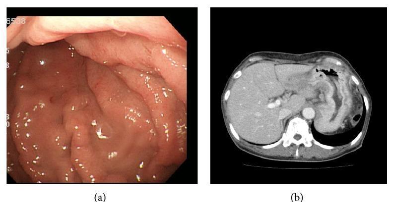
Finding Sources
Finding Sources
Finding Sources

Topic 1: Colon & Rectum Anatomy and Blood Supply (The Foundation)

Just like with the stomach, we need the map before we can understand the diseases. This topic is the single most important foundation for colorectal surgery exams, because almost every operation is named and designed around blood supply.

Step 1: The Basic Layout

Picture the large bowel as a frame around your abdomen, starting in the right lower corner and ending in the pelvis:
Caecum → Ascending colon → Hepatic flexure → Transverse colon → Splenic flexure → Descending colon → Sigmoid colon → Rectum → Anus
Real-life example: Think of it like a race track shaped like a picture frame - the "car" (stool) starts in the right lower corner (caecum), goes up the right side, across the top, down the left side, through an S-bend (sigmoid), and finishes at the rectum.

Step 2: Blood Supply - The Exam-Critical Divide

This is where surgery exams focus heavily, because knowing this divide tells you exactly how much bowel to remove in any operation.
Two main arteries split the colon into two territories:
TerritoryArterySegments Supplied
Midgut territorySuperior mesenteric artery (SMA)Caecum → ascending colon → hepatic flexure → most of the transverse colon (via ileocolic, right colic, and middle colic arteries)
Hindgut territoryInferior mesenteric artery (IMA)Distal transverse colon → splenic flexure → descending colon → sigmoid colon (via left colic and sigmoid branches)
Real-life example: Think of the SMA and IMA like two separate water pipelines supplying two halves of a garden. The SMA pipeline waters the right side of the garden (right colon), and the IMA pipeline waters the left side (left colon). Right at the border between the two pipelines (splenic flexure), the water pressure is weakest - this is exactly why that spot is vulnerable.

The Marginal Artery of Drummond

Running along the inner border of the entire colon is a continuous arterial arcade called the marginal artery of Drummond, which connects the SMA and IMA systems together. It's the safety net that keeps blood flowing even if one major vessel is compromised - and it's the vessel surgeons rely on to keep a bowel anastomosis (joined ends) alive after resection.

Watershed Areas (High Yield - Ischemic Colitis Topic Overlap)

Because the marginal artery is often thin/variable at certain points, two areas are especially vulnerable to poor blood flow (ischemia) if blood pressure drops or a vessel is occluded:
  1. Splenic flexure (Griffith's point) - border zone between SMA and IMA territory
  2. Rectosigmoid junction (Sudeck's point) - border zone between IMA branches themselves
Exam Pearl: These "watershed areas" are the classic sites for ischemic colitis, especially in elderly patients with low blood pressure, shock, or after aortic surgery. If a question describes an elderly patient with bloody diarrhea and abdominal pain after a hypotensive episode (e.g., post-cardiac surgery), think ischemic colitis at the splenic flexure.

Step 3: The Rectum - A Special Case

The rectum is about 15 cm long, divided into lower, middle, and upper thirds, and it has its own distinct blood supply that's a favorite exam topic in its own right:
VesselOriginNotes
Superior rectal arteryDirect continuation of the IMAMain supply to the rectum
Middle rectal arteryFrom internal iliac arteryVariable, supplies lower/middle rectum
Inferior rectal arteryFrom internal pudendal arterySupplies the anal canal region
Key structural point - the mesorectum: The rectal blood vessels and lymphatics travel within a fatty envelope called the mesorectum, which surrounds the rectum and is wrapped by the mesorectal fascia. This fascial envelope is exactly what surgeons must remove completely and intact during rectal cancer surgery (Total Mesorectal Excision, or TME) - we'll cover this in depth in the Rectal Cancer topic.
Real-life example: Think of the mesorectum like an orange peel wrapped tightly around the rectum, carrying the blood vessels and lymph channels inside the "peel." A rectal cancer growing outward will hit the peel (mesorectal fascia) before it can escape further, which is why removing the peel completely and intact (TME) is the key surgical principle for controlling local recurrence.

Step 4: Lymphatic Drainage - Follows the Arteries (Golden Rule)

This is one of the most useful shortcuts in the entire topic: lymphatic drainage of the colon and rectum follows the blood supply.
  • Right colon cancer → drains along ileocolic/right colic/middle colic nodes → toward the SMA root
  • Left colon cancer → drains along left colic/sigmoid nodes → toward the IMA root
  • Rectal cancer → drains mainly upward along the superior rectal vessels within the mesorectum (this is why the mesorectum must be removed with the tumor)
Why this matters practically: When a surgeon resects a colon cancer, they don't just remove the tumor - they remove the entire arterial territory feeding that segment, all the way back to the origin of the artery, because that's where the lymph nodes travel too. This single concept explains why a "right hemicolectomy" removes a large chunk of bowel even for a small caecal tumor - you're chasing the lymphatic territory, not just the tumor itself.

Quick Recap Table

ConceptKey Point
SMA territoryCaecum to distal transverse colon
IMA territoryDistal transverse colon to sigmoid
Marginal artery of DrummondConnects SMA and IMA systems, supports anastomoses
Watershed areasSplenic flexure (Griffith's point), rectosigmoid junction (Sudeck's point) - vulnerable to ischemia
MesorectumFatty envelope carrying rectal vessels/lymphatics, removed intact in TME
Golden ruleLymphatic drainage follows arterial supply

Test Your Understanding:

  1. An elderly patient develops sudden bloody diarrhea and left-sided abdominal pain after a major cardiac surgery complicated by low blood pressure. Based on watershed anatomy, where is the ischemic injury most likely located, and why?
  2. Why does a right hemicolectomy for a small caecal tumor involve removing such a large length of bowel, rather than just the small area around the tumor?
  3. Why is it surgically important to remove the mesorectum completely and intact (not just the rectum itself) when operating on rectal cancer?
Answer these, and we'll move to Topic 2: Colorectal Polyps next - covering polyp types and the adenoma-carcinoma sequence, which builds directly on this anatomy.

Continue to Topic 2: Colorectal Polyps and adenoma-carcinoma sequence

Finding Sources
Finding Sources
Finding Sources
Finding Sources

Topic 2: Colorectal Polyps and the Adenoma-Carcinoma Sequence

Step 1: What Is a Polyp? (The Basic Concept)

A polyp is simply any growth projecting from the bowel wall lining into the lumen. Think of it like a small bump or growth on a smooth carpet (the bowel mucosa) - most are harmless, but some types are dangerous because they can slowly transform into cancer over years.

Step 2: The 4 Main Types of Polyps (Classify First, Worry Later)

TypeMalignant PotentialKey Feature
Hyperplastic polypsEssentially none (except large ones in serrated pathway - advanced topic)Most common type found on colonoscopy, usually small, distal (rectosigmoid)
Adenomatous polypsThe dangerous ones - true precursors to cancerArise from glandular epithelium, subtypes below
Hamartomatous polypsLow, except in specific syndromesSeen in Peutz-Jeghers syndrome, juvenile polyposis (Topic 3)
Inflammatory polyps (pseudopolyps)None directlySeen in IBD (ulcerative colitis) - result of healing/inflammation, not a distinct growth
Real-life example: Think of hyperplastic polyps like harmless skin tags - annoying, common, but essentially never turn into cancer. Adenomatous polyps are more like a pre-cancerous mole - most stay benign, but a subset will progress to something serious if left alone.
Exam Pearl: Not all polyps are equal. When a question describes "a polyp," always ask "what type?" because only adenomatous polyps matter for cancer risk in the general population.

Step 3: The Adenoma-Carcinoma Sequence (The Central Concept of This Whole Topic)

This is the single most important idea in colorectal pathology: most colorectal cancers do not appear suddenly - they develop over 7-15 years from a benign adenomatous polyp, through a stepwise buildup of genetic mutations.
The classic genetic pathway (APC pathway - tested constantly):
  1. APC gene mutation (tumor suppressor, chromosome 5q) - the "first hit," often inherited in FAP or acquired sporadically → triggers early polyp formation. APC normally controls beta-catenin; when APC is lost, beta-catenin builds up and drives abnormal crypt cell proliferation.
  2. KRAS mutation (oncogene) - drives progression from small polyp to larger, more dysplastic adenoma
  3. p53 mutation (tumor suppressor, chromosome 17p) - the final hit that pushes the adenoma into full carcinoma
  4. DCC/DPC4 loss (chromosome 18q) - associated with more advanced disease
Real-life example: Think of this like a car slowly failing its safety inspections one system at a time. First the brakes go bad (APC) - the car is still driveable but risky. Then the steering gets worse (KRAS) - now it's clearly dangerous. Finally the engine fails completely (p53) - now it's a total breakdown (cancer). Each mutation is a separate "system failure," and it takes several failures accumulating together before you get a fully malignant cancer, not just one bad mutation.
Why this matters practically: This slow, stepwise process is exactly why colonoscopic screening works - if you find and remove the adenoma (polypectomy) before it accumulates all the mutations, you prevent the cancer from ever happening. Screening colonoscopy isn't just early detection - it's genuine cancer prevention.

Step 4: What Makes an Adenoma "High Risk"? (The 3 Factors - Very High Yield)

Not every adenomatous polyp carries equal risk. Three factors determine malignant potential, and this is one of the most frequently tested lists in surgery exams:
1. Histological subtype (architecture):
SubtypeMalignant Potential
Tubular adenoma~5% (lowest risk, most common type - about 75-85% of adenomas)
Tubulovillous adenoma~22% (intermediate)
Villous adenoma~40% (highest risk)
Real-life example: Think of tubular adenomas like a compact, well-organized bundle of straws - simple, contained. Villous adenomas are like a shaggy, frond-like carpet with finger-like projections - more surface area, more disorganized growth, much higher cancer risk. In fact, large villous adenomas in the rectum can secrete so much mucus and potassium that they cause a classic complication: hypokalemic, hypoproteinemic diarrhea (McKittrick-Wheelock syndrome) - a fantastic exam pearl connecting pathology to electrolyte disturbance.
2. Size:
  • <1 cm → low risk (<2%)
  • 1-2 cm → intermediate risk
  • >2 cm → high risk (can approach 10-40% depending on other features)
3. Degree of dysplasia:
  • Low-grade dysplasia → lower immediate risk
  • High-grade dysplasia → much closer to actual carcinoma, needs complete excision and often closer follow-up
Simple memory rule: Bigger, more villous, more dysplastic = more dangerous. All three factors point the same direction - the more disorganized and larger the growth, the more likely it harbors or will progress to cancer.

Step 5: Surveillance Concept (High Yield for Practical Management)

Once a polyp is removed at colonoscopy, follow-up timing depends on what was found:
  • 1-2 small (<1cm) tubular adenomas, low-grade dysplasia → repeat colonoscopy in longer interval (e.g., 7-10 years, per most current guidelines)
  • 3 or more adenomas, or any adenoma ≥1cm, villous features, or high-grade dysplasia → shorter surveillance interval (e.g., 3 years)
  • Malignant polyp with adverse features (poor differentiation, lymphovascular invasion, involved margin) → may need formal surgical resection rather than just endoscopic follow-up

Quick Recap Table

ConceptKey Point
Only dangerous polyp typeAdenomatous (tubular, tubulovillous, villous)
Genetic sequenceAPC → KRAS → p53 (classical pathway)
Highest risk histologyVillous (~40% malignant potential)
Risk factorsSize >2cm, villous architecture, high-grade dysplasia
Why screening worksRemoves polyp before mutations accumulate to cancer
Villous adenoma complicationMcKittrick-Wheelock syndrome (hypokalemic, mucus-secreting diarrhea)

Test Your Understanding:

  1. A colonoscopy report describes a 3 cm rectal polyp with a "shaggy, frond-like" surface. The patient also has chronic watery diarrhea and is found to have low potassium. What type of polyp is this most likely, and how does it explain the electrolyte abnormality?
  2. Why is colonoscopic polypectomy considered cancer prevention rather than just early cancer detection?
  3. Between a 0.5 cm tubular adenoma and a 2.5 cm villous adenoma, which one needs closer surveillance, and name the three factors used to decide this?
Answer these, and we'll move to Topic 3: Polyposis Syndromes next - FAP, Lynch syndrome, and Peutz-Jeghers, which builds on the genetics we just covered.

Move on to the next topic

Finding Sources
Finding Sources
Finding Sources

Topic 3: Polyposis Syndromes

These are the inherited conditions that dramatically raise colorectal cancer risk. They're tested heavily because each syndrome has a distinct gene, distinct extra-intestinal features, and a distinct management strategy. Let's build them one at a time.

Step 1: The Big Split - Adenomatous vs Hamartomatous Syndromes

Before memorizing individual syndromes, sort them into two buckets:
BucketPolyp TypeCancer RiskExamples
Adenomatous polyposis syndromesTrue adenomas (dysplastic, premalignant)Very high, essentially 100% by middle age if untreatedFAP, Gardner syndrome, Turcot syndrome, attenuated FAP, MUTYH-associated polyposis
Hamartomatous polyposis syndromesHamartomas (disorganized normal tissue, not truly dysplastic)Lower, but still elevatedPeutz-Jeghers syndrome, Juvenile polyposis syndrome
Real-life example: Think of adenomatous polyps like a house built with genuinely faulty wiring (dysplastic cells) - it's a matter of time before a fire (cancer) starts. Hamartomatous polyps are more like a house built with mismatched, disorganized furniture - it looks messy and abnormal, but the "wiring" itself isn't inherently faulty, so the fire risk is lower (though not zero).

Step 2: Familial Adenomatous Polyposis (FAP) - The Classic

  • Inheritance: Autosomal dominant
  • Gene: APC gene mutation (chromosome 5q) - the same gene from our adenoma-carcinoma sequence in Topic 2, but here it's inherited in every cell from birth, not acquired sporadically in one polyp
  • Diagnostic criterion: More than 100 colonic adenomas, typically developing around puberty
  • Natural history: If left untreated, colorectal cancer is essentially inevitable by the 40s
Real-life example: If sporadic adenoma-carcinoma sequence is like one house randomly getting faulty wiring, FAP is like an entire housing estate built by the same contractor using the same faulty wiring blueprint - every house (every polyp) has the same underlying defect, so cancer isn't a matter of "if" but "when," and it happens across hundreds of polyps simultaneously.
Management (Exam Pearl): Because malignant transformation is essentially guaranteed, prophylactic surgery (total colectomy, usually with ileal pouch-anal anastomosis) is indicated once polyps are identified, typically in the teens to twenties - before cancer develops, not after.
Important extra-colonic point: FAP patients also develop polyps around the duodenal ampulla (periampullary region), which is why they need lifelong upper GI endoscopic surveillance even after colectomy - duodenal/ampullary cancer becomes a leading cause of death in FAP patients who've had their colon removed.

Step 3: FAP Variants - Gardner and Turcot Syndrome (High Yield Buzzwords)

These are the same APC gene mutation as FAP, just with different extra-intestinal manifestations layered on top - a favorite "spot the syndrome" exam question.
VariantExtra Features (beyond colonic polyps)
Gardner syndromeOsteomas (jaw/skull bone tumors), epidermoid cysts, desmoid tumors, dental abnormalities
Turcot syndromeColonic polyps + CNS tumors (medulloblastoma or glioblastoma)
Real-life example: Think of Gardner and Turcot as FAP wearing different costumes. Same underlying APC defect (same "actor"), but Gardner syndrome shows up dressed with bone tumors and skin cysts, while Turcot syndrome shows up dressed with a brain tumor alongside the colon polyps.

Step 4: Lynch Syndrome (HNPCC) - The Non-Polyposis One

This name is tricky - "hereditary non-polyposis colorectal cancer" - because unlike FAP, patients don't have hundreds of polyps. Instead, the few adenomas they do form progress to cancer abnormally fast.
  • Inheritance: Autosomal dominant
  • Genes: Mutations in DNA mismatch repair genes - MLH1, MSH2, MSH6, PMS2
  • Mechanism: Mismatch repair genes normally proofread and fix DNA copying errors. When they're broken, errors accumulate rapidly, especially in repetitive DNA sequences, producing a signature called microsatellite instability (MSI) - found in about 85-90% of Lynch syndrome tumors
  • Cancer pattern: Colorectal cancer, but classically right-sided and occurring at a younger age than sporadic cancer
Real-life example: If FAP is like a housing estate with a faulty wiring blueprint, Lynch syndrome is like a construction crew that has no quality inspector at all. Mistakes (DNA errors) happen at the same normal rate as everywhere else, but nobody catches and corrects them (broken mismatch repair), so problems accumulate and cause failures (cancer) far faster than expected.
Associated cancers (Exam Pearl - Lynch syndrome is NOT just colorectal): Endometrial cancer (the most common extra-colonic cancer in Lynch), ovarian, gastric, urinary tract, small bowel, and biliary tract cancers. A young woman with endometrial cancer and a strong family history of colorectal cancer should raise suspicion for Lynch syndrome.
Management: Earlier and more frequent colonoscopic surveillance (often starting in the 20s), and consideration of prophylactic hysterectomy/oophorectomy in women once childbearing is complete, given the endometrial/ovarian cancer risk.

Step 5: Peutz-Jeghers Syndrome - The Hamartomatous One With Skin Signs

  • Inheritance: Autosomal dominant (STK11/LKB1 gene)
  • Polyps: Hamartomatous, can occur throughout the GI tract (not just colon - stomach, small bowel, colon)
  • Classic exam clue: Mucocutaneous pigmentation - dark brown/black macules around the lips, buccal mucosa, and fingers
  • Complications: Small bowel polyps can cause intussusception (a surgical emergency in a young patient) and chronic GI bleeding/anemia
  • Cancer risk: Increased risk of GI cancers plus certain extra-intestinal cancers (breast, ovarian, pancreatic, testicular)
Real-life example: Think of a young patient with freckle-like dark spots around the lips who then develops sudden colicky abdominal pain - this combination should immediately make you think Peutz-Jeghers with intussusception from a small bowel polyp acting as the "lead point."

Quick Recap Table

SyndromeGenePolyp TypeSignature Feature
FAPAPCAdenomatous (100+)Prophylactic colectomy needed, duodenal/ampullary risk
GardnerAPCAdenomatous+ Osteomas, epidermoid cysts, desmoids
TurcotAPCAdenomatous+ CNS tumors
Lynch (HNPCC)Mismatch repair genes (MLH1, MSH2 etc.)Few adenomas, rapid progressionRight-sided CRC, young age, MSI, endometrial cancer
Peutz-JeghersSTK11/LKB1HamartomatousPerioral pigmentation, intussusception risk

Test Your Understanding:

  1. A 16-year-old with a strong family history of colorectal cancer is found to have over 150 colonic adenomas on colonoscopy. What is the diagnosis, what gene is involved, and why is prophylactic colectomy recommended rather than just ongoing surveillance?
  2. A 32-year-old woman is diagnosed with endometrial cancer. Her mother had colon cancer at age 45. What syndrome should you suspect, what is the underlying molecular mechanism, and why is the colorectal cancer risk described as "non-polyposis" despite still being hereditary?
  3. A teenager presents with dark pigmented spots around his lips and sudden colicky abdominal pain. What syndrome do you suspect, and what surgical emergency should you be worried about?
Answer these, and next we'll move to Topic 4: Colorectal Carcinoma - Etiology & Pathology, which ties together everything from polyps and polyposis syndromes into the bigger picture of how sporadic colorectal cancer develops.

Move on to the next topic

Finding Sources
Finding Sources
Finding Sources
Finding Sources

Topic 4: Colorectal Carcinoma - Etiology & Pathology

We've covered polyps and inherited polyposis syndromes. Now let's zoom out to the bigger picture: most colorectal cancer is sporadic (not inherited), and this topic covers why it happens and how right-sided versus left-sided disease differs - a favorite comparison in exams.

Step 1: Risk Factors - The Big Categories

CategoryExamples
AgeThe single biggest factor - around 60% of cases occur in patients 70 years or older
DietLow fiber, high red/processed meat, high fat
LifestyleObesity, smoking, lack of physical exercise, alcohol
Inflammatory bowel diseaseUlcerative colitis (and Crohn's colitis) - chronic inflammation drives dysplasia
Genetic/family historyFAP, Lynch syndrome (Topic 3), or simply a first-degree relative with sporadic CRC
Previous adenomatous polypsDirect precursor lesion (Topic 2)
Real-life example: Think of risk factors like layers of stress on a bridge. Age is the slow, inevitable wear of decades of use. Diet and lifestyle are like extra heavy traffic loading the bridge daily. IBD is like a bridge that's already structurally damaged from chronic vibration (inflammation) - it fails faster than a normal bridge under the same load. Genetics (FAP/Lynch) is like a bridge built with a design flaw from day one.
Exam Pearl - IBD and cancer risk: Ulcerative colitis carries roughly 2.5 times higher risk of colorectal cancer compared to the general population, and this risk is directly tied to disease duration and extent (pancolitis carries higher risk than left-sided colitis alone) and severity of inflammation over time - not to the number of polyps, unlike sporadic disease. This is why UC patients need regular surveillance colonoscopy with random biopsies, looking specifically for dysplasia rather than discrete polyps, since UC-associated cancer often arises from flat, non-polypoid dysplasia.

Step 2: Beyond the Classical APC Pathway - The Serrated Pathway (Newer, Increasingly Tested)

In Topic 2, we covered the classical APC → KRAS → p53 adenoma-carcinoma sequence (called the chromosomal instability, or CIN, pathway). But there's a second, distinct route to cancer that's now heavily tested:
The Serrated Pathway:
  • Arises from serrated polyps (sessile serrated adenomas/polyps), typically found in the right colon
  • Driven by BRAF mutations (instead of APC) and CpG island methylator phenotype (CIMP) - meaning genes get silenced by abnormal DNA methylation rather than classic mutation
  • This methylation can silence the hMLH1 mismatch repair gene, producing sporadic microsatellite instability (MSI) - so a tumor can end up looking "Lynch-like" (MSI-high) without actually being inherited Lynch syndrome
Real-life example: Think of the classical pathway like a broken key that can't open a lock (a direct structural mutation), while the serrated/CIMP pathway is like wax poured over the lock (methylation silencing the gene) - different physical mechanism, but the same end result: the gene can't do its job.
Why this matters practically: Serrated polyps are flatter, harder to see on colonoscopy, and more common on the right side - this is part of why right-sided cancers can be trickier to catch on screening, and why colonoscopists now pay special attention to subtle flat lesions in the proximal colon, not just obvious polypoid growths.

Step 3: Right-Sided vs Left-Sided Colorectal Cancer (Very High Yield Comparison)

This is one of the most frequently tested comparisons in the entire colorectal topic, and it connects directly back to the anatomy from Topic 1 (remember: wide lumen + liquid stool on the right vs narrow lumen + solid stool on the left).
FeatureRight-Sided CancerLeft-Sided Cancer
Luminal caliberWideNarrow
Stool consistency thereLiquidSolid/formed
Typical presentationIron deficiency anemia, occult bleeding, vague abdominal discomfort, mass (tumor grows large and silent before causing symptoms)Altered bowel habit, obstruction, colicky pain, rectal bleeding, tenesmus
Growth patternOften bulky, polypoid, ulcerating massOften annular, "napkin-ring" constricting lesion
Molecular pathwayMore associated with serrated pathway, BRAF, MSIMore associated with classical CIN pathway (APC/KRAS/p53)
Prognosis (early stage)Slightly better in early stage-
Prognosis (advanced stage)Worse in stage III/IVBetter in stage III/IV
Response to biologic therapy (advanced disease)Bevacizumab tends to perform betterCetuximab tends to perform better
Real-life example: Think of the right colon like a wide highway with soft, flowing traffic (liquid stool) - a tumor can grow quite large there without ever blocking the road, so it stays silent until it erodes a vessel and causes slow bleeding (leading to anemia, found incidentally on blood tests). The left colon is like a narrow single-lane road with solid traffic (formed stool) - even a small tumor causes a noticeable traffic jam (obstruction, altered bowel habit), so it tends to announce itself earlier with symptoms.
Exam Pearl: "An elderly patient presents with unexplained iron-deficiency anemia and no other symptoms" is the classic vignette for a right-sided colon cancer. Always think colon cancer as a cause of unexplained iron deficiency anemia in an older adult, especially men or post-menopausal women, until proven otherwise.

Step 4: Putting It All Together - The Two Main Roads to Colorectal Cancer

PathwayGenesPolyp TypeTypical Location
Chromosomal instability (classical)APC → KRAS → p53Conventional adenoma (tubular/villous)More even distribution, classic left-sided predominance historically
Serrated/CIMP pathwayBRAF, CIMP, sporadic MSISerrated polypsRight colon predominance
Lynch syndrome (inherited MSI)Germline mismatch repair genesFew adenomas, rapid progressionRight-sided, young age

Quick Recap Table

ConceptKey Point
Biggest risk factor overallAge (60% of cases in patients 70+)
UC cancer risk driverDuration + extent + severity of inflammation, not polyp count
Classical pathwayAPC → KRAS → p53 (chromosomal instability)
Serrated pathwayBRAF + CIMP, right colon, can cause sporadic MSI
Right-sided presentationAnemia, occult bleeding, silent growth
Left-sided presentationObstruction, altered bowel habit, visible bleeding

Test Your Understanding:

  1. A 68-year-old man is found to have unexplained iron-deficiency anemia on routine blood work, with no GI symptoms. Which side of the colon is a cancer more likely to be on, and why does that side present this way rather than with obstruction?
  2. A tumor is found to have sporadic microsatellite instability (MSI) but the patient has no family history and doesn't meet criteria for Lynch syndrome. What molecular mechanism explains this "Lynch-like" appearance without actual inherited mutation?
  3. Why do patients with long-standing pancolitis from ulcerative colitis need surveillance colonoscopy focused on random biopsies for dysplasia, rather than just looking for discrete polyps like in the general population?
Answer these, and next we'll move to Topic 5: Clinical Presentation & Screening, followed by Topic 6: Investigations & Staging - covering how these cancers are actually diagnosed and staged in practice.

Move on to the next topic

Finding Sources
Finding Sources
Finding Sources

Topic 5: Clinical Presentation & Screening

Now let's cover how colorectal cancer actually shows up in patients, and how screening catches it before symptoms even start.

Step 1: Clinical Presentation - Location Determines Symptoms (Building Directly on Topic 4)

This reinforces what we just covered, now from the "patient in front of you" angle rather than the pathology angle.
Right-sided (proximal) colon cancer:
  • Iron deficiency anemia (fatigue, pallor) - often the only clue, found on routine bloods
  • A palpable mass in the right iliac fossa
  • Vague abdominal discomfort
  • Rarely causes obstruction (wide lumen, liquid stool)
Left-sided (distal) colon cancer:
  • Change in bowel habit - increased frequency, looser stool, or alternating constipation/diarrhea
  • Rectal bleeding (often mixed with stool, unlike hemorrhoidal bleeding which is typically fresh blood on the surface/after passing stool)
  • Colicky abdominal pain, progressive obstruction
Rectal cancer specifically - its own symptom triad (Exam Pearl, very frequently tested):
  • Bleeding per rectum
  • Tenesmus (a constant sensation of needing to defecate, even after the bowel is empty - caused by the tumor mass in the rectum tricking the nerve endings into signaling "there's still stool here")
  • Early morning diarrhea (mucus and loose stool passed first thing, sometimes forcing the patient to wake early)
Real-life example: Think of tenesmus like a fire alarm that won't stop ringing even after the fire is out - the rectal wall's stretch receptors are being irritated by the tumor mass itself, so they keep signaling "empty me" even when there's nothing left to pass. This is a classic symptom that should make you think rectal mass, not just simple constipation.
Other important but less specific features across all sites:
  • Weight loss, anorexia (advanced disease)
  • Abdominal mass (any site, if large enough)
  • Acute presentation with obstruction or perforation (Topic 9 territory)
  • Metastatic disease at presentation (liver mets causing hepatomegaly/jaundice, or the eponymous signs like we discussed for gastric cancer - though less classically taught for CRC, liver metastasis is by far the most common site)

Step 2: Screening - Why It Exists and How It Works

Remember from Topic 2: colonoscopic polypectomy is prevention, not just detection, because most CRC arises slowly from adenomas over 7-15 years. Screening exploits this long window.
Average-risk screening (Exam Pearl - age matters, and it has changed in recent guidelines):
  • Begin at age 45 (lowered from 50 in recent US guidelines, reflecting rising CRC incidence in younger adults)
  • Options include:
    • Colonoscopy every 10 years (gold standard - visualizes and can remove polyps in the same sitting)
    • FIT (fecal immunochemical test) annually - detects hemoglobin in stool using antibodies, more sensitive and specific than the older guaiac-based test
    • Flexible sigmoidoscopy every 5 years - limitation: misses proximal (right-sided) polyps and cancers, since it only reaches the sigmoid/descending colon
    • CT colonography, FIT-DNA as alternative options in some guidelines
Exam Pearl on FIT vs guaiac (gFOBT): FIT uses antibodies specific to human hemoglobin, so it isn't affected by dietary red meat or vitamin C the way the older guaiac test was - this makes FIT more accurate and now the preferred stool-based test.
Key logic point: If any stool-based test comes back positive, the patient must go on to colonoscopy - a positive FIT is not a diagnosis, it's a trigger for further investigation.
High-risk screening (differs from average-risk, ties back to Topic 3 and 4):
  • FAP - screening starts in the teens with annual sigmoidoscopy/colonoscopy
  • Lynch syndrome - colonoscopy starting in the 20s, repeated every 1-2 years
  • Ulcerative colitis - surveillance colonoscopy with random biopsies starting 8-10 years after diagnosis of pancolitis, then repeated at shorter intervals
  • First-degree relative with CRC - screening starts earlier than 45, often 10 years before the relative's age at diagnosis
Real-life example: Think of screening strategy like smoke detector placement. In a normal house (average risk), one detector per floor checked every 10 years (colonoscopy) is enough. In a house with a known history of electrical faults (FAP, Lynch, UC), you need detectors installed much earlier and checked far more frequently, because the "fire risk" (cancer risk) escalates faster and starts younger.

Step 3: Putting Presentation and Screening Together - A Common Exam Scenario

A classic vignette: "A 68-year-old man presents with fatigue. Blood tests show iron deficiency anemia. He has no GI symptoms." This is the textbook right-sided colon cancer presentation - the tumor bled slowly and silently for months before anemia became apparent, and now colonoscopy is mandatory before assuming any other cause (like diet) for the anemia.
Contrast with: "A 55-year-old woman reports 3 months of rectal bleeding, tenesmus, and feeling like she can't fully empty her bowels." This is the textbook rectal cancer triad, and demands urgent lower GI investigation, not reassurance that it's "probably just hemorrhoids."

Quick Recap Table

ConceptKey Point
Right-sided presentationIron deficiency anemia, mass, silent
Left-sided presentationChange in bowel habit, visible bleeding, obstruction
Rectal cancer triadBleeding, tenesmus, early morning diarrhea
Average-risk screening startAge 45
Screening optionsColonoscopy (10yr), FIT (annual), flex sig (5yr, misses proximal lesions)
Positive FITAlways needs follow-up colonoscopy
High-risk screeningStarts much earlier (teens for FAP, 20s for Lynch, 8-10yr post-diagnosis for UC)

Test Your Understanding:

  1. A patient undergoes flexible sigmoidoscopy as their screening test and it's reported as normal. Can you be reassured there's no colorectal cancer? Why or why not?
  2. A 60-year-old woman reports feeling like she constantly needs to pass stool even right after using the bathroom, along with some blood mixed in her stool. What is this sensation called, and what does it suggest about the cancer's location?
  3. Why does a patient with ulcerative colitis need surveillance colonoscopy to start 8-10 years after diagnosis rather than starting at age 45 like the average-risk population?
Answer these, and we'll move to Topic 6: Investigations & Staging next - covering colonoscopy, imaging, CEA, and the TNM/Dukes staging systems used to plan treatment.

Move onto next topic

Finding Sources
Finding Sources
Finding Sources

Topic 6: Investigations & Staging

Now that we know how colorectal cancer presents and how it's screened for, let's cover how a confirmed or suspected case is actually worked up and staged before treatment planning.

Step 1: Diagnosis - Getting the Tissue

Colonoscopy with biopsy is the gold standard for diagnosis - it directly visualizes the lesion, allows biopsy for histological confirmation, and can detect synchronous lesions (a second separate cancer or polyps elsewhere in the colon, which occurs in a meaningful minority of patients - this is exactly why a full colonoscopy, not just a sigmoidoscopy, is mandatory once cancer is suspected).
Digital rectal examination (DRE) is essential for any suspected rectal cancer - many low rectal tumors can be felt directly on a simple finger exam, giving immediate information about size, mobility, and distance from the anal sphincter (which matters hugely for surgical planning, covered in Topic 8).
Alternative visualization if colonoscopy is incomplete or unavailable: CT colonography or barium enema.

Step 2: Staging - Finding Out How Far It Has Spread

Once cancer is confirmed, the next question is always: has it spread, and if so, where? This determines whether surgery alone will cure the patient or whether additional treatment (chemotherapy, radiotherapy) is needed.
Standard staging workup:
  • CT chest, abdomen, and pelvis with contrast - now the standard method for staging colorectal cancer, looking for lymph node involvement, liver metastases (by far the most common distant site, since venous drainage of the colon goes to the portal vein first), and lung metastases
  • MRI pelvis - specifically required for rectal cancer (not routine colon cancer) - this is because MRI can assess the mesorectal fascia and local invasion in fine detail (remember the mesorectum from Topic 1), which CT simply cannot resolve well in the pelvis
  • Endorectal ultrasound (EUS) - an alternative/complementary tool for assessing depth of rectal tumor invasion, particularly useful for early-stage lesions being considered for local excision
Real-life example: Think of CT as a wide-angle camera good for scanning the whole body for obvious spread (liver, lungs, distant nodes), while MRI of the pelvis is a zoom lens macro shot specifically built to examine the fine anatomical layers around the rectum - you need the zoom lens here because the decision of "can we save the sphincter" depends on millimeter-level detail that the wide-angle camera can't capture.
Exam Pearl: This CT-for-body, MRI-for-rectum split is one of the most commonly tested practical points - a question describing rectal cancer staging that only mentions CT (with no MRI) should raise a red flag that the workup is incomplete.

Step 3: CEA - The Tumor Marker (Important Nuance)

Carcinoembryonic antigen (CEA) is the blood tumor marker associated with colorectal cancer, but it has a specific, limited role that's frequently misunderstood in exams:
  • NOT used for screening or diagnosis - it lacks sensitivity and specificity (can be raised in smokers, other cancers, and various benign conditions; can be normal even with cancer present)
  • Used for:
    1. Baseline measurement before surgery - establishes each patient's own reference level
    2. Postoperative follow-up/surveillance - a rising CEA after apparently curative surgery is a red flag for recurrence, often before it's visible on imaging
    3. Prognostic information - a very high preoperative CEA can indicate more advanced or aggressive disease
Real-life example: Think of CEA like a car's check engine light - it's not diagnostic of any single specific problem, and you can't use it alone to diagnose what's wrong. But once you know a patient's baseline reading, watching it trend upward over time after treatment is a useful early warning that something has gone wrong, prompting further workup (imaging) to find out what.

Step 4: Staging Systems - Dukes vs TNM (Very High Yield, Frequently Tested Together)

Dukes' Classification (older, originally for rectal cancer, still referenced clinically and in exams for its simplicity):
Dukes StageExtent of Disease
AConfined to the bowel wall (mucosa/submucosa, not through muscularis)
BSpread through the bowel wall, but no lymph node involvement
CLymph node metastases present
DDistant metastases (added later, not in the original description)
TNM Staging (modern, more detailed, now the standard - AJCC system):
ComponentWhat It Measures
T (Tumor)Depth of invasion through the bowel wall layers (T1 = submucosa, T2 = muscularis propria, T3 = through the wall into pericolic/perirectal tissue, T4 = invades adjacent organs)
N (Node)Number of regional lymph nodes involved
M (Metastasis)Presence of distant metastasis (liver, lung, peritoneum, etc.)
How they roughly correspond (a classic exam matching exercise):
TNM StageRoughly Equivalent DukesApprox. 5-year Disease-Free Survival
Stage IDukes AExcellent (>90%)
Stage IIDukes B~85%
Stage IIIDukes CLower, drops further with more nodes involved
Stage IVDukes DPoor, depends heavily on resectability of metastases
Exam Pearl: TNM has now largely superseded Dukes for formal reporting because it captures much more granular detail (exact T substage, exact N substage, specific M sites), but Dukes classification is still frequently quoted in older papers, viva questions, and quick clinical shorthand because of its simplicity - so you need to know both and how they map to each other.

Quick Recap Table

ConceptKey Point
DiagnosisColonoscopy + biopsy (full colonoscopy to rule out synchronous lesions)
Colon cancer staging imagingCT chest/abdomen/pelvis
Rectal cancer staging imagingCT (distant) + MRI pelvis (local, mesorectal fascia)
CEA roleNOT for diagnosis/screening; used for baseline + postoperative surveillance
Dukes A/B/C/DWall only / through wall / nodes / distant mets
TNMT = depth, N = nodes, M = metastasis; now standard system

Test Your Understanding:

  1. A patient is diagnosed with rectal cancer via colonoscopy and biopsy. The surgeon orders a CT chest/abdomen/pelvis but no MRI. Is the staging workup complete? Why or why not?
  2. A patient's CEA level is normal before colorectal cancer surgery, and it stays normal afterward. Six months later it starts rising steadily. What does this suggest, and what would you do next?
  3. A tumor has invaded through the muscularis propria into the perirectal fat but has not spread to any lymph nodes. What Dukes stage and roughly what TNM stage does this correspond to?
Answer these, and we'll move to Topic 7: Surgical Management next - covering right hemicolectomy, left hemicolectomy, anterior resection, and APR, which is where all this staging information gets put into practice.

Move onto next topic

Finding Sources
Finding Sources
Finding Sources

Topic 7: Surgical Management of Colorectal Cancer

This is where all the anatomy (Topic 1) and staging (Topic 6) knowledge comes together into actual operations. The golden rule from Topic 1 - resection follows the arterial and lymphatic territory, not just the visible tumor - drives every decision here.

Step 1: The Core Principle - Match the Operation to the Blood Supply

Remember: lymphatic drainage follows arteries. So the extent of resection for any colon cancer is decided by which artery feeds that segment, because you must remove the entire lymphatic drainage basin along with the tumor, tracing back to the artery's origin.
Real-life example: Think of it like removing a contaminated branch of a river system. You don't just scoop out the dirty water where you see it - you trace the branch back to its main tributary junction and remove the whole branch, because contamination (cancer cells) could be anywhere along that entire branch's drainage.

Step 2: Colon Cancer Operations by Location

Tumor LocationOperationVessels Ligated
Caecum, ascending colon, hepatic flexureRight hemicolectomyIleocolic artery, right colic artery (± branches of middle colic)
Proximal/mid transverse colonExtended right hemicolectomyAs above, plus the middle colic artery is taken further, sometimes taking down the splenic flexure
Splenic flexure/distal transverse colonLeft hemicolectomy (or extended right, surgeon preference)Left branch of middle colic + inferior mesenteric artery branches
Descending colon/sigmoidSigmoid colectomy / left hemicolectomyIMA (or its sigmoid branches), often IMA taken at its origin from the aorta for full lymphadenectomy
Exam Pearl: The splenic flexure is a genuine gray zone in exams - because it sits at the SMA/IMA watershed (Griffith's point, remember Topic 1), some surgeons treat it with an extended right hemicolectomy and others with a left hemicolectomy. Both are acceptable; the key teaching point is understanding why this location is uniquely ambiguous - it's the anatomical border between two vascular territories.

Step 3: Rectal Cancer Operations - The Sphincter Question

Rectal cancer surgery has one central decision that dominates the whole topic: can the anal sphincter be preserved, or does the patient need a permanent stoma?
Anterior Resection (AR) - sphincter-preserving
  • Removes the diseased rectum (and often sigmoid colon), then joins the remaining colon to the lower rectal stump or anal canal
  • High anterior resection - for upper rectal tumors, anastomosis higher up
  • Low anterior resection (LAR) - for lower rectal tumors, anastomosis closer to the anal canal, sometimes with a temporary defunctioning stoma (loop ileostomy) to protect a low, more fragile join while it heals
  • Includes Total Mesorectal Excision (TME) - see Step 4 below
Abdominoperineal Resection (APR)
  • Removes the entire rectum, anal canal, and anus, with a permanent colostomy
  • Reserved for tumors too low to achieve an adequate margin while preserving the sphincter, or where the tumor directly invades the sphincter complex
Real-life example: Think of the rectum like a garden hose with a nozzle (the anal sphincter) at the end. If the damaged section of hose is higher up, you can cut it out and reconnect the remaining hose to the nozzle (anterior resection - sphincter preserved). But if the damage extends all the way down into the nozzle itself, you have to remove the nozzle entirely and create a brand-new opening elsewhere (APR - permanent colostomy) because there's nothing left to reconnect to.
Exam Pearl - the historical shift: Decades ago, APR (permanent colostomy) was the default operation for most rectal cancers. Two advances changed this dramatically:
  1. Stapling devices - allow very low anastomoses that would be technically impossible to hand-sew
  2. Neoadjuvant chemoradiotherapy - shrinks (downstages) the tumor before surgery, sometimes converting a tumor that would have needed APR into one small enough for sphincter-sparing surgery
This is why the proportion of rectal cancer patients needing permanent colostomy has fallen substantially over recent decades - a great exam discussion point on "why has APR become less common."

Step 4: Total Mesorectal Excision (TME) - The Modern Gold Standard for Rectal Cancer

Introduced by Professor Bill Heald, TME revolutionized rectal cancer surgery by formalizing exactly what we discussed in Topic 1: removing the mesorectum completely and intact, following the mesorectal fascia as a "holy plane" of dissection, rather than bluntly dissecting close to the tumor.
Why it matters: Before TME became standard, local recurrence rates after rectal cancer surgery were unacceptably high (up to 30-40% in some series), because cancer cells deposited in the mesorectal fat were left behind. TME reduced local recurrence dramatically by ensuring the entire lymphatic/fatty envelope comes out with the specimen, undamaged.
Modern techniques for performing TME: Open, laparoscopic, robotic, and transanal TME (taTME) - approaching the dissection from below via the anus as well as from above, useful for very low or technically difficult tumors, though it has generated some controversy (a Norwegian moratorium was placed on taTME after concerns about unusual recurrence patterns in some centers - a nuanced, higher-level exam/viva point if asked about controversies in rectal cancer surgery).

Quick Recap Table

ConceptKey Point
Guiding principleResection follows arterial/lymphatic territory
Right hemicolectomyCaecum to hepatic flexure
Left hemicolectomySplenic flexure to descending colon
Splenic flexureWatershed gray zone, either extended right or left hemicolectomy
Anterior resectionSphincter-preserving, includes TME
APRTumor too low/invading sphincter, permanent colostomy
Why APR rates fellStapling devices + neoadjuvant chemoradiotherapy downstaging
TMEComplete, intact mesorectal fascia removal, reduced local recurrence dramatically

Test Your Understanding:

  1. A tumor is found at the splenic flexure. Why is this location considered a surgical gray zone, and what two operations might reasonably be chosen?
  2. A patient with a very low rectal tumor invading the anal sphincter complex is told they will need a permanent colostomy. What operation is this, and why can't the sphincter be preserved in this case?
  3. Why did the introduction of Total Mesorectal Excision dramatically reduce local recurrence rates compared to older blunt dissection techniques?
Answer these, and we'll move to Topic 8: Rectal Cancer Specifics next - covering neoadjuvant chemoradiotherapy in more depth and the decision-making around sphincter preservation.

Continue to Topic 8: Rectal Cancer Specifics (neoadjuvant therapy and sphincter-preservation decisions)

Finding Sources
Finding Sources
Finding Sources

Topic 8: Rectal Cancer Specifics - Neoadjuvant Therapy and Sphincter-Preservation Decisions

This topic pulls together everything from Topics 6 and 7 into the modern decision-making framework for rectal cancer - one of the richest areas for exam questions because it blends imaging, oncology, and surgical judgment.

Step 1: The Circumferential Resection Margin (CRM) - The Single Most Important Concept Here

Before we talk about neoadjuvant therapy, you need to understand why it's given, and that comes down to one measurement: the circumferential resection margin (CRM).
What it is: The distance between the outer edge of the tumor (or the deepest point of spread through the bowel wall/mesorectum) and the mesorectal fascia - the "orange peel" surface we discussed in Topic 1 that forms the outer boundary of the surgical specimen in a TME.
Why it matters: If the tumor is close to or touching this fascial margin, there's a high risk that surgery alone will leave microscopic cancer behind at that margin, leading to local recurrence. Recall from Topic 7: an involved CRM and incomplete mesorectal excision were the two dominant historical causes of local recurrence after rectal cancer surgery.
How it's assessed: MRI pelvis (from Topic 6) is the tool used to predict CRM status before surgery even happens - this is exactly why MRI staging is mandatory for rectal cancer but not routine colon cancer.
Real-life example: Think of the CRM like the safety clearance distance around a controlled demolition. If the building being demolished (tumor) is far from the surrounding structures (fascial margin), it's safe to proceed with demolition alone. If it's right up against a neighboring building, you need to do something first to create safe clearance, before you can demolish - that "something first" is neoadjuvant therapy.

Step 2: Neoadjuvant Therapy - Treating Before Surgery

The goal: Shrink (downstage) the tumor and its local extension before surgery, to achieve a clear CRM and reduce local recurrence risk - sometimes converting a tumor that would otherwise need an APR into one small enough for sphincter-sparing surgery.
When it's used - based on MRI staging:
  • Early-stage rectal cancer (favorable T1-T2, node-negative, clear predicted CRM): Surgery alone (TME) is often sufficient
  • Locally advanced disease (T3/T4, threatened or involved CRM, extramural venous invasion, or node-positive): Neoadjuvant chemoradiotherapy (or newer intensified regimens - "total neoadjuvant therapy," combining chemo and radiotherapy before surgery) is given first
Real-life example: Think of neoadjuvant therapy like weeding and clearing overgrown brush before building a fence exactly on a property line. If you tried to build precisely on the line while thick brush (the locally advanced tumor) is still there, you'd inevitably damage the neighboring property (leave cancer cells behind, positive margin). Clearing the brush first (shrinking the tumor with chemoradiotherapy) lets you build the fence (do the resection) cleanly and exactly where it needs to go.
Exam Pearl: Chemoradiotherapy in rectal cancer is used almost exclusively for local control (reducing local recurrence and enabling clear margins), while any survival/distant metastasis benefit is more modest and debated - contrast this with adjuvant chemotherapy after surgery, which is primarily aimed at eliminating micrometastatic distant disease. This distinction (local control vs systemic control) is a classic exam differentiation point.

Step 3: Complete Clinical Response and "Watch and Wait"

Here's where the topic gets genuinely advanced and is now a major theme in modern rectal cancer management.
What happens: After neoadjuvant chemoradiotherapy, up to 20% (and in some more intensive regimens, higher) of patients show no evidence of residual cancer on clinical examination, endoscopy, and MRI - this is called a complete clinical response (cCR).
The "Watch and Wait" approach (Habr-Gama protocol): Instead of proceeding to radical surgery (TME ± APR) in these complete responders, selected patients can be managed with intensive surveillance alone - regular MRI, endoscopy, and biopsy - reserving surgery only if the tumor recurs. This allows some patients to completely avoid major surgery and a permanent stoma.
Real-life example: Think of it like a fire that appears to have been fully extinguished after firefighters treated it (chemoradiotherapy). Traditionally, you'd still demolish the whole building afterward "just to be safe" (routine surgery for everyone). The watch-and-wait approach is more like using thermal cameras and repeated inspections (MRI, endoscopy) to confirm there's truly no hidden embers left, and only demolishing if a hotspot reappears - sparing many buildings (patients' sphincters/organs) unnecessary demolition.
Exam Pearl and the honest caveat: This is an evolving, somewhat controversial area - a meaningful proportion of patients with an apparent complete clinical response can still develop local regrowth, so watch and wait requires very disciplined, frequent surveillance and careful patient selection, and it isn't offered universally. If a question asks about organ preservation for rectal cancer, watch and wait is the concept being tested, but you should note it's still considered a specialized approach requiring expert centers, not the default management.

Step 4: Putting the Whole Decision Pathway Together

  1. MRI pelvis assesses T-stage, N-stage, CRM status, extramural venous invasion
  2. If early, favorable, clear CRM → straight to TME surgery (anterior resection or APR depending on distance from sphincter, as in Topic 7)
  3. If locally advanced or threatened CRMneoadjuvant chemoradiotherapy first, then reassess
  4. After neoadjuvant therapy:
    • Good response, tumor shrunk enough → proceed to sphincter-preserving surgery where previously an APR might have been needed
    • Complete clinical response → consider watch and wait in appropriate patients/centers
    • Poor response or progression → proceed to surgery (which may still be APR if the tumor remains too low/extensive)

Quick Recap Table

ConceptKey Point
CRMDistance from tumor to mesorectal fascia; assessed on MRI
Why neoadjuvant therapy is givenThreatened/involved CRM, locally advanced (T3/T4, node+) disease
Main goal of chemoradiotherapyLocal control (downstaging, clear margins), not primarily distant control
Complete clinical responseNo detectable residual cancer after neoadjuvant therapy (~20%+)
Watch and waitSurveillance instead of surgery in selected complete responders, avoids stoma
Risk of watch and waitLocal regrowth possible, needs intensive follow-up, expert centers only

Test Your Understanding:

  1. An MRI shows a rectal tumor with a predicted involved circumferential resection margin. Why would this finding change the treatment plan from "straight to surgery" to "neoadjuvant chemoradiotherapy first"?
  2. A patient with a rectal tumor initially requiring an APR undergoes neoadjuvant chemoradiotherapy and the tumor shrinks significantly. How might this change the planned surgery, and why?
  3. What is a "complete clinical response," and why is it controversial to manage these patients with watch and wait instead of proceeding straight to surgery?
Answer these, and we'll move to Topic 9: Complications & Emergency Presentations next - covering acute large bowel obstruction and perforation, which is where colorectal cancer often first presents in the emergency setting.
This is a shared conversation. Sign in to Orris to start your own chat.