
Cardiac disease in pregnancy Who grading
- WHO I: No detectable increased risk of maternal mortality; small or no increase in morbidity
- WHO II: Small increased risk of maternal mortality or moderate morbidity
- WHO III: Significantly increased risk; expert counseling required, intensive specialist care needed
- WHO IV: Extremely high risk; pregnancy contraindicated
Modified WHO Classification of Maternal Cardiovascular Risk in Pregnancy
Principles of the mWHO Classification
| Class | Risk Description |
|---|---|
| mWHO I | No detectable increased risk of maternal mortality and no/mild increase in morbidity |
| mWHO II | Small increased risk of maternal mortality or moderate increase in morbidity |
| mWHO III | Significantly increased risk of maternal mortality or severe morbidity. Expert counseling required. If pregnancy is decided upon, intensive specialist cardiac and obstetric monitoring needed throughout pregnancy, childbirth, and the puerperium |
| mWHO IV | Extremely high risk of maternal mortality or severe morbidity. Pregnancy is contraindicated. If pregnancy occurs, termination should be discussed. If pregnancy continues, care as for Class III |
Application: Conditions Under Each Class
mWHO Class I (Maternal cardiac event rate: ~2.5-5.9%)
- Small/mild pulmonary stenosis
- Patent ductus arteriosus (uncomplicated)
- Mitral valve prolapse
- Successfully repaired simple lesions (ASD, VSD, PDA, anomalous pulmonary venous drainage)
- Isolated atrial or ventricular ectopic beats
mWHO Class II (Maternal cardiac event rate: ~5.7-10.5%)
- Unoperated ASD or VSD
- Repaired Tetralogy of Fallot
- Most supraventricular arrhythmias
- Turner syndrome without aortic dilation
mWHO Class III (Maternal cardiac event rate: ~10-19%)
- Mild left ventricular impairment (EF <45%)
- Hypertrophic cardiomyopathy
- Native or tissue valve disease not in WHO I or IV (mild mitral stenosis, moderate aortic stenosis)
- Marfan or other heritable thoracic aortic disease (HTAD) without aortic dilation
- Aorta <45 mm in bicuspid aortic valve pathology
- Repaired cyanotic heart disease
- Mechanical valve
- Systemic right ventricular (mild to moderate) impairment
- Fontan circulation (uncomplicated)
- Unrepaired cyanotic heart disease
- Moderate mitral stenosis
- Moderate aortic dilation (40-45 mm in Marfan/HTAD; 45-50 mm in bicuspid aortic valve; Turner syndrome)
- Vascular Ehlers-Danlos
mWHO Class IV - Pregnancy Contraindicated (Maternal cardiac event rate: ~19-27%)
- Pulmonary arterial hypertension (maternal mortality up to 50%)
- Severe systemic ventricular dysfunction (EF <30% or NYHA class III-IV)
- Previous peripartum cardiomyopathy with any residual LV impairment
- Severe mitral stenosis
- Severe symptomatic aortic stenosis
- Systemic right ventricle with moderate or severely decreased function
- Severe aortic dilation (>45 mm in Marfan/HTAD; >50 mm in bicuspid aortic valve; tetralogy of Fallot >50 mm)
- Severe coarctation of the aorta
- Fontan with any complication
Follow-Up Frequency by Class
| Class | Minimum Visits in Pregnancy | Setting |
|---|---|---|
| I | Once or twice | Local hospital |
| II | Once per trimester | Local hospital |
| III | Bi-monthly | Referral hospital |
| IV | Monthly or bi-monthly | Expert center for pregnancy and cardiac disease |
Key Points for Exam & Clinical Practice
-
mWHO IV = Pregnancy contraindicated - the "Big Three" to remember: pulmonary arterial hypertension, severe LV dysfunction (EF <30% or NYHA III-IV), and peripartum cardiomyopathy with residual LV impairment.
-
Mechanical valves = mWHO III (not IV) unless complicated - but carry very high anticoagulation management complexity.
-
Marfan syndrome risk depends on aortic root size: without dilation = mWHO III; with severe dilation (>45 mm) = mWHO IV.
-
ROPAC registry data confirmed that mWHO categories are strongly associated with maternal, obstetric, and fetal outcomes. Overall maternal death rate was ~1% in women with CVD, compared to 0.007% in the general population.
-
Other risk scores (CARPREG II, ZAHARA) provide similar predictive value but are more complex to apply. The mWHO system is preferred for clinical use.
Regurgitation ar and mr
Aortic Regurgitation (AR) & Mitral Regurgitation (MR) in Pregnancy
Core Principle
"The pregnancy-induced decrease in systemic vascular resistance reduces the risk of cardiac failure with these conditions, especially in those with chronic lesions."
- Harrison's Principles of Internal Medicine, 22nd Edition
Pathophysiology in Pregnancy
| Factor | Effect on Regurgitation |
|---|---|
| SVR decreases | Reduces regurgitant flow, promotes forward cardiac output |
| Heart rate increases | Shortens diastole (especially beneficial in AR - less time for backflow) |
| Blood volume increases | Increases preload (may worsen LV dilation if severe) |
| Postpartum SVR normalization | Can unmask/worsen heart failure as volume load persists |
Mitral Regurgitation (MR) in Pregnancy
Etiology
- Young women: Rheumatic or congenital disease, mitral valve prolapse (MVP) (most common cause in developed countries)
- Older women: Hypertension, ischemia, idiopathic myocardial disease, infective endocarditis
- Rheumatic disease is the most common cause worldwide
Tolerance
- Mild to moderate MR: well tolerated - managed conservatively (reduced activity, salt restriction, low-dose diuretics if needed)
- Heart failure occurs in 20-25% of women with moderate to severe MR
WHO Classification
| Severity | mWHO Class |
|---|---|
| Mild-moderate MR, preserved LV function | II |
| Severe MR with LV dysfunction | III-IV |
| Severe MR + greatly enlarged, hypokinetic ventricle | IV - advise against pregnancy |
Management
- Mild/moderate: Reduced physical activity, salt restriction, diuretics (furosemide) if needed
- If already pregnant with severe MR: Maximum medical therapy; aim to carry to term; delay surgery if possible
- Diuresis + hydralazine + nitrates: First-line for heart failure in pregnancy (ACE inhibitors and ARBs are contraindicated)
- Surgical valve replacement: Avoid during pregnancy - high risk of fetal loss with cardiopulmonary bypass
- Valvuloplasty/repair: Best undertaken before pregnancy
MVP with MR
- MVP alone (without significant MR): mWHO I - pregnancy, labor, and delivery are safe
- MVP with significant MR: Treat as MR (higher risk class)
Aortic Regurgitation (AR) in Pregnancy
Etiology
- Rheumatic heart disease (often with coexisting mitral stenosis)
- Bicuspid aortic valve (BAV)
- Marfan syndrome (aortic annular dilation)
- Infective endocarditis
- Systemic lupus erythematosus (SLE)
- Coarctation of the aorta
Tolerance
- AR imposes a volume overload (not pressure overload) on the LV - generally well tolerated in pregnancy
- Both pregnancy and AR contribute to hypervolemia and peripheral vasodilation (synergistic effects)
- Chronic AR: prolonged course without decompensation; once heart failure appears, course may progress rapidly
Diagnosis
- High-pitched, blowing diastolic murmur
- Quantified by Doppler echocardiography
WHO Classification
| Clinical Status | mWHO Class |
|---|---|
| AR with preserved LV function | II-III |
| AR with LV dysfunction / heart failure | IV |
| AR + Marfan syndrome or coarctation | IV (risk of aortic rupture) |
Key Warning - Aortic Root/Dissection Risk
"Special care must be taken to rule out aortic aneurysm or dissection, especially if aortic regurgitation is associated with Marfan syndrome or coarctation of the aorta, because these conditions can result in aortic rupture and constitute strong reasons to advise against pregnancy."
- Creasy & Resnik's Maternal-Fetal Medicine
Management
- If LV dysfunction and heart failure absent: carefully supervised pregnancy is appropriate
- Aortic valve replacement needed: advise against pregnancy; complete family before cardiac dysfunction necessitates surgery
- Medical therapy in pregnancy: diuretics + hydralazine/nitrates (avoid ACE inhibitors/ARBs/ARNIs - teratogenic)
- Avoid surgical replacement during pregnancy
Side-by-Side Comparison: AR vs MR in Pregnancy
| Feature | Mitral Regurgitation (MR) | Aortic Regurgitation (AR) |
|---|---|---|
| Common etiology | MVP, rheumatic, ischemic | BAV, Marfan, rheumatic, SLE |
| Type of LV load | Volume overload | Volume overload |
| General tolerance | Well tolerated (mild-moderate) | Well tolerated (if LV preserved) |
| Effect of SVR decrease | Beneficial - less regurgitant flow | Beneficial - less regurgitant flow |
| Specific pregnancy risk | HF in 20-25% with moderate-severe | Aortic rupture if Marfan/coarctation |
| Postpartum watch | SVR normalization - watch for HF | Same + aortic monitoring |
| mWHO class | II (mild-mod) to IV (severe+LVD) | II-III (preserved) to IV (LVD/Marfan) |
| Acute onset | Exception - poorly tolerated | Exception - poorly tolerated |
| Key contraindication to pregnancy | Severe MR + dilated hypokinetic LV | AR + Marfan + aortic dilation >45mm |
Postpartum Considerations (Both AR and MR)
- Women with regurgitant lesions are at increased risk for heart failure postpartum
- Mechanism: postpartum normalization of SVR in the face of a continued volume load
- Monitor closely; treat with diuretics if heart failure develops
- Women who develop AF during pregnancy are at high risk for embolic events - anticoagulation should be considered
Steps of labour
cardinal movements of labor fetal head engagement descent flexion rotation extension
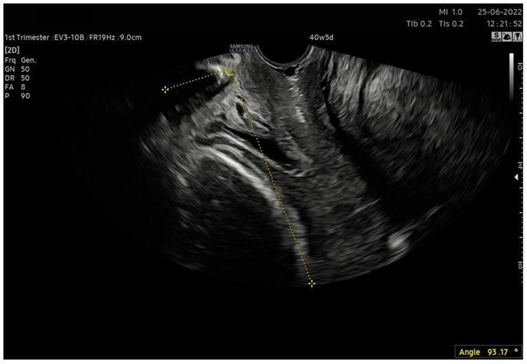
This diagnostic image is a 2D sagittal transvaginal ultrasound scan used in obstetrics to assess fetal engagement. The primary focus is the measurement of the Angle of Progression (AoP), a quantitative marker of fetal head descent in the birth canal. The image shows the pubic symphysis as a hyperechoic structure at the superior aspect. Two yellow dotted caliper lines are superimposed on the anatomy: the first line follows the long axis of the pubic symphysis, and the second line is drawn tangential to the leading bony edge of the fetal skull. The intersection of these lines defines the AoP, which is measured here at 93.17 degrees. Soft tissue structures of the cervix and pelvic floor are visible with intermediate echogenicity. This measurement is clinically significant for evaluating fetal station and predicting the success of labor induction or vaginal delivery, particularly when the fetal head is not yet fully engaged. The scan is labeled at 40 weeks and 5 days of gestation, indicating a term pregnancy assessment.
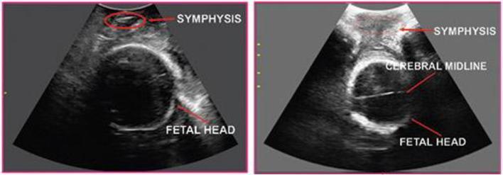
This diagnostic image consists of two side-by-side trans-labial ultrasound views showing anatomical landmarks used to monitor fetal descent and labor progress. Both images display the symphysis pubis as a superior, hyperechoic (bright) bony landmark. The fetal head is visible inferior to the symphysis as a hypoechoic circular structure with a distinct echogenic skull boundary. In the left panel, the longitudinal orientation focuses on the relationship between the maternal pubic bone and the fetal skull, relevant for measuring the Angle of Progression (AoP) and fetal head station. The right panel shows the probe rotated 90 degrees to a transverse plane, revealing the cerebral midline as a horizontal, echogenic linear structure within the fetal cranium. This view is clinically used to determine fetal head position and the rotation angle (RA) during the second stage of labor. These imaging techniques provide objective, real-time quantitative monitoring to assist in clinical decision-making regarding the mode of delivery, such as predicting the likelihood of successful vaginal birth versus the need for cesarean section.
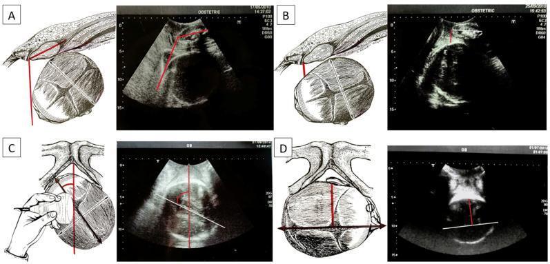
This figure presents a multi-panel comparison between anatomical illustrations and corresponding translabial ultrasound images to define four key parameters for assessing fetal head descent and orientation during labor. Panel A illustrates the Angle of Progression (AoP), showing the angle between the long axis of the pubic symphysis and a tangent line to the fetal skull. Panel B demonstrates the Head-Symphysis Distance (HSD), representing the shortest distance between the lower margin of the symphysis pubis and the fetal skull surface. Panel C depicts the Midline Angle (MLA), which measures the angle between the fetal head midline (the echogenic line between cerebral hemispheres) and the anteroposterior diameter of the maternal pubis. Panel D displays the Asynclitism Degree (AD), showing the lateral tilt of the fetal head by measuring the distance between the midline and the presenting parietal bone. These parameters serve as objective sonographic markers for determining fetal station, rotation, and potential dystocia, transitioning from traditional digital vaginal examinations to standardized intrapartum ultrasonography.
Stages of Labour
STAGE 1 - Cervical Dilation
Two Phases:
Latent Phase
- Infrequent, irregular, moderately uncomfortable contractions
- Gradual cervical effacement (thinning), softening, and anterior repositioning
- Little active dilation occurs
- Duration: up to 20 hours (nullipara) | up to 14 hours (multipara)
- Best managed at home; clear liquids encouraged
- Prolonged latent phase: can be treated with morphine, which often accelerates transition to active phase
Active Phase
- Begins at 3-4 cm dilation
- Rapid, progressive cervical dilation
- Rate: ≥1.2 cm/hr (nullipara) | ≥1.5 cm/hr (multipara)
- Contractions become regular, stronger, and more frequent
- Fetal monitoring: fetal heart auscultation every 30 minutes (low-risk) or 15 minutes (high-risk)
- Depends on: strength/frequency of contractions + fetal head size and position + bony pelvis dimensions
| First Stage Total | Latent Phase | Active Phase | |
|---|---|---|---|
| Nullipara | ~12 hours | <20 hours | Dilates ≥1.2 cm/hr |
| Multipara | ~7 hours | <14 hours | Dilates ≥1.5 cm/hr |
STAGE 2 - Expulsion of the Fetus
- Average: 50-54 minutes (nullipara) | 20 minutes (multipara)
- Alert for possible CPD (cephalopelvic disproportion) if pushing >2 hrs (no epidural) or >3 hrs (with epidural) in nullipara
- Fetal heart auscultated every 15 minutes (low-risk) or 10 minutes (high-risk)
The 7 Cardinal Movements of Labour (Vertex/Cephalic Presentation)
| # | Movement | Description |
|---|---|---|
| 1 | Engagement | Biparietal diameter (BPD) passes through pelvic inlet. Fetus at 0 station. Occurs last 2 weeks in primipara; at onset of labour in multipara |
| 2 | Flexion | Head flexes to minimize presenting diameter (suboccipito-bregmatic becomes the presenting diameter). Occurs passively. Necessary for engagement and descent |
| 3 | Descent | Downward passage through birth canal. Gradual and progressive (not necessarily continuous). Driven by uterine + abdominal contractions |
| 4 | Internal Rotation | Occiput rotates from transverse to anteroposterior position under symphysis pubis (occiput anterior = OA, most common). Necessary to traverse ischial spines |
| 5 | Extension | As head reaches introitus, it extends around the pubic symphysis. Occiput, bregma, forehead, nose, mouth, chin deliver in sequence. Head drops down after delivery (chin over anal region) |
| 6 | External Rotation (Restitution) | Head returns to anatomic position relative to shoulders. Head rotates back to transverse position it occupied at engagement. Passive movement |
| 7 | Expulsion | Shoulders and rest of body delivered. Anterior shoulder under symphysis first → posterior shoulder over perineum → body follows easily |
"The fetus will follow the path of least resistance by adaptation of the smallest achievable diameter of the presenting part to the most favorable dimensions and contours of the birth canal."
- Roberts and Hedges' Clinical Procedures in Emergency Medicine
STAGE 3 - Placental Stage
- Duration: ~15 minutes in 90% of pregnancies
- Active intervention usually not needed until 30 minutes (unless hemorrhage)
- Retained placenta = not expelled within 60 minutes
Mechanism of Placental Separation
- Uterus retracts - reduces area of placental attachment
- Hematoma forms deep to placenta - separates it from uterine wall
- Placenta separates through the spongy layer of decidua basalis
- Expulsion through vaginal canal
Signs of Placental Separation
- Lengthening of the umbilical cord
- A gush of blood
- Change in contour of the uterine fundus (becomes globular and firm)
Management
- Allow spontaneous separation
- Gentle traction on cord + maternal expulsive efforts
- Oxytocin (20-40 units/L IV, or 20 units IM) to maintain uterine contractility
- Uterine massage + immediate breastfeeding aids uterine contraction
STAGE 4 - The Recovery Stage
- Monitor for: excessive vaginal bleeding, boggy fundus, hypotension
- Uterine fundal massage to maintain contraction
- If bleeding persists:
- Oxytocin IV/IM
- Methylergonovine (Methergine) 0.2 mg IM (if not hypertensive)
- PGF₂α (Hemabate) 250 μg IM q15-20 min (up to 3 doses)
- Misoprostol 200-1000 μg (oral/sublingual/vaginal/rectal)
- Inspect for cervical and vaginal lacerations after placental delivery
- Repair episiotomy/lacerations
True vs False Labour (Braxton Hicks)
| Feature | True Labour | False Labour |
|---|---|---|
| Contraction rhythm | Regular | Irregular |
| Intervals | Gradually shorten | Unchanged |
| Intensity | Gradually increases | Unchanged |
| Discomfort location | Back and abdomen | Lower abdomen only |
| Effect of sedation | No effect | Usually relieved |
| Cervical dilation | Yes | No |
Mr and ar severe in preg management guidelines
Severe MR & Severe AR in Pregnancy - Management Guidelines
Overarching Principle
Pre-Pregnancy Counseling & Intervention
Indications to Intervene Before Attempting Pregnancy
| Condition | Threshold for Pre-Pregnancy Surgery |
|---|---|
| Severe MR | Symptoms or early LV dysfunction (EF declining, LV dilation) |
| Severe MR + EF <40% | Strongly advise against pregnancy - refer for surgery first |
| Severe MR + greatly enlarged hypokinetic LV | Advise against pregnancy (WHO IV) - fetal risk >50% |
| Severe AR + LV dysfunction/heart failure | Intervene before conception |
| AR + Marfan syndrome / coarctation | Advise against pregnancy (risk of aortic rupture) |
| Severe AR + need for valve replacement | Advise to complete family before replacement needed |
"Surgical treatment is best undertaken before pregnancy. After successful mitral valve repair with significantly improved ventricular function, pregnancy may be undertaken successfully."
- Creasy & Resnik's Maternal-Fetal Medicine
Key Pre-Pregnancy Note on Prophylactic Valve Replacement
- Mechanical valve: maternal mortality ~1.4%, only 58% event-free pregnancies
- Bioprosthetic valve: maternal mortality ~1.5%, only 79% event-free pregnancies
WHO Risk Classification
| Severity | mWHO Class |
|---|---|
| Severe MR + preserved LV (EF ≥45%) | III |
| Severe MR + mild LV dysfunction (EF 30-45%) | III-IV |
| Severe MR + EF <30% or NYHA III-IV | IV - contraindicated |
| Severe AR + preserved LV | III |
| Severe AR + LV dysfunction / heart failure | IV |
| Any severe symptomatic AS/AR/MR | High risk (IV if symptomatic) |
Management During Pregnancy
General Principles (Both MR and AR)
- Multidisciplinary team - cardiologist + obstetrician + anaesthesiologist at a referral/expert centre
- Serial echocardiographic monitoring throughout pregnancy and postpartum
- Avoid ACE inhibitors, ARBs, ARNIs (sacubitril/valsartan) - all teratogenic/fetotoxic
- Avoid surgical valve replacement during pregnancy - high fetal loss risk with cardiopulmonary bypass
- Monitor closely for arrhythmias (especially AF)
Medical Management of Severe MR in Pregnancy
| Clinical Status | Management |
|---|---|
| Mild-moderate MR, stable | Reduced activity, salt restriction, low-dose diuretics PRN |
| Severe MR, preserved LV, asymptomatic | Close monitoring + conservative measures; aim to carry to term |
| Severe MR developing heart failure | Diuretics (furosemide) + hydralazine + nitrates (afterload reduction) |
| Severe MR + AF | Rate control + anticoagulation (high embolic risk) |
| Severe MR + EF <40% | Strongly advise against further pregnancies; valve surgery before next conception |
| Severe MR + dilated hypokinetic LV (WHO IV) | Counsel for termination; if continuing → intensive care as per class III |
- Diuretics - furosemide (loop diuretic) - safe in pregnancy
- Hydralazine - vasodilator (afterload reduction) - safe in pregnancy
- Nitrates (isosorbide di/mononitrate) - safe in pregnancy
- Digoxin - rate control + mild inotropy
- Beta-blockers - for rate control if tachycardia
- ACE inhibitors (captopril, enalapril) - fetotoxic
- ARBs - fetotoxic
- Sacubitril/valsartan (ARNI) - contraindicated
- Spironolactone - avoid (anti-androgenic effects on fetus)
Medical Management of Severe AR in Pregnancy
| Clinical Status | Management |
|---|---|
| Severe AR, preserved LV, no symptoms | Carefully supervised pregnancy; monitor echocardiographically each trimester |
| Severe AR + developing symptoms | Diuretics + hydralazine + nitrates |
| AR + Marfan / HTAD, no aortic dilation | mWHO III; beta-blockers to limit aortic wall stress |
| AR + Marfan with aorta >45 mm | mWHO IV - pregnancy contraindicated |
| AR + heart failure | Same as MR heart failure protocol (diuretics + hydralazine/nitrates) |
| AF develops | Anticoagulate (high embolic risk) |
- Rule out aneurysm/dissection at outset
- Serial echo for aortic root diameter every trimester
- If Marfan: beta-blockers throughout pregnancy regardless of symptoms
- Operative vaginal delivery (forceps/ventouse) to limit Valsalva stress if aortic root 40-45 mm
Mode of Delivery
| Risk Class | Preferred Mode | Notes |
|---|---|---|
| mWHO I-II | Vaginal delivery | First choice; caesarean only for obstetric indications |
| mWHO III | Vaginal delivery preferred at referral centre | Assisted second stage (avoid prolonged Valsalva) |
| mWHO IV | Vaginal or caesarean based on individual assessment | Expert centre mandatory; multidisciplinary team present |
| Severe AR + aortic dilation 40-45 mm | Operative vaginal delivery | To limit Valsalva/aortic wall stress |
| AR + Marfan aorta >45 mm | Elective caesarean | Prevent dissection risk |
- Epidural analgesia preferred (reduces cardiovascular stress of pain)
- Avoid fluid overload during labour
- Left lateral tilt to avoid aortocaval compression
- Assisted second stage (forceps/ventouse) to shorten pushing phase
- Have cardiac team available at delivery
Postpartum Management (Critical Period)
- SVR normalises rapidly after delivery
- But volume load (from uterine involution/autotransfusion of ~500 mL) persists
- This combination can precipitate acute pulmonary oedema
- Close monitoring for first 24-48 hours
- IV loop diuretics (furosemide) to reduce redistribution pulmonary oedema risk - especially for moderate-severe regurgitation
- Echocardiographic follow-up at 4-6 weeks postpartum
- Counsel regarding future pregnancies and planned valve surgery
- Can now restart ACE inhibitors/ARBs postpartum (caution if breastfeeding - enalapril considered safest)
Summary: Key Decision Points
Severe MR/AR - PRECONCEPTION
├── EF <40% + symptoms → Surgery BEFORE pregnancy
├── EF ≥45%, asymptomatic → Pregnancy possible with close monitoring (mWHO III)
└── EF <30% or NYHA III-IV → Advise against pregnancy (mWHO IV)

Severe MR/AR - DURING PREGNANCY (if presenting already pregnant)
├── Asymptomatic, preserved LV → Conservative: monitoring + salt restriction + diuretics PRN
├── Developing HF → Furosemide + Hydralazine + Nitrates
├── AF → Rate control + anticoagulation
├── Refractory HF, <22 weeks → Discuss termination
├── Refractory HF, >22 weeks → Maximise medical therapy; aim for 28-34 weeks then deliver
└── Valve surgery in pregnancy → Last resort; extremely high fetal loss risk
Mechanical ven in covid ards
Mechanical Ventilation in COVID-19 ARDS
Pathophysiology of COVID-19 ARDS
- "Silent/happy hypoxemia" - hypoxemia disproportionate to perceived breathlessness (possibly due to SARS-CoV-2 effect on carotid bodies or heterogeneous individual responses)
- Hypercoagulability - in situ microthrombosis + macrovascular PE contribute to V/Q mismatch
- Immune dysregulation - cytokine storm worsens alveolar damage beyond initial viral injury
- Heterogeneous lung injury (mix of recruitable and non-recruitable lung)
Pre-Intubation: Escalating Oxygen Support
Step-up Approach Before Invasive Ventilation
| Step | Intervention | Notes |
|---|---|---|
| 1 | Conventional low-flow O₂ | Nasal cannula/facemask; SpO₂ target ≥94% |
| 2 | High-Flow Nasal Cannula (HFNC) | Reduces intubation need in severe pneumonia; observational data shows possible mortality benefit in COVID ARDS |
| 3 | Awake prone positioning | Often improves oxygenation, well tolerated, reduces intubation risk on HFNC |
| 4 | NIV/CPAP | Evidence conflicting; risk of aerosol generation |
| 5 | Invasive mechanical ventilation | When HFNC/NIV fails or patient deteriorates rapidly |
"Placing patients in the prone position often improves their oxygenation, is generally well tolerated, and reduces the risk of intubation among patients on HFNC."
- Current Surgical Therapy, 14th Ed.
Indications for Intubation & Invasive MV
- Worsening hypoxemia despite HFNC (SpO₂ <92% on FiO₂ ≥0.6 or flow ≥40 L/min)
- Increasing work of breathing / respiratory distress
- Altered consciousness / inability to protect airway
- Haemodynamic instability
- PaO₂/FiO₂ <150 despite non-invasive support
Lung-Protective Ventilation Strategy (ARDSNet Protocol)
Core Ventilator Settings
| Parameter | Target | Rationale |
|---|---|---|
| Mode | Volume-Assist Control (AC-VC) | Only mode proven to improve survival in ARDS |
| Tidal Volume (Vt) | 6 mL/kg predicted body weight (PBW) | Prevent volutrauma; use PBW (not actual weight - actual ≈20% heavier due to oedema/fat) |
| Plateau Pressure (Pplat) | < 30 cm H₂O | Prevent barotrauma; check by inspiratory hold |
| Driving Pressure (ΔP) | < 15 cm H₂O | ΔP = Pplat - PEEP; best predictor of ARDS mortality among all ventilator variables |
| PEEP | Per PEEP-FiO₂ table (see below) | Prevent atelectrauma; titrate to oxygenation |
| SpO₂ target | 88-95% | Avoid hyperoxia; accept permissive hypoxaemia |
| PaO₂ target | 55-80 mmHg | |
| Respiratory Rate | 6-35 breaths/min | Titrate to achieve pH goal |
| pH target | 7.30-7.45 | Permissive hypercapnia accepted |
| FiO₂ | Minimum to achieve SpO₂/PaO₂ target; aim <0.60 | Prevent hyperoxic lung injury |
| I:E ratio | 1:1 to 1:3 |
ARDSNet PEEP-FiO₂ Table
| FiO₂ | 0.3 | 0.4 | 0.4 | 0.5 | 0.5 | 0.6 | 0.7 | 0.7 | 0.7 | 0.8 | 0.9 | 0.9 | 1.0 |
|---|---|---|---|---|---|---|---|---|---|---|---|---|---|
| PEEP | 5 | 5 | 8 | 8 | 10 | 10 | 10 | 12 | 14 | 14 | 14 | 16 | 18-24 |
| FiO₂ | 0.3 | 0.3 | 0.3 | 0.3 | 0.3 | 0.4 | 0.4 | 0.5 | 0.5-0.8 | 0.8 | 0.9 | 1.0 | 1.0 |
|---|---|---|---|---|---|---|---|---|---|---|---|---|---|
| PEEP | 5 | 8 | 10 | 12 | 14 | 14 | 16 | 16 | 18 | 20 | 22 | 22 | 24 |
PEEP Titration Principles
- Titrate to minimize driving pressure (ΔP) - if increasing PEEP reduces ΔP → beneficial (recruiting lung); if ΔP rises → reducing PEEP
- Avoid high PEEP recruitment manoeuvres (PEEP 25-35 cm H₂O) - associated with increased mortality (55% vs 49% control in one RCT)
- Esophageal manometry-guided PEEP (transpulmonary pressure) - theoretically elegant but not shown to improve outcomes over standard PEEP-FiO₂ table
Permissive Hypercapnia
- Accept rising PaCO₂ as long as pH ≥7.30
- Contraindications to permissive hypercapnia:
- Raised intracranial pressure (trauma, mass lesion)
- Acute cerebrovascular disease / stroke
- Acute/chronic myocardial ischaemia
- Severe pulmonary hypertension
- Right ventricular failure
- Severe uncorrected metabolic acidosis
- Sickle cell anaemia
Prone Positioning - KEY Intervention
Indications
- PaO₂/FiO₂ < 150 mmHg despite adequate PEEP
- Particularly severe COVID ARDS with diffuse consolidation pattern
Protocol
- Duration: ≥16 hours/day (shorter duration does not confer mortality benefit)
- Continue prone positioning until PaO₂/FiO₂ improves to >150 on PEEP ≤10 and FiO₂ ≤0.6
Evidence
- PROSEVA trial (>500 patients): 16% reduction in mortality + ~4 extra ventilator-free days
- Meta-analysis of 6 RCTs paired with lung-protective ventilation: RR of death 0.74 (95% CI 0.59-0.95)
- Benefit attenuated without concurrent lung-protective ventilation
- In COVID-19: universally applied - applies to both intubated and awake (pre-intubation) patients
Physiologic Mechanism
- Repositions heart off posterior lung - reduces compression atelectasis
- Redistributes ventilation more uniformly - reduces overdistension of ventral units
- Improves V/Q matching in dorsal (most affected) lung regions
Neuromuscular Blockade (NMB) / Sedation
| Scenario | Recommendation |
|---|---|
| Early severe COVID ARDS | Deep sedation + NMB required to prevent ventilator dyssynchrony and VILI |
| Established, improving ARDS | Wean NMBs + deep sedation as soon as feasible |
| Risk of prolonged NMB | ICU-acquired weakness, delirium, prolonged ventilation - wean early |
| Agent | Cisatracurium (ACURASYS trial) or vecuronium; 48-hour course |
"Paralytics and deep sedation should be weaned as soon as feasible since they contribute to prolonged weakness and delirium."
- Current Surgical Therapy, 14th Ed.
Adjunct Pharmacologic Therapies in COVID ARDS
Proven to Reduce Mortality
| Drug | Evidence | Indication |
|---|---|---|
| Dexamethasone 6 mg/day × 10 days | RECOVERY trial: 1/3 reduction in mortality in ventilated patients | All mechanically ventilated COVID-19 patients |
| Tocilizumab (anti-IL-6R) | RECOVERY + REMAP-CAP: reduced 28-day mortality; benefits additive to steroids | Hypoxia + CRP >75 mg/L; within first 24 hours of MV |
| Baricitinib (JAK inhibitor) | COV-BARRIER: 1/3 reduction in 60-day mortality added to standard care | HFNC/NIV or early mechanical ventilation; benefit maximal before intubation |
Anticoagulation
- Prophylactic LMWH: All hospitalized COVID-19 patients (VTE prevention)
- Therapeutic anticoagulation: Benefits hospitalized patients on low-flow O₂ (reduces progression to organ support) BUT NOT in critically ill/mechanically ventilated patients (increases bleeding without benefit - ATTAC/ACTIV-4A/REMAP-CAP trial)
Not Recommended
- Recruitment manoeuvres with very high PEEP (≥25 cm H₂O) - increased mortality
- Routine liquid ventilation - no benefit, possible harm
- Partial liquid ventilation with perfluorocarbons - no benefit
ECMO (Extracorporeal Membrane Oxygenation)
- PaO₂/FiO₂ <80 mmHg despite optimal MV, prone positioning, and high PEEP
- Or unacceptable hypercapnia/acidosis despite best MV
- EOLIA trial: 28% RR reduction in 60-day mortality (not statistically significant as standalone but significant in meta-analysis)
- Must be at a centre with ECMO expertise
- VV-ECMO (veno-venous) for respiratory failure
Monitoring on Mechanical Ventilation
| Parameter | Target | How |
|---|---|---|
| Plateau pressure | <30 cm H₂O | Inspiratory hold manoeuvre |
| Driving pressure | <15 cm H₂O | Pplat - PEEP |
| SpO₂ | 88-95% | Continuous pulse oximetry |
| PaO₂/FiO₂ ratio | Monitor trend | Serial ABGs |
| Auto-PEEP | None/minimal | Expiratory hold |
| Cardiac output / haemodynamics | Maintain perfusion in prone | Arterial line, echo |
Weaning & Liberation from MV
- Daily spontaneous breathing trials (SBT) once improving
- Target: FiO₂ ≤0.4, PEEP ≤8, PaO₂/FiO₂ >150, haemodynamically stable
- Analgesia-first sedation strategy once acute phase resolved
- SAT (Spontaneous Awakening Trial) + SBT bundle reduces ventilator days
Post-ICU Considerations
- Fatigue, dyspnea, exertional intolerance (months)
- Impaired diffusion capacity in 1/3 to 1/2 at 6 months
- Ground-glass opacities and fibrosis on CT
- Cognitive impairment, anxiety, depression
- Aggressive physical rehabilitation + multidisciplinary follow-up recommended