
Types of murmur sound in heart
Reading File
Finding Sources
Searching PubMed
"Heart Murmurs"[MeSH Terms]
Searching the Web
heart murmur types systolic diastolic continuous American Heart Association
Reading File
Reading File
Reading File
Heart murmurs are sounds caused by turbulent blood flow. They are classified mainly by timing in the cardiac cycle:
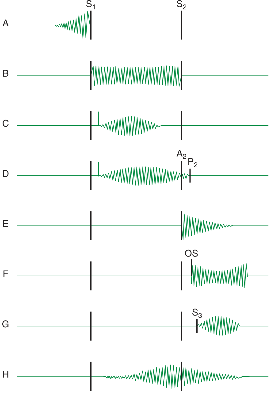
-
Systolic murmurs: occur between S1 and S2, when the ventricles contract.
- Ejection systolic, crescendo-decrescendo: aortic stenosis, pulmonary stenosis, innocent flow murmur.
- Pansystolic / holosystolic: same intensity throughout systole; mitral regurgitation, tricuspid regurgitation, ventricular septal defect.
- Late systolic: mitral valve prolapse, often with a midsystolic click.
-
Diastolic murmurs: occur between S2 and the next S1, while ventricles relax and fill. These generally need clinical evaluation.
- Early diastolic, decrescendo blowing murmur: aortic regurgitation or pulmonary regurgitation.
- Mid-diastolic, low-pitched rumble: mitral stenosis or tricuspid stenosis.
- Presystolic accentuation: louder just before S1 in mitral stenosis when sinus rhythm is present.
-
Continuous murmurs: heard in both systole and diastole, continuing through S2.
- Classic example: patent ductus arteriosus, often described as a “machinery” murmur.
-
To-and-fro murmurs: separate systolic and diastolic murmurs with a gap around S2, often from combined stenosis and regurgitation of the same valve.
Murmurs are also described by site, radiation, pitch, quality, shape, and intensity. Intensity is graded I to VI; a palpable vibration called a thrill usually accompanies grade IV or louder murmurs. The American Heart Association overview notes that timing, loudness, location, and sound quality help identify the likely cause.
Angina
Reading File
Finding Sources
Searching PubMed
"Angina Pectoris"[MeSH Terms]
Searching the Web
angina symptoms emergency signs diagnosis treatment National Heart Lung and Blood Institute
Reading File
Reading File
Reading File
Angina pectoris is chest discomfort caused by an imbalance between the heart muscle’s oxygen demand and its blood supply, most often due to coronary artery disease.
Typical symptoms
- Pressure, tightness, heaviness, squeezing, burning, or “indigestion” in the chest
- May spread to the left arm, shoulder, neck, jaw, back, or upper abdomen
- Can include breathlessness, sweating, nausea, or unusual fatigue
- In women, older adults, and people with diabetes, symptoms may be less typical.
Main types
-
Stable angina
Predictable discomfort with exertion, emotional stress, cold exposure, or heavy meals. It usually settles within a few minutes with rest or prescribed nitroglycerin.
Lippincott Illustrated Reviews: Pharmacology, p. 413. -
Unstable angina
New chest pain, pain at rest, pain that is increasing in frequency/severity, lasts longer, or fails to improve with rest or prescribed medication. This is part of acute coronary syndrome and needs emergency assessment. -
Vasospastic (Prinzmetal or variant) angina
Usually occurs at rest, often at night or early morning, due to temporary spasm of a coronary artery. -
Microvascular angina
Related to dysfunction of the heart’s small vessels. It may occur with exertion or at rest and is more commonly recognized in women.
Important
If chest pressure/pain is new, severe, persistent for more than a few minutes, happens at rest, or occurs with sweating, breathlessness, fainting, or nausea, call emergency services immediately. Do not assume it is only indigestion. NHLBI guidance advises urgent action for chest pain that does not go away.
What tests are used to evaluate suspected angina?
Reading File
Finding Sources
Finding Sources
Searching PubMed
"Angina Pectoris"[MeSH Terms] AND diagnosis
Searching the Web
2024 guideline evaluation suspected chronic coronary syndrome angina ECG troponin stress imaging CT coronary angiography
Tests are selected based on whether symptoms suggest acute coronary syndrome or stable/chronic angina.
Initial evaluation
- Clinical history and physical examination: character of pain, relation to exertion, duration, associated breathlessness/sweating, and cardiac risk factors.
- 12-lead ECG: looks for ischemic ST-T changes, prior myocardial infarction, arrhythmia, or left ventricular hypertrophy.
- Blood tests:
- High-sensitivity cardiac troponin, repeated over time if acute or recent prolonged pain is possible. Raised troponin suggests myocardial injury such as NSTEMI rather than unstable angina.
- Full blood count, glucose/HbA1c, lipid profile, kidney function, and sometimes thyroid testing to identify risk factors or contributors such as anemia.
Tests to confirm coronary disease or inducible ischemia
- Exercise ECG stress test (treadmill test): assesses symptoms, ECG changes, exercise capacity, and blood-pressure response during exertion.
- Stress echocardiography: uses exercise or medication-induced stress to detect new regional wall-motion abnormalities.
- Nuclear myocardial perfusion imaging: SPECT or PET assesses blood flow to the heart muscle during stress and rest.
- Stress cardiac MRI: evaluates myocardial perfusion and scar without ionizing radiation.
- Coronary CT angiography (CCTA): a non-invasive scan showing coronary plaques and narrowing. It is often used when obstructive coronary disease is suspected.
- Invasive coronary angiography: catheter-based imaging, usually for high-risk patients, persistent symptoms despite treatment, strongly abnormal non-invasive tests, or when revascularization may be needed. Pressure-wire measures such as FFR/iFR can determine whether a narrowing is actually limiting blood flow.
Additional tests in selected cases
- Echocardiography at rest: assesses heart pumping function, wall-motion abnormalities, valve disease, and alternative causes of symptoms.
- Ambulatory ECG monitoring: if symptoms could be due to intermittent arrhythmia or vasospastic angina.
- Coronary vasoreactivity and microvascular testing: may be considered when symptoms persist but CCTA/angiography shows no major blockage, to assess vasospastic or microvascular angina.
The 2024 ESC approach begins with history, examination, resting ECG, and blood tests, then uses CCTA or functional stress imaging to establish the diagnosis; invasive angiography is generally reserved for higher-risk situations or when intervention is likely. ESC 2024 guidance
Emergency note: chest pain at rest, worsening/new chest pain, pain lasting more than a few minutes, or pain with sweating, fainting, breathlessness, or nausea needs urgent emergency assessment with ECG and serial troponins.
Ischemia
Reading File
Finding Sources
Searching PubMed
"Myocardial Ischemia"[MeSH Terms]
Searching the Web
myocardial ischemia symptoms causes emergency evaluation National Heart Lung Blood Institute
Ischemia means inadequate blood flow and oxygen delivery to a tissue compared with its needs.
Myocardial ischemia
In the heart, myocardial ischemia occurs when coronary blood flow cannot meet the oxygen requirement of the heart muscle. The usual cause is narrowing or blockage of a coronary artery due to atherosclerotic plaque. It can also result from coronary spasm, small-vessel dysfunction, anemia, low blood pressure, or a fast heart rate.
- Angina is the common symptom of myocardial ischemia.
- Ischemia may also be silent, with no chest pain, especially in some people with diabetes or older adults.
- If ischemia is severe or prolonged, heart muscle injury can occur, producing a myocardial infarction (heart attack).
Common features
- Central chest pressure, tightness, heaviness, or burning
- Discomfort spreading to an arm, shoulder, neck, jaw, back, or upper abdomen
- Shortness of breath, sweating, nausea, dizziness, or fatigue
- Often triggered by exertion or stress in stable coronary disease, but may occur at rest in acute coronary syndrome.
How it is detected
- ECG, including possible ST-segment or T-wave changes
- Serial high-sensitivity troponin tests if acute injury is suspected
- Exercise ECG or stress imaging
- Coronary CT angiography or invasive coronary angiography when indicated
Textbook sources describe ischemia as an oxygen supply-demand mismatch that can range from silent ischemia to angina, myocardial infarction, or sudden cardiac death. Goldman-Cecil Medicine International Edition, Pathobiology, pp. 1431-1435. The NCBI clinical review similarly emphasizes prompt ECG and cardiac biomarker evaluation for suspected acute myocardial ischemia.
Seek emergency care now for new or persistent chest pressure, pain at rest, breathlessness, sweating, fainting, nausea, or pain radiating to the arm/jaw, particularly if symptoms last more than a few minutes.
This is a shared conversation. Sign in to Orris to start your own chat.