
Necrotising fasciitis
Reading File
Now let me search the medical library and PubMed in parallel.
Finding Sources
Searching PubMed
"necrotizing fasciitis"[MeSH Terms] AND management
Good - Bailey and Love's has the most comprehensive sections. Let me read all relevant blocks in parallel.
Reading File
Reading File
Reading File
Searching PubMed
"necrotizing fasciitis"[MeSH Terms]
Let me also read the summary box from block1 and get the radiology section, plus fetch images and search for the LRINEC score details.
Reading File
Reading File
Reading File
Finding Sources
Loading Image
Let me now get the LRINEC score details and the clinical images showing progression.
Finding Sources
Loading Image
Loading Image
Let me get the LRINEC table details from Tintinalli's and Mulholland, plus the bullae image.
Reading File
Reading File
Loading Image
I now have comprehensive information from multiple authoritative textbooks. Let me compile the full answer.
Necrotising Fasciitis
Necrotising fasciitis (NF) is a rapidly progressing, life-threatening deep soft tissue infection causing necrosis of the subcutaneous tissues, fascia, and eventually overlying skin. Without aggressive treatment, mortality approaches 70%.
Classification
| Type | Microbiology | Details |
|---|---|---|
| Type I | Polymicrobial (most common) | Mixed aerobic + anaerobic organisms |
| Type II | Monomicrobial | Group A beta-haemolytic Streptococcus (GAS), ± S. aureus |
| Type III | Gram-negative (marine) | Vibrio vulnificus - associated with seawater exposure |
| Type IV | Fungal | Candida spp. - immunocompromised patients |
Named variants:
- Fournier's gangrene - perineal/scrotal involvement
- Meleney's gangrene - abdominal wall; synergistic bacterial gangrene
Microbiology
Common organisms include:
- Streptococcus pyogenes (group A Streptococcus) - most feared monomicrobial cause
- Staphylococcus aureus (including MRSA)
- Escherichia coli, Pseudomonas, Proteus, Klebsiella
- Anaerobes: Bacteroides, Clostridium (gas-forming)
Clostridial infection produces rapid, gas-forming, necrotic infection (gas gangrene / clostridial myonecrosis) and carries especially high mortality.
Pathophysiology
Organisms enter via trauma, surgery, or a skin breach and spread along fascial planes, causing:
- Acute inflammatory infiltrate in subcutaneous tissue
- Thrombosis of microvascular supply → ischaemia
- Tissue necrosis spreads contiguously along fascial planes (occasionally "skip lesions" that later coalesce)
- Toxin release + systemic sepsis → septic shock, multiorgan failure
The ischaemia explains the hallmark disproportionate pain and the relative absence of lymphangitis (lymphatics traverse the dermis, which is initially spared).
Risk Factors / Predisposing Conditions
- Diabetes mellitus (most common comorbidity)
- Penetrating trauma or recent surgery
- Immunosuppression (steroids, chemotherapy, HIV)
- Intravenous drug use
- Obesity, peripheral vascular disease
- Perineal infection (perianal abscess, Bartholin's cysts)
- Pressure sores, bites, boils, varicella
Up to 30% of patients may have no identifiable comorbidities. 80% have a history of prior trauma/infection and >60% of cases begin in the lower extremities.
Clinical Features
Early (may resemble cellulitis)
- Erythema, warmth, swelling, pain
- Fever, tachycardia
Classical signs
- Disproportionate pain relative to visible skin changes (early hallmark)
- Oedema extending beyond the visible erythema
- Woody-hard subcutaneous texture - inability to distinguish fascial planes
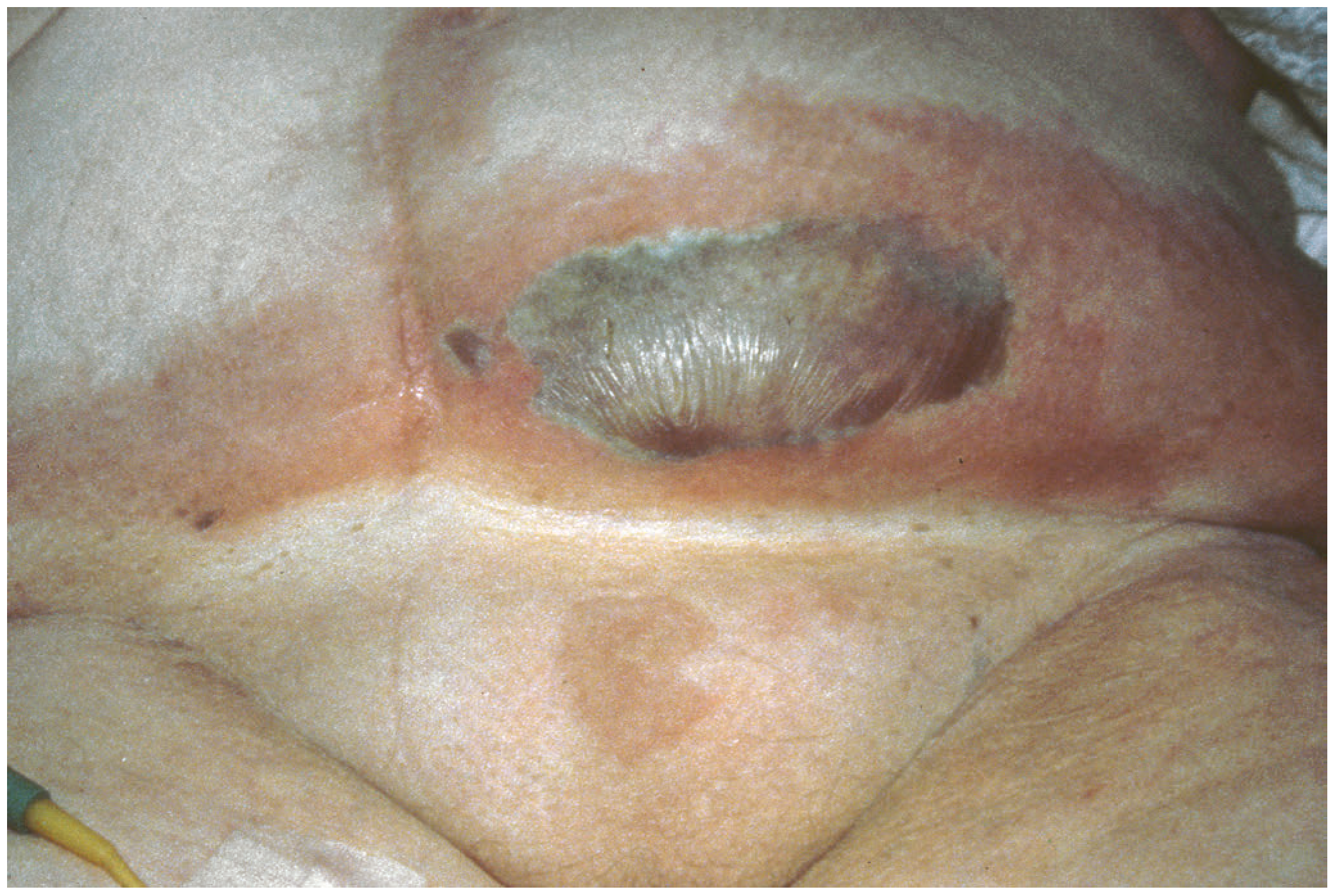
- Skin turns dusky-blue/purple-black (progressive thrombosis)
- Skin vesicles and bullae (Figure below) - contain dark purple/haemorrhagic fluid
- Soft-tissue crepitus (gas-forming organisms)
- "Dishwater" (grey/brown) drainage from wound
- Fixed skin staining
Late / severe
- Cutaneous gangrene and frank skin necrosis
- Hypotension, septic shock
- Renal failure (hypovolaemia + cardiovascular collapse)
- Absence of lymphangitis
"Hard" signs (crepitus, skin necrosis, bullae, hypotension, gas on X-ray) are present in less than half of patients at presentation - Tintinalli's Emergency Medicine
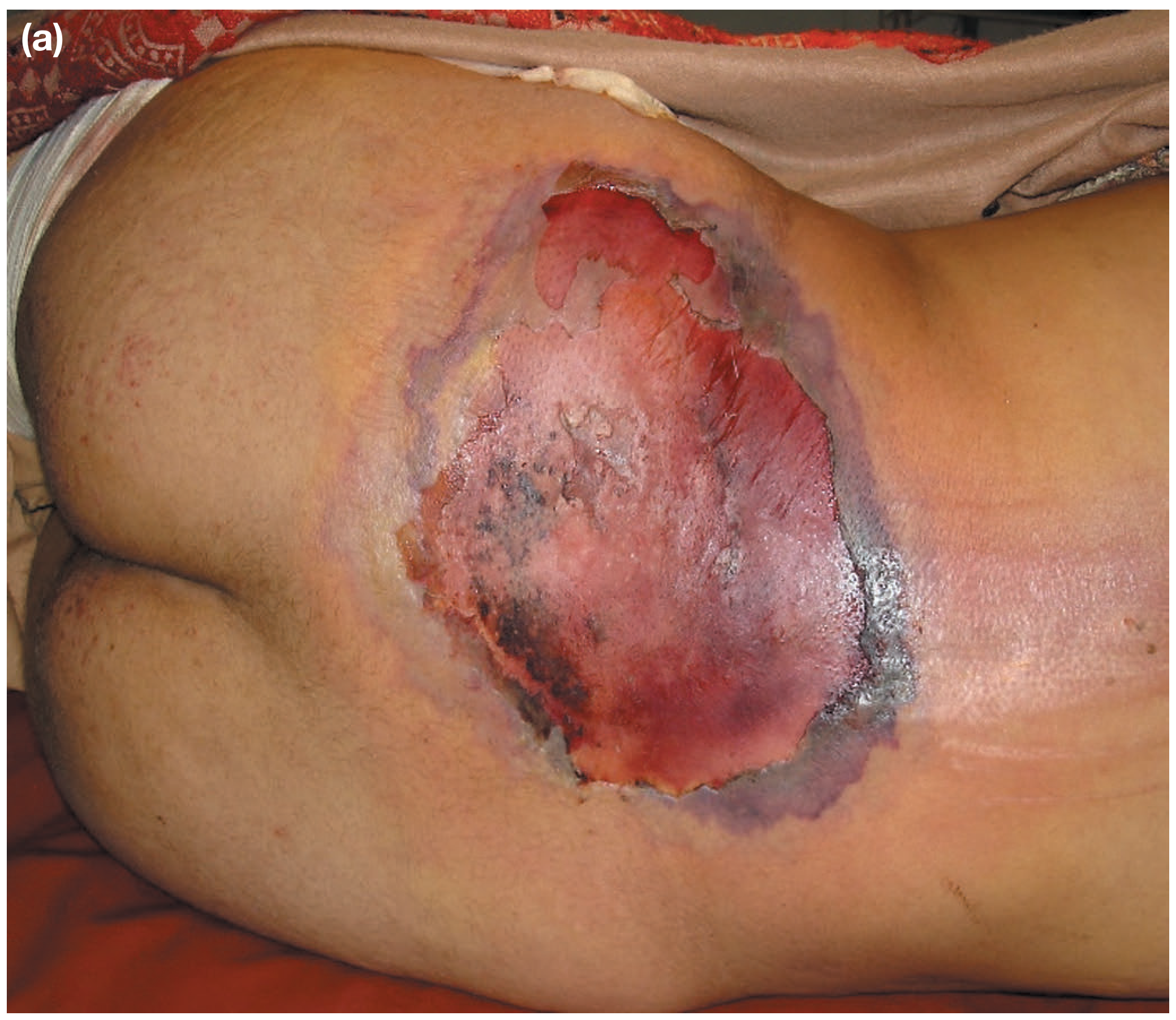
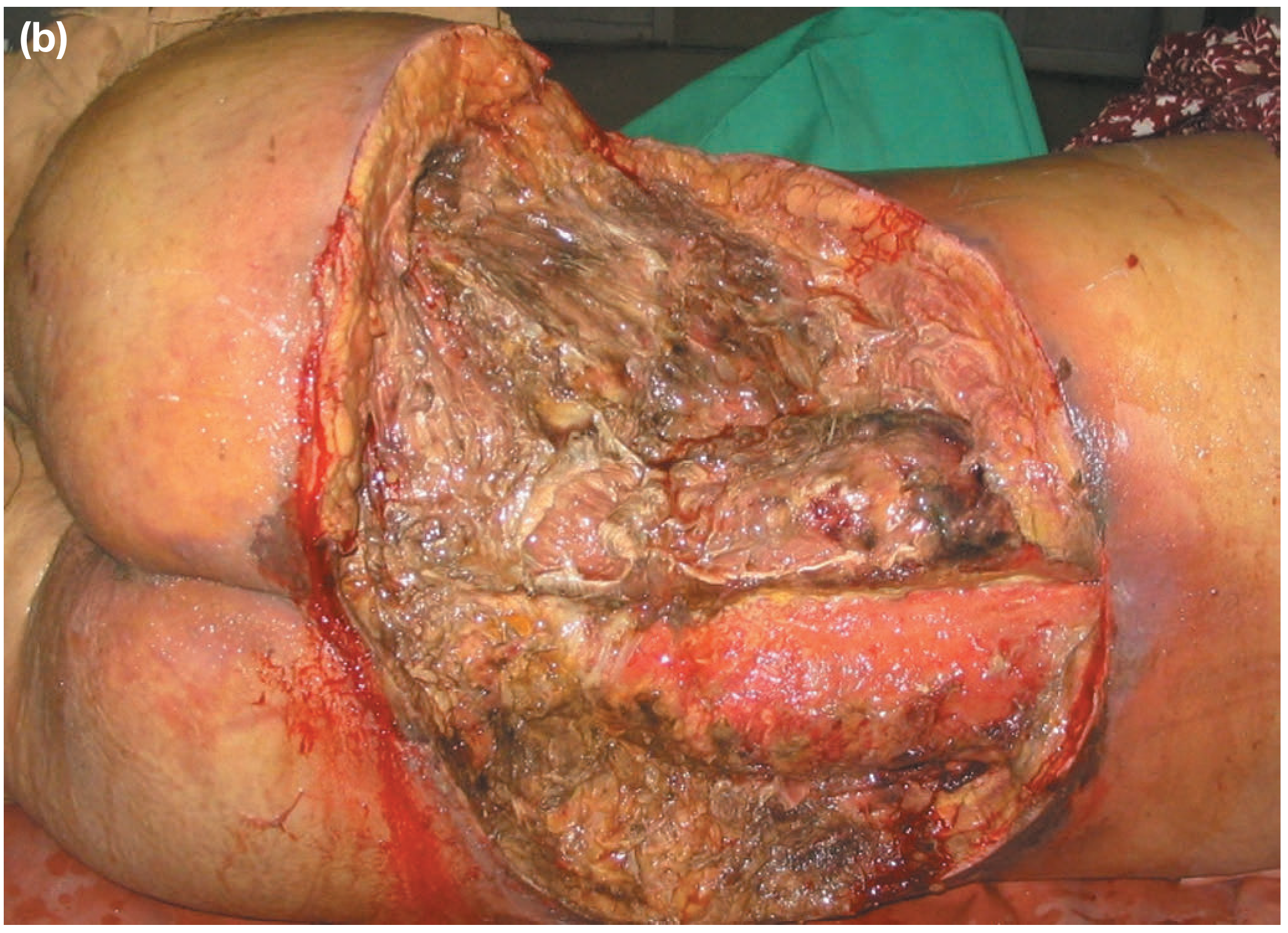
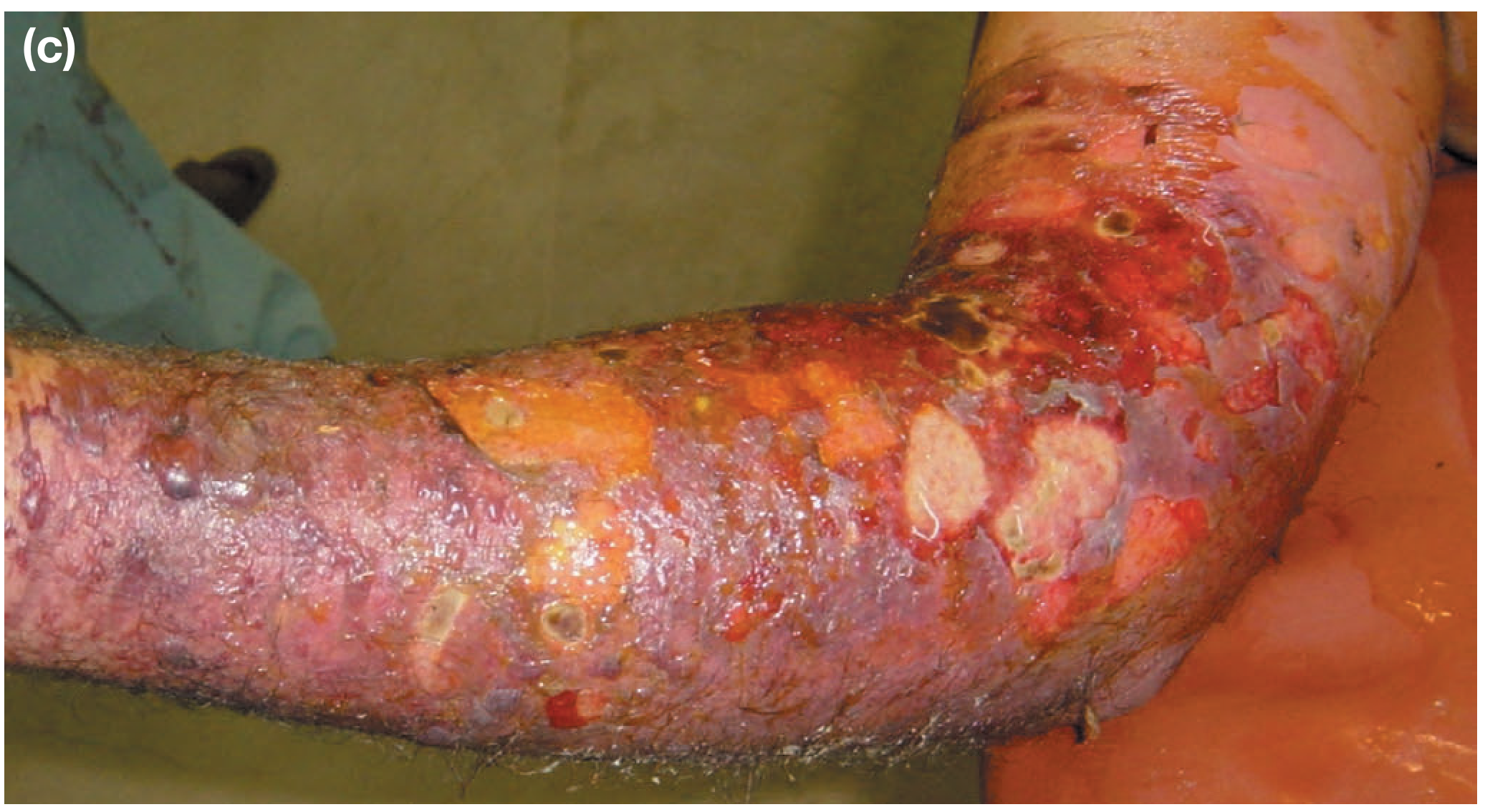
Diagnosis
This is primarily a clinical diagnosis. Do not delay surgical treatment if suspicion is high.
Investigations
Laboratory (LRINEC Score):
The LRINEC (Laboratory Risk Indicator for Necrotising Fasciitis) score uses routine labs:
| Variable | Value | Points |
|---|---|---|
| CRP (mg/L) | <150 | 0 |
| ≥150 | +4 | |
| WBC (×10³/mm³) | <15 | 0 |
| 15-25 | +1 | |
| >25 | +2 | |
| Haemoglobin (g/dL) | >13.5 | 0 |
| 11-13.5 | +1 | |
| <11 | +2 | |
| Sodium (mmol/L) | ≥135 | 0 |
| <135 | +2 | |
| Creatinine (µmol/L) | ≤141 | 0 |
| >141 | +2 | |
| Glucose (mmol/L) | ≤10 | 0 |
| >10 | +1 |
Maximum score: 13
- Score ≥6: PPV 92%, NPV 96% - raise suspicion, carefully evaluate
- Score ≥8: Strongly predictive of NF
- Caution: LRINEC ≥6 misses many cases (especially Vibrio species and neck infections). It should NOT be used in isolation to exclude the diagnosis. - Tintinalli's Emergency Medicine
Other lab findings: Elevated creatinine kinase (often enormously elevated), hyponatraemia, thrombocytopaenia (especially Vibrio), lactic acidosis, elevated band count.
Imaging
- Plain X-ray: May show gas in soft tissues - do NOT delay surgery for this
- CT: Useful for showing gas tracking along fascial planes, but MRI is preferred
- MRI: Best modality - superior soft-tissue contrast, shows fascial plane involvement without radiation; can map extent for surgical planning
- Ultrasound: Can detect abnormal muscle echo texture; useful in children and resource-limited settings
"Relevant imaging must therefore be performed immediately" - emergency surgical debridement cannot wait - Grainger & Allison's Diagnostic Radiology
Operative diagnosis
Intraoperative findings confirm the diagnosis:
- "Finger test": Lack of resistance to blunt finger dissection along fascial planes
- Absence of bleeding (devascularised tissue)
- Grey/necrotic fascia that peels easily from underlying muscle
- "Dishwater" fluid
Biopsy of fascial layers confirms the histological diagnosis.
Management
1. Resuscitation (simultaneous with surgical planning)
- Aggressive IV fluid resuscitation
- Haemodynamic monitoring (ICU admission)
- Oxygen supplementation; intubation if airway compromised
- Correct electrolyte derangements
2. Antibiotics (IV, broad-spectrum, started immediately)
- High-dose penicillin G + third-generation cephalosporins (e.g., cefotaxime) + metronidazole (anaerobic cover)
- Add clindamycin for GAS (inhibits toxin production via ribosomal mechanism - "Eagle effect")
- Cover MRSA if suspected (vancomycin/linezolid)
- Adjust based on wound cultures and sensitivities
3. Surgery (cornerstone of treatment - must not be delayed)
"This is a surgical emergency and the diseased area should be debrided as soon as possible until viable, healthy, bleeding tissue is reached." - Bailey and Love's Surgery
- Radical surgical debridement: Remove all devitalised tissue generously, going beyond the zone of induration
- Wound is lightly packed open with gauze/dressings
- Planned re-look in 24-48 hours - necrosis is prone to spread; multiple debridements are typically required (daily until healthy bleeding tissue is reached)
- Vacuum-assisted closure (VAC) dressings after initial debridement
- Early skin grafting in selected cases to minimise protein/fluid losses
4. Adjuncts
- Hyperbaric oxygen (HBO): Increasingly used; reduces mortality from 30-50% (without HBO) to 9-20% (with HBO). Mechanism: enhances neutrophil killing of anaerobes, promotes wound healing
- IVIG may be considered for streptococcal toxic shock syndrome
Prognosis / Mortality
| Setting | 30-day Mortality | 1-year Mortality |
|---|---|---|
| Without aggressive treatment | up to 70% | - |
| With treatment | 26-40% | up to 50% |
| With HBO | 9-20% | - |
Independent predictors of mortality: Age >60, WBC >30,000, creatinine >2 mg/dL, cardiac disease, septic shock, clostridial infection, high ASA class, dialysis dependence.
Limb loss occurs in ~26% of extremity cases. Independent predictors: cardiac disease, shock at admission, clostridial infection.
Key Summary Points
(Bailey and Love's Surgery, 28th Ed.)
- Surgical emergency - primarily a clinical diagnosis
- Polymicrobial synergistic infection (or monomicrobial with GAS)
- 80% have a history of previous trauma or infection
- Rapid progression to septic shock
- Treat with urgent resuscitation, IV broad-spectrum antibiotics, and radical surgical debridement
- LRINEC score is a useful adjunct but must not replace clinical judgement
- Re-debridement planned at 24-48 hours; multiple procedures are the norm
- Mortality 30-50% even with treatment
This is a shared conversation. Sign in to Orris to start your own chat.