


Explain the case and how did they diagnose it's tonsilitis
acute bacterial tonsillitis enlarged erythematous tonsils throat examination
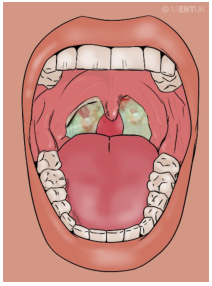
Summary : This illustration shows the inside of a human mouth with a focus on the throat, depicting inflamed tonsils with white-greenish patches, suggestive of tonsillitis or a similar infection. photo: Scene Overview : • Main subject is the open mouth and throat, viewed from the front. • The tonsils are visibly enlarged and covered with white-greenish exudate. • The uvula is centered and appears slightly swollen. • The tongue is prominent in the lower half of the image. • Teeth are visible along the top and bottom edges of the mouth. • The background is a solid, muted color, emphasizing the oral cavity. Technical Details : • No scale bar or magnification indicated. • No staining or medical imaging modality used; this is a digital illustration. • No on-image text except a faint watermark in the upper right corner. Spatial Relationships : • Tonsils are located on either side of the uvula, at the back of the throat. • White-greenish patches are localized to the tonsils, not extending to other oral structures. • The tongue occupies the foreground, with the throat and tonsils in the background. Analysis : • The key visual message is the presence of inflamed, exudate-covered tonsils, which is characteristic of acute tonsillitis or pharyngitis. • The illustration highlights the typical appearance of infected tonsils, useful for educational or diagnostic reference.
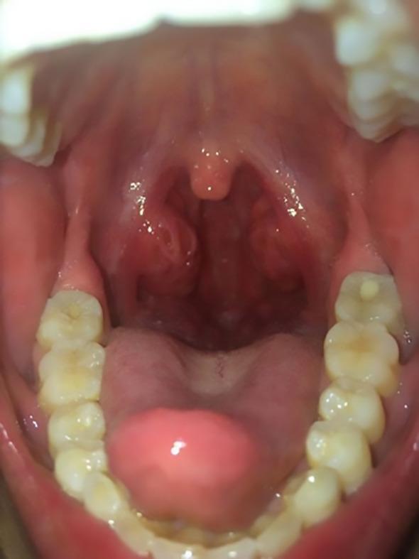
This clinical photograph displays an intraoral view of the oropharynx, highlighting a case of severe acute tonsillitis. The primary findings include significant bilateral tonsillar hypertrophy, with the palatine tonsils appearing markedly swollen and erythematous. Multiple small, white-to-yellowish purulent exudates are visible within the tonsillar crypts, a hallmark of bacterial infection. The surrounding oropharyngeal mucosa and soft palate exhibit generalized hyperemia. The uvula is midline and appears slightly edematous but not deviated. Other visible anatomical landmarks include the tongue and the maxillary and mandibular dentition. This visual representation is characteristic of infectious processes such as Lemierre syndrome precursors or streptococcal pharyngitis, demonstrating clinical signs that can lead to airway narrowing or systemic inflammatory response syndrome (SIRS).
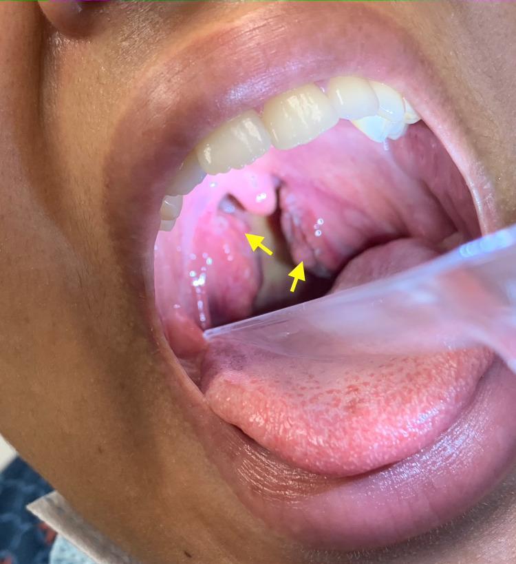
This clinical photograph displays an intraoral view of the oropharynx, captured using a clear plastic tongue depressor. The image shows significant bilateral tonsillar hypertrophy, with the palatine tonsils appearing erythematous and markedly enlarged, nearly meeting at the midline. Yellow arrows highlight the presence of white-to-grayish follicular exudates localized on the tonsillar surfaces. The surrounding pharyngeal mucosa exhibits diffuse erythema, consistent with an acute inflammatory process. These visual findings are classic clinical indicators of acute exudative tonsillitis, which can be seen in conditions such as streptococcal pharyngitis or infectious mononucleosis. The anatomical focus includes the soft palate, uvula, palatine tonsils, and the posterior pharyngeal wall, demonstrating typical signs of infectious oropharyngeal pathology.
Case Summary - Akshay (Case 12)
| Detail | Info |
|---|---|
| Name | Akshay |
| Age | 12 years |
| Gender | Male |
| Weight | 29 kg |
| Date of Admission | 26/7/26 |
| Past Medical History | None (k/c/o: None) |
Chief Complaints
- Sore throat for 3 days
- Fever for 2 days
- Reduced appetite and difficulty swallowing (odynophagia) for 2 days
Vitals Over 3 Days (Trending Downward = Improving)
| Parameter | Day 1 | Day 2 | Day 3 |
|---|---|---|---|
| Temp | 101.8°F | 100.4°F | 99.2°F |
| Pulse Rate | 96 bpm | 94 bpm | 88 bpm |
| Resp Rate | 22 cpm | 21 cpm | 20 cpm |
| BP | 100/65 mmHg | 102/66 | 104/60 |
How Was Tonsillitis Diagnosed?
1. Subjective Findings (Patient's Symptoms)
- Sore throat, fever, difficulty swallowing, reduced appetite - all classic features pointing toward pharyngotonsillitis.
2. Objective Finding - Throat Examination
- "Enlarged erythematous tonsillitis" was documented on throat exam.
- This means the tonsils were visibly swollen (enlarged) and red/inflamed (erythematous), the primary physical sign of tonsillitis.
3. Lab Investigations - Why They Point to BACTERIAL (Not Viral) Cause
| Test | Akshay's Result | Normal Range | Interpretation |
|---|---|---|---|
| Hb | 12.5 g/dL | 11.5-16.5 | Normal |
| TC (Total Leukocyte Count) | 14,500 cells/mm³ | 4,000-11,000 | HIGH - Leukocytosis |
| RBC | 4.5 mill/cumm | 3.5-5.5 | Normal |
| Neutrophils (N) | 78% | 40-60% | HIGH - Neutrophilia |
| Eosinophils (E) | 3% | 1-4% | Normal |
| Lymphocytes (L) | 18% | 20-40% | Slightly low (relative) |
| ESR | 25 mm/hr | <15 (child) | Mildly elevated - active inflammation |
Final Diagnosis
- Clinical presentation (sore throat + fever + dysphagia)
- Throat exam showing enlarged, erythematous tonsils
- Lab findings showing leukocytosis with neutrophilia (bacterial pattern)
Treatment Plan
In-hospital (D1-D3):
| Drug | Generic | Dose | Route | Frequency |
|---|---|---|---|---|
| Syr. Nasamox | Amoxicillin | 250 mg/mL | Oral | TID (3x/day) |
| Syr. Calpol | Paracetamol | 250 mg/5mL | Oral | SOS (as needed for fever) |
| Syr. Cetzine | Cetirizine HCl | 5 mg | Oral | OD (once daily) |
| Warm saline gargles | - | 100 mL | Gargle | TID |
Discharge Medications:
- Syr. Amoxicillin - 250 mg - oral - TID - 7 days (to complete the antibiotic course)
- Syr. PCM (Paracetamol) - 250 mg - oral - SOS - 3 days (for fever/pain relief)
- Syr. Cetirizine - 5 mg - oral - OD - 3 days (antihistamine for throat irritation/inflammation)