
Full details about myopia, types, causes treatment
myopia nearsightedness refractive error diagram eye
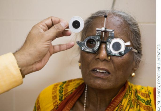
Clinical photograph of a patient undergoing a subjective refraction and visual acuity assessment. The patient is wearing an adjustable metal trial frame, an ophthalmic device featuring multiple slots designed to hold trial lenses for determining a patient's refractive error (myopia, hyperopia, or astigmatism). The examiner's hand is visible holding a pinhole occluder toward the patient's right eye. This specific tool is used in clinical practice to determine if a reduction in visual acuity is due to a refractive error—which typically improves when viewing through a small aperture—or an underlying organic pathology. The image demonstrates key steps in an optometric or ophthalmological examination, focusing on the diagnostic equipment used for determining the best-corrected visual acuity (BCVA). The setting is an eye care clinic, highlighting the procedural interaction between the clinician and the patient during vision screening.
![<table><thead><tr><th>Condition</th><th>Frequency</th></tr></thead><tbody><tr><td>Refractive errors</td><td></td></tr><tr><td> Myopia (−0.75 D or more in eye with lesser refractive error)</td><td>0.7%–9.2%<sup>16, 17,18</sup> (prevalence in children aged 5–17 years)</td></tr><tr><td> Myopia (more than -2.0 D)</td><td>0.2%–2%<sup>19</sup> (prevalence in children aged 3–5 years)</td></tr><tr><td> Hyperopia (+3.0 D or more in eye with lesser refractive error)</td><td>4%–9%<sup>16, 17</sup> (prevalence in children aged 5–17 years)</td></tr><tr><td> Hyperopia (more than +3.25 D)</td><td>6%–7%<sup>19</sup> (prevalence in children aged 3–5 years)</td></tr><tr><td> Astigmatism (worse eye cylinder power 3.0 D or more)</td><td>0.5%–3%<sup>16, 20</sup> (prevalence in children aged 5–17 years)</td></tr><tr><td> Astigmatism (cylinder power more than 1.5 D)</td><td>4%–11%<sup>19</sup> (prevalence in children aged 3–5 years)</td></tr><tr><td>Amblyopia</td><td>0.8%–3%<sup>21–25</sup> (prevalence in children aged 6–72 months)</td></tr><tr><td></td><td>0.08%–4.6%<sup>19, 21–25</sup> (prevalence in children aged 6–72 months)</td></tr><tr><td>Strabismus</td><td>1.2%–6.8% (prevalence in children aged 6–17 years)<sup>26–32</sup></td></tr><tr><td>Cerebral visual impairment, including traumatic brain injury</td><td>Accurate prevalence or incidence data are lacking</td></tr><tr><td></td><td>0.02%<sup>33, 34</sup> (prevalence in children aged 0–1 year)</td></tr><tr><td></td><td>0.1%<sup>24</sup> (prevalence in children aged 6 months to 6 years)</td></tr><tr><td>Cataract</td><td>0.42%<sup>35</sup> (prevalence in children aged 6 to 15 years)</td></tr><tr><td></td><td>8.6%–9.2%<sup>36–38</sup> (incidence of severe ROP in cohorts 1000–1250 g [mean] at birth)</td></tr><tr><td>ROP</td><td>15.2%–18.3%<sup>39, 40</sup> (incidence of severe ROP in cohorts 800–999 g [mean] at birth)</td></tr><tr><td>Congenital glaucoma</td><td>0.0015%–0.0054%<sup>41, 42</sup> (prevalence in newborns)</td></tr><tr><td></td><td>0.0011%–0.0013%<sup>43–46</sup> (yearly incidence in children aged <5 years)</td></tr><tr><td>Retinoblastoma</td><td>0.00036%–0.00041%<sup>47, 48</sup> (yearly incidence in children aged <15 years)</td></tr><tr><td>Pediatric uveitis</td><td>Incidence 0.004%<sup>49</sup> (yearly incidence in children aged <16 years)</td></tr></tbody></table>
D = diopter; g = grams; ROP = retinopathy of prematurity.](https://cdn.orris.care/cdss_images/GLGCA_10674_1766768343352_bc83d9e0-a63b-4f49-8863-6fdf14f1c7a5_c81eeb05-ffd0-4cc8-a677-8ba73319caef.png)
<table><thead><tr><th>Condition</th><th>Frequency</th></tr></thead><tbody><tr><td>Refractive errors</td><td></td></tr><tr><td> Myopia (−0.75 D or more in eye with lesser refractive error)</td><td>0.7%–9.2%<sup>16, 17,18</sup> (prevalence in children aged 5–17 years)</td></tr><tr><td> Myopia (more than -2.0 D)</td><td>0.2%–2%<sup>19</sup> (prevalence in children aged 3–5 years)</td></tr><tr><td> Hyperopia (+3.0 D or more in eye with lesser refractive error)</td><td>4%–9%<sup>16, 17</sup> (prevalence in children aged 5–17 years)</td></tr><tr><td> Hyperopia (more than +3.25 D)</td><td>6%–7%<sup>19</sup> (prevalence in children aged 3–5 years)</td></tr><tr><td> Astigmatism (worse eye cylinder power 3.0 D or more)</td><td>0.5%–3%<sup>16, 20</sup> (prevalence in children aged 5–17 years)</td></tr><tr><td> Astigmatism (cylinder power more than 1.5 D)</td><td>4%–11%<sup>19</sup> (prevalence in children aged 3–5 years)</td></tr><tr><td>Amblyopia</td><td>0.8%–3%<sup>21–25</sup> (prevalence in children aged 6–72 months)</td></tr><tr><td></td><td>0.08%–4.6%<sup>19, 21–25</sup> (prevalence in children aged 6–72 months)</td></tr><tr><td>Strabismus</td><td>1.2%–6.8% (prevalence in children aged 6–17 years)<sup>26–32</sup></td></tr><tr><td>Cerebral visual impairment, including traumatic brain injury</td><td>Accurate prevalence or incidence data are lacking</td></tr><tr><td></td><td>0.02%<sup>33, 34</sup> (prevalence in children aged 0–1 year)</td></tr><tr><td></td><td>0.1%<sup>24</sup> (prevalence in children aged 6 months to 6 years)</td></tr><tr><td>Cataract</td><td>0.42%<sup>35</sup> (prevalence in children aged 6 to 15 years)</td></tr><tr><td></td><td>8.6%–9.2%<sup>36–38</sup> (incidence of severe ROP in cohorts 1000–1250 g [mean] at birth)</td></tr><tr><td>ROP</td><td>15.2%–18.3%<sup>39, 40</sup> (incidence of severe ROP in cohorts 800–999 g [mean] at birth)</td></tr><tr><td>Congenital glaucoma</td><td>0.0015%–0.0054%<sup>41, 42</sup> (prevalence in newborns)</td></tr><tr><td></td><td>0.0011%–0.0013%<sup>43–46</sup> (yearly incidence in children aged <5 years)</td></tr><tr><td>Retinoblastoma</td><td>0.00036%–0.00041%<sup>47, 48</sup> (yearly incidence in children aged <15 years)</td></tr><tr><td>Pediatric uveitis</td><td>Incidence 0.004%<sup>49</sup> (yearly incidence in children aged <16 years)</td></tr></tbody></table> D = diopter; g = grams; ROP = retinopathy of prematurity.
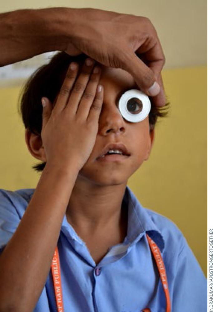
This clinical photograph depicts a pediatric vision screening or eye examination being conducted on a school-aged child in a community setting. The image demonstrates a monocular visual acuity test procedure. The child is manually occluding his right eye with his palm, while an examiner uses an ophthalmic tool, likely a pinhole occluder or a specialized viewing aperture, over the left eye. The examiner’s hand is visible, stabilizing the tool and lifting the child's eyelid to ensure an unobstructed view of the ocular surface and pupil. This visual highlights public health efforts for early detection of refractive errors, such as myopia, in school-aged populations. The setting appears to be a school, indicated by the child's uniform and lanyard. Key educational concepts illustrated include clinical examination techniques for pediatric ophthalmology, the use of simple diagnostic occluders, and vision screening protocols for refractive error identification in low-resource environments.
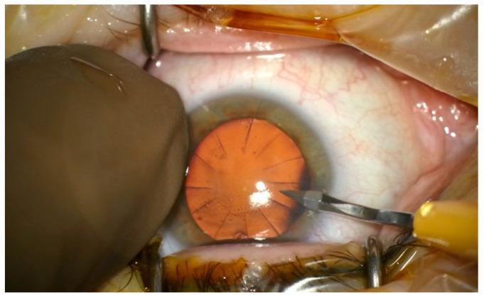
This intraoperative clinical photograph captures the anterior segment of a human eye during a cataract surgery following a historical radial keratotomy (RK). The cornea demonstrates multiple distinct radial incisions, approximately 12 to 16 in number, which extend from the central pupillary zone toward the limbus, a hallmark of prior refractive surgery for myopia. These scars appear as linear, fibrotic tracks on the corneal surface. A surgical micro-blade with a yellow handle is visible on the right, performing a lateral clear corneal incision between two existing RK scars. On the left, a blunt stabilizer or surgical gloved finger provides globe immobilization. Speculums are in place to retract the eyelids, exposing the bulbar conjunctiva, which shows mild surgical hyperemia. The red reflex is visible through the pupil, highlighting the radial arrangement of the old incisions. This image illustrates the surgical challenge of creating new cataract incisions while avoiding intersection with unstable previous RK scars to prevent corneal dehiscence or irregular astigmatism.
"myopia"[MeSH Terms] AND "treatment"[MeSH Terms]
Myopia (Nearsightedness) - Full Clinical Overview
What is Myopia?
- The axial length of a normal eye is 20-24 mm
- In myopia, axial length is elongated (often >25 mm; can reach 40 mm in severe cases)
- Measured in negative diopters (D)
Types of Myopia
1. Simple (Physiological / School) Myopia
- Most common form
- Develops during childhood (typically ages 6-14), usually progresses until age 20-25 years
- Results from normal biological variation in eye growth - the eye is slightly too long or the cornea slightly too curved
- Typically -0.5 D to -6.0 D
- Fully correctable with glasses, contact lenses, or LASIK to 20/40 or better in ≥95% of patients
2. High Myopia
- Defined as refractive error of more than -6.0 diopters, with axial length usually >26 mm
- Affects >2% of adult Western European/American populations; up to 10% in East Asians
- Associated with posterior segment complications (see below)
- Can be associated with lens changes: posterior subcapsular opacity and early-onset nuclear sclerosis
3. Pathological / Degenerative Myopia
- Progressive, characterized by anteroposterior elongation of the scleral envelope with mechanical stretching of ocular tissues
- Develops posterior staphyloma - localized deformation where there is an abrupt increase in posterior radius of curvature
- A significant cause of legal blindness, with maculopathy as the most common cause of visual loss
- Associated with lacquer cracks, CNV (choroidal neovascularization), retinal detachment, and hemorrhage
- Partly heritable condition
4. Progressive Myopia
- High pathologic myopia / staphyloma / globe elongation
- Diabetes mellitus (reversible metabolic lens changes)
- Cataract development
- Corneal ectasia (keratoconus, post-refractive surgery)
- Medications: miotic drops, sulfa drugs, tetracycline
- Physiological childhood progression
Causes and Risk Factors
Optical/Structural Causes
| Mechanism | Description |
|---|---|
| Axial myopia | Eye too long (most common) |
| Refractive myopia | Cornea or lens has excess curvature/power |
| Index myopia | Increased refractive index of lens (e.g., nuclear sclerosis in aging/diabetes) |
Genetic Factors
- Strong hereditary component - both parents myopic = significantly increased risk
- Associated syndromes with high myopia:
- Stickler syndrome (most common)
- Marfan syndrome
- Down syndrome
- Prematurity
- Noonan syndrome
- Ehlers-Danlos syndrome
- Pierre-Robin syndrome
- Cornelia de Lange syndrome
Environmental / Behavioral Factors
- Strong evidence for a causative association with long-term intensive near-visual activity (reading, computers, screens)
- Reduced time outdoors - sunlight exposure and distance viewing are protective
- Urban living, higher education levels correlate with higher prevalence
Symptoms
- Blurred distance vision (unable to read a blackboard, driving difficulty)
- Near vision is normal or near-normal
- Discovered in children during school screening
- In high/pathological myopia: floaters, flashing lights (from vitreous traction/retinal changes), reduced vision in adulthood
Diagnosis / Signs
- Refraction: Negative spherical power required to correct vision
- Axial length measurement by A-scan ultrasound or optical biometry
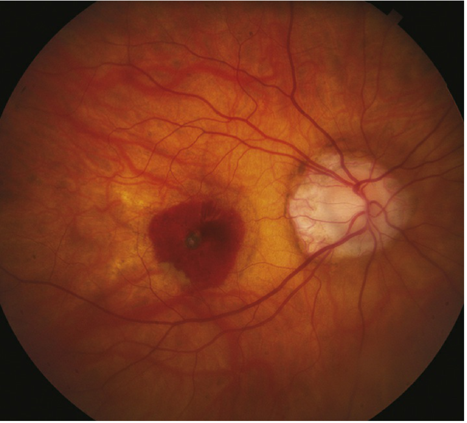
- Fundus findings in degenerative myopia:
- Tessellated (tigroid) fundus - diffuse RPE attenuation exposing large choroidal vessels
- Myopic crescent - white sclera or choroidal vessels adjacent to disc
- Posterior staphyloma - localized scleral ectasia at the posterior pole
- Lacquer cracks - fine irregular yellow lines at posterior pole (RPE-Bruch-choriocapillaris ruptures, in ~5% of highly myopic eyes)
- Fuchs spots - hyperpigmented macular spots
- Anomalous/tilted optic disc
- Lattice degeneration, peripheral retinal thinning
- Risk for CNV (choroidal neovascularization) and RRD (rhegmatogenous retinal detachment)
Treatment
1. Optical Correction
- Spectacles (glasses): Concave (diverging) lenses - first-line for all ages
- Soft contact lenses: Standard correction for mild-moderate myopia
- Rigid gas-permeable (RGP) lenses: Better optics for high myopia
2. Myopia Control (Slowing Progression in Children)
| Intervention | Detail |
|---|---|
| Low-dose atropine 0.01% | Instilled at bedtime; significantly slows progression in children aged 5-15 years progressing >1 D/year |
| Orthokeratology (Ortho-K) | Rigid lenses worn overnight to reshape cornea temporarily |
| Multifocal contact lenses | High-add (+2.50D) centre-distance soft multifocals in ages 7-11 with <5D myopia (BLINK trial shows dose-dependent slowing) |
| Time outdoors | Sunlight exposure is protective - children should be encouraged to spend more time outdoors |
3. Refractive Surgery (Permanent Correction)
| Procedure | Indication |
|---|---|
| LASIK (Laser in situ keratomileusis) | Moderate to high myopia; depends on corneal thickness |
| PRK / Surface ablation | Low-moderate myopia; preferred if thin cornea |
| SMILE (Refractive lenticule extraction) | Small-incision technique for myopia and myopic astigmatism |
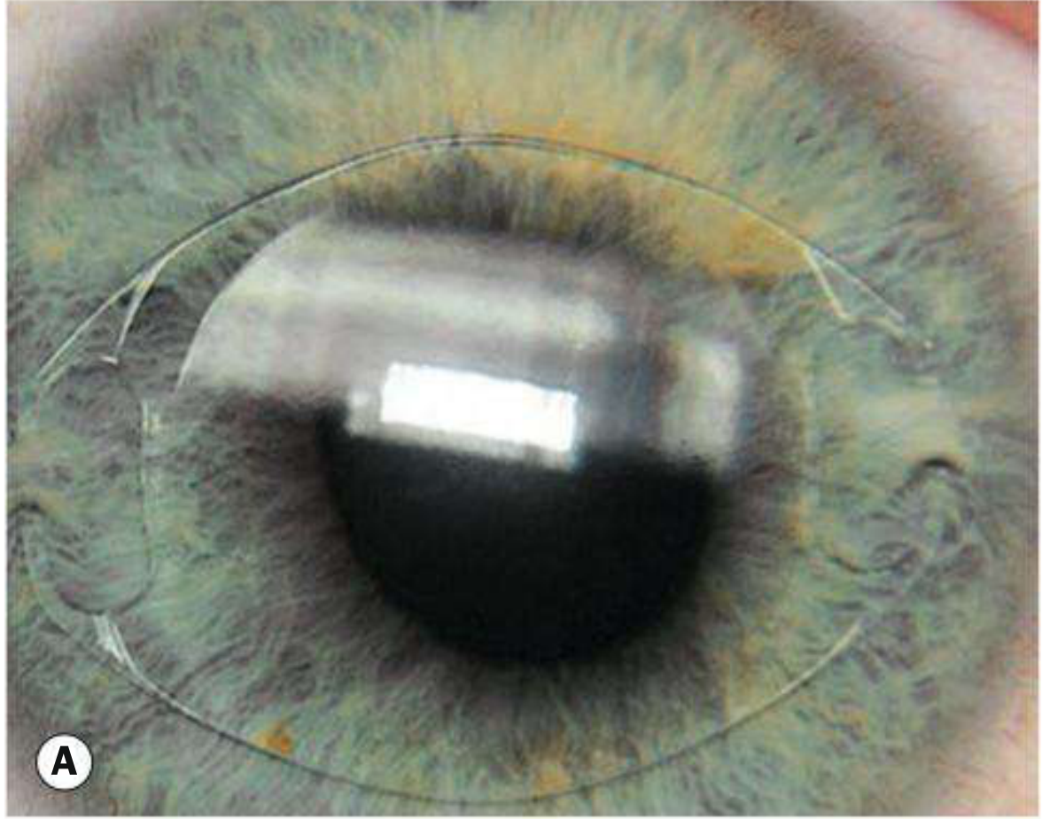
| Phakic ICL (Implantable Collamer Lens) | Very high myopia (-3 D to -20.5 D); inserted behind iris, in front of crystalline lens |
| Iris-clip (lobster claw) implant | Anterior chamber phakic implant attached to iris |
| Clear lens exchange | Very high myopia; carries small risk of retinal detachment |
| Radial keratotomy (RK) | Historically used; now predominantly of historical interest only |
4. Treatment of Complications (Degenerative Myopia)
- Choroidal neovascularization (CNV): Anti-VEGF intravitreal injections (same agents as for wet AMD)
- Retinal holes/detachment: Laser photocoagulation, cryotherapy, pneumatic retinopexy, scleral buckle, vitrectomy
- Cataracts: Phacoemulsification and IOL implantation
- Pathologic myopia is otherwise treated palliatively with glasses/contacts; refractive surgery is less successful due to severe errors and posterior segment abnormalities
Key Clinical Points
- Rapidly progressing myopia after age 25 requires evaluation for diabetes, cataract, or corticosteroid use
- Normal myopia correctable to 20/40 or better in ≥95% of patients with LASIK
- High myopia requires regular fundus surveillance for retinal complications
- Axial length >25 mm (normal 20-24 mm) defines pathologic range; severe cases may reach 40 mm
- Kanski's Clinical Ophthalmology, 10th ed. - Chapters on Degenerative Myopia and Correction of Myopia
- Goldman-Cecil Medicine, International Ed. - Chapter 391
- Wills Eye Manual, 8th ed. - Sections 11.22 and Progressive Myopia
Simple or pathological myopia me kya difference hai
pathological myopia fundus posterior staphyloma lacquer cracks retinal changes
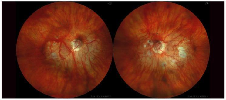
Clinical photograph of the ocular fundus of both eyes (OD and OS) from a single patient, demonstrating advanced pathological changes associated with high myopia and symmetric Type I posterior staphyloma according to Curtin’s classification. Both images show a characteristic 'tessellated' appearance due to retinal pigment epithelium thinning. The optic discs in both eyes appear abnormally large and tilted with significant excavation (cupping), surrounded by extensive peripapillary chorioretinal atrophy. Multiple patches of white-to-pale yellow lacquer cracks and chorioretinal atrophic lesions are visible, particularly in the macula and peripapillary regions. The retinal vasculature exhibits stretching and straightening, with visible underlying choroidal vessels. The overall presentation illustrates a posterior staphyloma where the posterior pole is abnormally ectatic, leading to significant thinning of the retina and choroid. This educational material is relevant for ophthalmology, specifically for studying the progression and clinical manifestations of pathologic myopia and staphyloma classification.
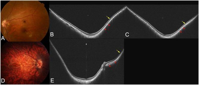
This composite figure illustrates the clinical and diagnostic imaging findings of pathologic myopia and macular staphyloma using fundus photography and wide-field optical coherence tomography (WF-OCT). (A, D) Fundus photographs demonstrate characteristic myopic changes: Image A shows peripapillary diffuse chorioretinal atrophy (PDCA), while Image D reveals more advanced diffuse chorioretinal atrophy with multiple lacquer cracks and prominent tessellation. (B, C, E) Cross-sectional WF-OCT scans highlight the morphological alterations of a staphyloma. Key features include posterior displacement of the sclera in the staphylomatous area and localized scleral inward protrusions (indicated by yellow arrows) marking the staphyloma edge. The choroid displays dynamic thickness changes, thinning as it approaches the staphyloma edge and re-thickening toward the posterior pole or optic nerve. Red arrowheads point to areas where the inner sclera is displaced posteriorly corresponding with choroidal re-thickening. These images serve as educational tools for identifying the structural boundaries and retinal-choroidal thinning associated with high axial length and posterior staphyloma progression.
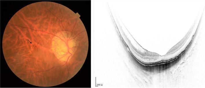
The visual content consists of a side-by-side comparison of a color fundus photograph and a cross-sectional optical coherence tomography (OCT) scan of a highly myopic eye. The fundus image on the left displays a classic 'tessellated' background, where thinning of the retinal pigment epithelium makes the underlying choroidal vessels prominently visible. A black arrow indicates 'lacquer cracks,' which represent linear ruptures in Bruch's membrane. A prominent peripapillary conus is also visible adjacent to the optic disc, characteristic of axial myopia. The OCT scan on the right demonstrates pathological changes in macular architecture, specifically inner foveoschisis characterized by intraretinal splitting or separation of the neurosensory retinal layers. The scan also reveals a significantly concave posterior staphyloma contour. This combined imaging set is essential for the diagnosis and staging of pathologic myopia, illustrating key complications like myopic traction maculopathy and degenerative Bruch's membrane changes. It serves as an educational tool for ophthalmology students and residents to differentiate between stages of myopic maculopathy using the ATN classification system.
Simple vs Pathological Myopia - Poora Farq
Ek Nazar Mein (Quick Comparison Table)
| Feature | Simple Myopia | Pathological Myopia |
|---|---|---|
| Dusra naam | Physiological / School myopia | Degenerative / Malignant myopia |
| Power | -0.5 D se -6.0 D tak | -6.0 D se zyada |
| Axial length | Thodi badi (24-26 mm) | >26 mm (extreme cases mein 40 mm tak) |
| Progression | 20-25 saal ki umra mein ruk jaati hai | Zindagi bhar badhti rehti hai |
| Cause | Normal biological variation | Posterior sclera ka progressive weakening |
| Aankhon ki structure | Normal rehti hai | Mechanical stretching se damage hoti hai |
| Glasses se correction | Poori tarah theek (95%+ patients) | Sirf palliative (aankh ka nuksaan nahi rokta) |
| Blindness ka risk | Nahi | Haan - legal blindness tak |
| LASIK | Bahut effective | Kam effective - posterior changes ki wajah se |
Simple Myopia - Detail Mein
Kya hota hai?
- Aankhein badhti hain growing age mein - ye normal growth ka variation hai
- Retina aur choroid bilkul normal rehte hain
- Aankhein 20-25 saal ki umra mein grow karna band kar deti hain
Numbers:
- Typically -0.5 D se -6.0 D
- Axial length 24-26 mm (normal 20-24 mm)
Symptoms:
- Door ki cheezein blur dikhti hain
- Paas ka kaam (padhna) bilkul theek
- Koi pain, floaters, ya flashes nahi
Treatment:
- Glasses / Contact lenses - poori correction milti hai
- LASIK - 95% se zyada patients mein 20/40 ya better vision milta hai
- Ek baar stable ho jaye toh regular follow-up kaafi hai
Pathological Myopia - Detail Mein
Kya hota hai?
- Aankh ka axial length badhta rehta hai - 40 mm tak bhi ja sakta hai
- Retina, choroid aur sclera par mechanical stretching hoti hai
- Tissues damage hote rehte hain
Fundus ke Signs (Ophthalmoscope se dikh the hain):
| Sign | Kya hota hai |
|---|---|
| Tessellated / Tigroid fundus | RPE patla ho jaata hai, neeche ki badi choroidal vessels dikhne lagti hain |
| Posterior staphyloma | Aankh ke peeche baahar ki taraf bulge banta hai - bahut khatranaak |
| Lacquer cracks | RPE-Bruch membrane mein cracks - ~5% cases mein; CNV ka risk |
| Myopic crescent | Disc ke paas sclera ya choroidal vessels dikhne lagte hain |
| Fuchs spots | Macula mein pigmented spots |
| Tilted optic disc | Optic nerve oblique angle pe insert hoti hai |
| Lattice degeneration | Peripheral retina mein thinning |
Complications (Khatarey):
Pathological Myopia
       │
       ├── Choroidal Neovascularization (CNV) → Anti-VEGF injection chahiye
       ├── Retinal hole → Laser photocoagulation
       ├── Retinal detachment → Surgery chahiye
       ├── Intraocular hemorrhage
       ├── Cataract (posterior subcapsular, early onset)
       └── Legal Blindness ← Maculopathy sabse common cause hai
Associated Systemic Conditions:
- Marfan syndrome, Stickler syndrome, Down syndrome, Ehlers-Danlos, prematurity
Sabse Important Farq - Ek Line Mein
Simple myopia mein sirf focus ka problem hai - glasses se theek ho jaata hai. Pathological myopia mein aankh ki structure hi damage hoti rehti hai - permanent vision loss ho sakta hai.
Treatment ka Farq
| Simple Myopia | Pathological Myopia | |
|---|---|---|
| Glasses | Poori correction | Sirf symptoms control |
| LASIK | Excellent results | Poor results - posterior changes limit success |
| Myopia control (atropine 0.01%) | Haan, progression rokne ke liye | Haan, children mein |
| Anti-VEGF injections | Nahi chahiye | CNV hone par zaroori |
| Retinal surgery | Nahi | Detachment ya holes pe zaroori |
| Regular fundus monitoring | Occasional | Frequent - complications ke liye |
- Kanski's Clinical Ophthalmology, 10th ed. - Chapter on Degenerative Myopia
- Goldman-Cecil Medicine - Chapter 391 (Pathologic vs Non-pathologic Myopia)
Simple moderate pr high myopia
myopia grades low moderate high diopters eye axial length comparison
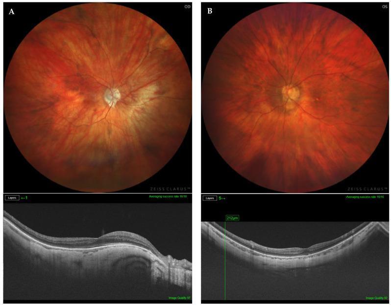
Educational diagnostic comparison of two highly myopic eyes (OD and OS) featuring fundus photography and structural Optical Coherence Tomography (OCT). Panel A displays the right eye (OD) of a 70-year-old patient with an axial length of 29.21 mm. The fundus image shows a tessellated pattern characteristic of high myopia, a pale, well-defined optic disc, and peripapillary atrophy. The corresponding OCT B-scan demonstrates a normal retinal profile despite the elongated globe. Panel B displays the left eye (OS) of a 62-year-old patient with an axial length of 29.75 mm. The fundus reveals a similar tessellated appearance and clear retinal vasculature. The sub-adjacent OCT shows preserved foveal architecture and retinal layer integrity, with a caliper measurement of 212µm indicating local retinal thickness. The images illustrate types of non-macular posterior staphyloma (PS) in high myopia where the macula remains structurally intact on OCT despite significant axial elongation and global pathology.
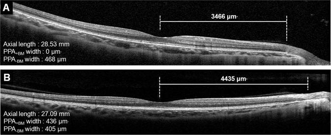
Educational diagnostic comparison using horizontal Spectral-Domain Optical Coherence Tomography (SD-OCT) B-scans of the retina in high myopia. The image displays two distinct cases, labeled A and B, focusing on the microstructural relationship between macular Bruch's membrane (BM) length and beta-parapapillary atrophy (β-PPA). Case A shows a myopic eye with an axial length of 28.53 mm, characterized by the presence of PPA without Bruch's membrane (PPA-BM = 468 μm) and the absence of PPA with Bruch's membrane (PPA+BM = 0 μm), resulting in a macular BM length of 3466 μm. Case B illustrates a myopic eye with a shorter axial length (27.09 mm) but a longer macular BM length (4435 μm), featuring both PPA+BM (436 μm) and PPA-BM (405 μm). Key anatomical landmarks visible include the foveal depression, retinal layers, retinal pigment epithelium (RPE), and the termination points of the BM. This comparison demonstrates that macular BM length can be longer in eyes with PPA+BM regardless of axial length, illustrating complex morphological changes in myopic globe elongation.
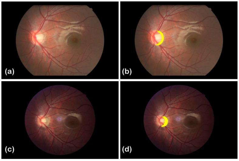
This clinical imaging set presents a longitudinal comparison of fundus photographs from a myopic patient, captured in 2012 (top row, a and b) and 2016 (bottom row, c and d). The images demonstrate the progression of peripapillary atrophy, a common feature in high myopia. Panels (a) and (c) show the raw fundus photography of the left eye, centered on the posterior pole including the optic disc, macula, and retinal vasculature. Panels (b) and (d) are annotated versions where the peripapillary atrophic arc is highlighted in yellow adjacent to the temporal margin of the optic disc. Clinically, the case tracks a transition from mild atrophy in 2012 (spherical equivalent -2.50 D, axial length 25.04 mm) to moderate atrophy in 2016 (spherical equivalent -5.50 D, axial length 27.47 mm). The visual comparison highlights changes in the morphology and extent of the atrophic region relative to the optic disc over a four-year interval, providing a visual record of myopic chorioretinal degeneration.
Low / Moderate / High Myopia - Teen Grades ka Poora Farq
Grade Classification (Diopters ke hisaab se)
LOW MYOPIA          MODERATE MYOPIA         HIGH MYOPIA
  0 to -3 D    →     -3 to -6 D      →      -6 D se zyada
| Feature | Low (Simple) | Moderate | High |
|---|---|---|---|
| Power | 0 to -3.00 D | -3.00 to -6.00 D | -6.00 D se zyada |
| Axial length | ~23-25 mm | ~25-26 mm | >26 mm |
| Door ka vision | Thodi blur | Kaafi blur | Bahut zyada blur |
| Paas ka vision | Normal | Normal | Normal (ya thodi takleef) |
| Progression | 20-25 saal mein ruk jaati | 20-25 saal mein ruk jaati | Ruk sakti hai ya badhti rehti hai |
| Retinal changes | Nahi | Normally nahi | Haan - serious risk |
| Blindness risk | Practically zero | Bahut kam | Haan - especially >-10 D |
| Fundus | Normal | Normal | Tessellated, staphyloma, lacquer cracks possible |
Low Myopia (0 to -3 D)
Kaun hote hain?
- Sabse zyada common grade
- Bachpan mein shuru, school age mein diagnose hoti hai
- Zyada tar 20-25 saal mein stable ho jaati hai
Symptoms:
- Door ki cheezein thodi blur (blackboard, TV)
- Raat ko driving mein thodi takleef
- Paas ka kaam (padhna, mobile) bilkul normal
Treatment:
- Thin glasses ya soft contact lenses - kaafi hai
- PRK / Surface ablation - laser surgery excellent results deti hai
- Low risk - zyada monitoring ki zaroorat nahi
Moderate Myopia (-3 to -6 D)
Kaun hote hain?
- Genetic + environmental factors dono
- Zyada padhne wale, screen time zyada wale bacchon mein common
- Bhi 20-25 saal mein stable hoti hai zyada tar
Symptoms:
- Door ki cheezein kaafi zyada blur - bina glasses ke kuch bhi saaf nahi dikhta
- Glasses ke bina roz ka kaam mushkil
- Raat ko halos ya glare ho sakta hai
Treatment:
- Glasses / Contact lenses - zaroori hai, correction achhi milti hai
- LASIK - moderate myopia ke liye excellent candidate - corneal thickness allow kare toh
- SMILE - LASIK jaisa hi, chhoti incision se
- Surface ablation (PRK) - thin cornea mein prefer karte hain
- Regular eye check (1-2 saal mein) adequate hai
High Myopia (-6 D se zyada)
Kaun hote hain?
- -6 D se zyada power
- Axial length >26 mm, severe cases mein 40 mm tak
- East Asians mein zyada prevalent (up to 10%)
- Genetic syndromes se associated: Marfan, Stickler, Down syndrome, Ehlers-Danlos
Symptoms:
- Door ki vision bahut zyada kharab
- Floaters (kaale dhabbe) aa sakte hain - vitreous changes se
- Flashing lights - retinal traction ka sign
- Adults mein vision loss bhi ho sakta hai
Fundus Signs (Aankhon ke andar kya dikhta hai):
| Sign | Matlab |
|---|---|
| Tessellated fundus | RPE patla, neeche ke choroidal vessels dikhte hain |
| Myopic crescent | Disc ke paas sclera visible |
| Lacquer cracks | Bruch's membrane mein cracks - ~5% cases |
| Posterior staphyloma | Peeche baahar bulge - bahut serious |
| Fuchs spots | Macula pe pigmented spots |
Complications:
- Choroidal neovascularization (CNV)
- Retinal detachment
- Subretinal hemorrhage
- Early cataract (posterior subcapsular)
- Legal blindness (maculopathy)
Treatment:
| Treatment | Detail |
|---|---|
| Glasses / Contact lenses | Thicker glasses, correction milti hai |
| LASIK | Corneal thickness pe depend karta hai - limited |
| ICL (Implantable Collamer Lens) | Best option for high myopia: -3 D se -20.5 D tak; iris ke peeche, lens ke aage daali jaati hai |
| Iris-clip (Lobster claw) implant | Anterior chamber mein iris pe attach hoti hai |
| Clear lens exchange | Bahut zyada power mein; cataract surgery jaisi; retinal detachment risk |
| Anti-VEGF injections | Agar CNV ho jaaye |
| Retinal surgery | Detachment ya holes pe |
| Regular fundus monitoring | Frequent - 6 mahine ya saal mein |
| Myopia control (children mein) | Atropine 0.01%, multifocal lenses, outdoors time |
Treatment ka Grade-wise Summary
Low Myopia          →  PRK / Surface Ablation  (best)
Moderate Myopia     →  LASIK / SMILE           (best)
High Myopia         →  ICL / Phakic implant    (best for very high)
Very High Myopia    →  Clear Lens Exchange      (last resort)
Ek Important Point
25 saal ke baad myopia tezi se badhe toh - diabetes, cataract, ya steroid ka side effect check karo. Normal myopia 25 saal ke baad nahi badhni chahiye.
- Kanski's Clinical Ophthalmology, 10th ed. - Correction of Myopia + Degenerative Myopia chapters
- Goldman-Cecil Medicine - Chapter 391