Make it look like an college assignment not a pdf presentation. Add more relevant images. I want images below the text not on the sides
hypertrophic cardiomyopathy pediatric echocardiogram
dilated cardiomyopathy pediatric heart pathology
left ventricular non-compaction LVNC echocardiogram trabeculations
arrhythmogenic right ventricular cardiomyopathy ARVC cardiac MRI epsilon wave ECG
restrictive cardiomyopathy biatrial enlargement cardiac MRI
Takotsubo stress cardiomyopathy apical ballooning left ventricle
Pompe disease Fabry disease metabolic storage cardiomyopathy cardiac
How do they perform usg/ ct biopsy
ultrasound CT guided percutaneous biopsy procedure technique steps
ultrasound guided core needle biopsy technique probe needle
Image-Guided Biopsy: USG and CT Techniques
What is it?
Pre-Procedure Steps (Common to Both USG and CT)
1. Case Selection & Contraindications
- Most biopsies are done under local anaesthesia ± sedation; GA may be used in children
- Contraindications: Uncorrected coagulopathy, no safe needle approach route
- Check for anticoagulant use — timing around anticoagulant cessation must be planned
- Obstructed organs (e.g. bile duct, kidney) should be decompressed first (biliary drainage / nephrostomy) before biopsy
- Vascular lesions carry higher bleeding risk — cross-matched blood may be arranged
2. Preprocedural Assessment
- Review imaging, plan approach, choose guidance modality
- Patient consent (written) + information leaflets
- Optimise patient position to identify safest needle route before starting
- Establish IV access; cross-match blood if high-risk
Needle Types: FNA vs Core Biopsy
| Feature | Fine Needle Aspiration (FNA) | Core Needle Biopsy |
|---|---|---|
| Needle gauge | 20–25 G (small) | 14–19 G (large) |
| Sample type | Cells → cytology | Tissue cylinder → histology |
| Advantage | Can transgress bowel or cross vascular structures safely | Preserves architecture; allows immunohistochemistry & receptor testing |
| Use | Superficial lesions, near critical vessels | Most tumour diagnoses, cancer staging |
Core Needle Devices
- Semiautomatic (e.g. Temno): the central notched stylet is advanced to the target, then the cutting sheath is fired over it — no extra forward excursion, safer near vessels
- Fully automatic (e.g. Achieve, BioPince, Bard Max-Core): both stylet and cutting sheath fire together with a preset "throw" (1–2 cm ahead of the tip) — more force for fibrous lesions, but the operator must ensure the throw doesn't exceed the lesion
Coaxial Technique
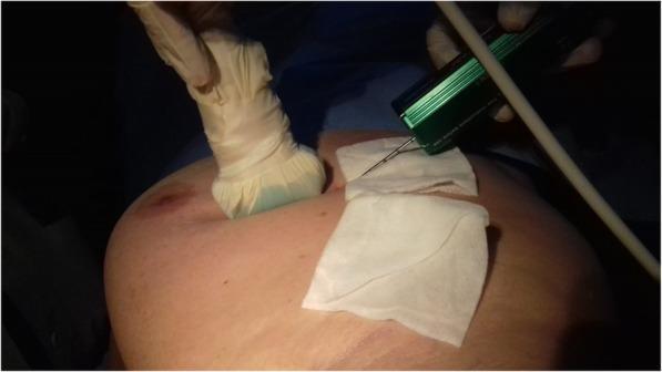
Ultrasound-Guided Biopsy
Technique — Step by Step
- Position the patient — optimise to bring the target as close to the transducer as possible
- Sterile prep — skin cleaned with povidone-iodine, sterile transducer cover and gel applied, sterile drapes placed
- Plan the approach — identify the lesion and safest needle path on USG, avoiding vessels, bowel, pleura
- Local anaesthesia — 1% lidocaine injected superficially (skin wheal, 25G) and then deeper along the planned needle track, under real-time USG guidance
- Skin nick — a small incision is made with a scalpel at the needle entry site (for core biopsy)
- Needle insertion — the needle is introduced in the same plane as the ultrasound beam ("in-plane" technique) so the entire needle shaft is visible as an echogenic line; needle guides (attached to the transducer) can assist alignment
- Real-time targeting — the needle tip is advanced to the lesion under continuous live imaging; tip visibility is improved by:
- Bevel facing upward
- Gently "jiggling" the needle
- Injecting a tiny volume of air or local anaesthetic
- Roughened/polymer-coated needle tips that scatter ultrasound
- Firing the device — core biopsy gun is fired; the sample is retrieved from the notch
- Multiple passes — typically 2–4 cores are taken; with coaxial technique, the outer needle stays in place between passes
- Haemostasis — firm pressure applied for 10+ minutes; bleeding assessed with colour Doppler
- Immediate post-procedure scan — to check for haematoma

CT-Guided Biopsy
Technique — Step by Step
- Scout CT scan — patient placed in CT gantry; a preliminary scan identifies the lesion and plans the needle path (angle, depth, level of entry)
- Grid / gantry angulation — a radiopaque grid or laser lines on the gantry mark the skin entry point; if needed, gantry tilt is used for oblique approaches
- Patient positioning — optimised (prone, supine, lateral) to get the safest angle to the target while avoiding critical structures (vessels, nerves, pleura)
- Sterile prep and local anaesthesia — same as USG; lidocaine infiltrated down to the target level
- Skin nick and needle insertion — the needle is advanced in the intended direction toward the target; breath-hold instruction reduces respiratory motion artefact for thoracic/abdominal lesions
- Confirmation CT — a limited scan (or CT fluoroscopy in real-time systems) is acquired to check needle position; adjustments are made and confirmed with repeat CT
- Coaxial approach — the outer coaxial needle is confirmed at the lesion margin; the inner biopsy needle is then fired through it — particularly important for lung to minimise pleural passes
- Sample acquisition — multiple cores taken; for centrally necrotic tumours, the periphery of the lesion is targeted (viable tissue, not necrotic centre)
- Post-procedure CT — a scan is taken immediately after to check for pneumothorax (lung biopsies), haemorrhage, or other complications
Modality Comparison at a Glance
| Feature | USG | CT |
|---|---|---|
| Real-time guidance | Yes (continuous) | No (intermittent) / CT fluoroscopy = near real-time |
| Radiation | None | Yes |
| Best targets | Soft tissue, liver, thyroid, breast, kidney, LN | Lung, bone, retroperitoneum, deep/obscured structures |
| Speed | ~15–30 min | ~45–60 min |
| Limitations | Poor through bone/gas | Radiation; no true real-time |
| Coaxial technique | Occasionally | Routinely (lung) |
Post-Procedure Care (Both Modalities)
- Observation for 4 hours (vital signs, pain, bleeding)
- Post-lung biopsy: CXR or CT to exclude pneumothorax
- Simple analgesia (NSAIDs) for pain
- Avoid strenuous activity for 24–48 hours
- No driving on the day of procedure
- Results typically available within a few days (histology lab)
How it is different from surgical biopsy
Image-Guided Biopsy vs Surgical Biopsy
The Core Concept
Types of Surgical Biopsy
| Type | What is done | When used |
|---|---|---|
| Incisional biopsy | A portion of the lesion is cut out (lesion left in place) | Large tumours (> 5 cm) where a needle cannot give enough tissue |
| Excisional biopsy | The entire lesion is removed with a margin of normal tissue | Small, superficial lesions (< 5 cm); done with curative intent |
Head-to-Head Comparison
| Feature | Image-Guided Biopsy (USG/CT) | Surgical (Open) Biopsy |
|---|---|---|
| Approach | Percutaneous — needle through intact skin | Open incision into skin, dissection to the lesion |
| Anaesthesia | Local ± sedation | Regional or general anaesthesia usually required |
| Setting | Radiology suite / day case | Operating theatre |
| Size of sample | Small core (14–19 G) or cells (FNA) | Large — can remove the entire lesion |
| Tissue architecture | Core biopsy preserves architecture; FNA does not | Fully preserved — best for lymphoma, complex lesions |
| Sampling error | Yes — 19–44% of atypical ductal hyperplasia on core are actually carcinoma on excision | Minimal with excisional biopsy; entire lesion in the pot |
| Diagnostic accuracy | ~95–99% for accessible, well-targeted lesions | Near 100% with excision |
| Complication rate | < 1% (bleeding, infection, pneumothorax for lung) | Higher — bleeding, wound infection, anaesthesia risks, scarring |
| Morbidity | Very low | Significant — operative recovery needed |
| Cost | Much lower | Much higher (OR time, GA, admission) |
| Time | 15–60 min, usually same day | Hours (procedure + recovery); often inpatient |
| Tumour seeding risk | Low; sheathed needle + planned tract excision at definitive surgery | Higher if incision improperly placed or extensive dissection done |
| Repeat sampling | Easy to repeat | Re-operation is more morbid |
| Immunohistochemistry / molecular testing | Usually adequate with core biopsy | Always adequate |
| Lymphoma diagnosis | Inadequate — cannot assess nodal architecture | Entire node needed → open excision mandatory |
When Each is Chosen
Image-Guided (USG/CT) is preferred when:
- Lesion is accessible and can be visualised on imaging
- A tissue diagnosis is needed before planning definitive therapy (e.g. confirming metastasis to avoid unnecessary surgery)
- Patient is a poor surgical candidate (coagulopathy, comorbidities)
- For staging cancer — confirming nodal or distant disease
- Deep lesions (retroperitoneum, lung, liver, bone) not easily reached surgically without major dissection
Surgical Biopsy is used when:
- Core needle biopsy yields nondiagnostic or inconclusive results after adequate attempts
- The lesion is suspected lymphoma — architecture of the entire node is essential for subtype classification, flow cytometry, and molecular studies
- Very small superficial lesions where excisional biopsy = diagnosis + treatment in one step
- Technically impossible needle biopsy (no safe needle path)
- Surgeon needs to excise with clear margins from the outset (e.g. melanoma, small breast lesion)
Critical Surgical Biopsy Rules (when open is done)
- Incision orientation — for extremity tumours, incisions must be along the long axis (longitudinal), never transverse — a transverse incision contaminates a wider field and can compromise subsequent limb-sparing resection
- No undermining — avoid dissecting into surrounding subcutaneous layers; it spreads disease
- Meticulous haemostasis — a haematoma seeds tumour cells into tissue planes and makes follow-up physical examination very difficult
- Directly overlie the target — the incision must be directly over the lesion, not tunnelled from a distant site (tunnelling contaminates a larger field)
- No enucleation — never shell a sarcoma out of its pseudocapsule without a margin
- Orient the specimen — sutures/clips mark margins; pathologist inks them to determine if margins are clear
- Plan for re-excision — biopsy scar must be oriented so it can be excised if repeat surgery is needed
The Sampling Error Problem
Core needle biopsy samples only a tiny sliver of tissue. If that sliver is not representative (e.g. it samples the necrotic centre, a reactive area, or misses a small focus of malignancy), the result is false negative. This is especially relevant in:
- Atypical ductal hyperplasia of the breast (19–44% upgraded to carcinoma on excision)
- Heterogeneous tumours
- Very small lesions
Summary in One Sentence Each
- Image-guided biopsy = minimally invasive, low risk, fast, good for most solid tumours — but small sample, possible sampling error, inadequate for lymphoma
- Surgical biopsy = invasive, morbid, expensive — but maximum tissue, definitive architecture, needed when needle fails or lymphoma is suspected, and excisional biopsy can be simultaneously curative

Okay I need this type of format by mentioning time Patient name,age,BP,diagnosis,patient Complaint,clinical Findings and treatment Plan.The name should be the names if kyrgyz people because I'm studying mbbs in Kyrgyzstan.Then my posting is under ENT department ,so give me situations according to that