
Sciatica
Reading File
Now let me search the medical library and PubMed simultaneously for comprehensive sciatica coverage.
Finding Sources
Searching PubMed
sciatica lumbar radiculopathy treatment
Finding Sources
Reading File
Reading File
Reading File
Reading File
Reading File
Reading File
Reading File
Now let me fetch the MRI image of disk herniation that was referenced, and also get the Goldman-Cecil differential diagnosis table:
Loading Image
Reading File
I now have comprehensive information from multiple authoritative textbooks. Let me compile the full answer.
Sciatica
Definition
Sciatica is pain that radiates along the distribution of the sciatic nerve - from the lower back, through the buttock, down the posterolateral thigh, and typically into the leg, foot, and toes. It is a symptom, not a diagnosis; the term reflects irritation or compression of the L4, L5, or S1 nerve roots (or the sciatic nerve itself).
The lifetime prevalence of true sciatica is only 2-4%, even though low back pain affects 60-80% of people at some point. Up to 70% of acute sciatic episodes resolve within 3 months.
- Bailey and Love's Short Practice of Surgery, 28th Edition
Causes (Etiology)
The most common cause is lumbar disc herniation compressing a spinal nerve root. The majority of disc herniations occur at L4-L5 (compressing L5 root) or L5-S1 (compressing S1 root).
Other causes include:
Neurogenic - within the spine:
- Herniated nucleus pulposus (most common)
- Spinal canal stenosis (central, lateral recess, or foraminal)
- Spondylolisthesis
- Synovial cyst (from facet joint degeneration)
- Arachnoid or perineural (Tarlov) cysts
- Tumor: neurofibroma, ependymoma, or metastatic disease (prostate, breast, myeloma)
- Infection: epidural abscess, discitis, herpes zoster, Lyme disease
- Inflammatory radiculopathy (sarcoidosis, paraneoplastic)
Neurogenic - outside the spine:
- Lumbosacral plexopathy
- Sciatic neuropathy (e.g., piriformis syndrome)
- Peripheral neuropathy
Non-neurogenic mimics:
- Hip arthritis, trochanteric bursitis
- Sacroiliac joint disease
- Iliotibial band syndrome, hamstring tendinitis
- Peripheral vascular disease
Special causes:
-
Catamenial sciatica - endometriosis involving the sciatic nerve at the sciatic notch (linked temporally to premenstruation)
-
Piriformis syndrome - the sciatic nerve runs through or near the piriformis; hypertrophy/spasm can cause buttock pain and vague sciatic symptoms
-
Goldman-Cecil Medicine, Table 369-5 | Adams and Victor's Principles of Neurology, 12th Ed.
Clinical Presentation
- Unilateral leg pain radiating into the buttock, posterolateral thigh, leg, foot, and/or toes
- Often accompanied by low back pain
- Pain worsened by movement, coughing, sneezing, Valsalva
- Pain relieved by rest or recumbency
- Straight leg raise (Lasègue sign): pain reproduced when the ipsilateral leg is raised while supine - a hallmark test
Root-level localization:
| Root | Level | Pain/Sensory Loss | Motor Weakness | Reflex |
|---|---|---|---|---|
| L4 | L3-L4 disc | Anterior thigh/medial leg | Knee extension | Knee jerk |
| L5 | L4-L5 disc | Anterior leg, dorsal foot, great toe | Foot dorsiflexion, EHL, eversion | None (or tibialis posterior) |
| S1 | L5-S1 disc | Plantar/lateral foot | Plantar flexion, peroneals | Ankle jerk |
- Bradley and Daroff's Neurology in Clinical Practice
Imaging
MRI is the preferred imaging modality - it provides excellent visualization of disc herniations, nerve roots, and surrounding structures. CT myelography is an alternative. Plain radiographs are obtained if tumor, fracture, spondylolisthesis, or infection is suspected.
Key principle: Imaging findings must always be interpreted in clinical context - anatomical abnormalities are frequently incidental and not the source of symptoms.
Here is a lumbar MRI showing L4-L5 disc herniation with posterior displacement of the dura (straight arrows) and compression of cauda equina roots (curved arrows):
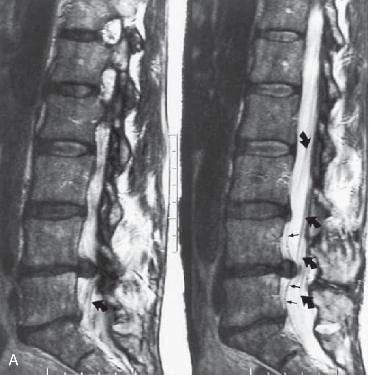
Fig. 104.30 A - Bradley and Daroff's Neurology in Clinical Practice
EDX (Electrophysiology): Needle EMG is the most sensitive electrodiagnostic tool. Fibrillation potentials in a segmental myotomal distribution with normal SNAP in the corresponding dermatome confirms radiculopathy. EMG and imaging are complementary.
Red Flags - Cauda Equina Syndrome (Emergency)
Bilateral or progressive sciatica with any of the following demands urgent MRI and neurosurgical consultation:
- Saddle anesthesia (perineal numbness)
- Urinary/fecal retention or incontinence
- Bilateral leg weakness
- Progressive neurological deficit
The most common cause is a very large L4-5 or L5-S1 disc herniation; others include tumor, epidural abscess, hematoma.
- Bailey and Love's Surgery, Table 37.1
Treatment
Conservative (First-line)
Most patients with acute sciatica recover within 6 weeks with simple nonoperative therapy:
- Activity: Early mobilization is better than bed rest; prolonged immobilization is detrimental
- Analgesics: NSAIDs are the primary pharmacologic option (though less effective for radicular than non-specific back pain); limited opioids for severe pain
- Physical therapy: Back-strengthening exercises, avoidance of straining maneuvers
- Spinal manipulation: Small reduction in pain up to 12 weeks
Drugs with limited or no proven benefit in sciatica:
-
Muscle relaxants - insufficient evidence
-
Antiepileptics (gabapentin - small study benefit; pregabalin - not helpful for acute or chronic sciatica per Goldman-Cecil)
-
Antidepressants - not recommended
-
Oral corticosteroids - no lasting measurable benefit
-
Tintinalli's Emergency Medicine | Goldman-Cecil Medicine | Bradley and Daroff's Neurology
Epidural Steroid Injection
Provides minor reduction in leg pain and sensory deficits vs. placebo at 2-6 weeks, but:
- No significant functional benefit
- Does not reduce need for surgery
- No proven longer-lasting value
- An option for moderately-to-severely symptomatic patients as follow-up (not an ED procedure)
Surgery
Indicated for:
- Severe or progressive neurological deficits
- Failure of nonoperative therapy after 6 weeks
- Cauda equina syndrome (urgent/emergent)
- Spinal instability
- Neurological deficits critical to the patient's vocation
Microsurgical discectomy (with minimal laminectomy) is the standard operation for disc herniation. About 90% of patients report excellent relief of neuropathic pain after surgery. However, benefits over conservative therapy diminish at longer follow-up. "Failed back surgery syndrome" (chronic pain after surgery) is more common when patients lack objective radiculopathy signs or corresponding imaging findings.
A 2025 systematic review and meta-analysis (Low et al., Eur Spine J 2025, PMID 40488872) confirmed that symptom duration affects outcomes of lumbar discectomy - supporting timely surgical decision-making when indicated.
A 2024 systematic review of clinical practice guidelines (Price et al., PM&R 2024, PMID 38629664) found heterogeneity across guidelines for medication recommendations in lumbosacral radiculopathy, reflecting the limited evidence base for most drug therapies.
Prognosis
- 70% of acute sciatic episodes resolve within 3 months
- Workers off work >6 months with back pain have a guarded prognosis for return to work
- Physicians should emphasize exercise, avoid deconditioning, and enable early return to graded work for pain lasting >4 weeks
Typhoid..at last pathophysiology in flowchart form
Reading File
Finding Sources
Finding Sources
Reading File
Reading File
Reading File
Reading File
Reading File
Reading File
I now have all the pathophysiology details needed. Let me build the comprehensive flowchart.
Typhoid Fever - Complete Overview with Pathophysiology Flowchart
Pathophysiology Flowchart
INGESTION OF S. Typhi / S. Paratyphi
(contaminated food or water, fecal-oral route)
          │
          ▼
GASTRIC ACID BARRIER
  ├─ Normal acid → most organisms killed
  └─ Achlorhydria / large inoculum → organisms survive
          │
          ▼
SMALL INTESTINE (Terminal Ileum)
S. Typhi penetrates intestinal epithelium via M cells
overlying Peyer's patches
          │
          ▼
UPTAKE BY MONONUCLEAR PHAGOCYTES
in the lamina propria (submucosal lymphoid tissue)
  • Unlike NTS: NO massive neutrophil infiltration
  • Mononuclear (macrophage) predominant response
  • Key virulence factor: Vi antigen (capsule) resists
    phagocytic killing → intracellular survival
          │
          ▼
PEYER'S PATCHES ENLARGE
Plateaulike elevations up to 8 cm in terminal ileum
          │
          ├──────────────────────────────────────────┐
          ▼                                          ▼
PRIMARY BACTEREMIA                        LYMPHATIC SPREAD
(1st-2nd week)                     Organisms travel to mesenteric
  • Organisms enter portal circulation     lymph nodes → reactive
  • Seeded into liver, spleen,             hyperplasia
    gallbladder, bone marrow              Bacteria-laden phagocytes
  • Blood cultures POSITIVE (90%)          accumulate
          │
          ▼
MULTIPLICATION INSIDE MACROPHAGES
(in liver, spleen, bone marrow, lymph nodes)
  • "Typhoid nodules" - small foci of parenchymal
    necrosis with macrophage aggregates in liver,
    bone marrow, lymph nodes
  • Spleen: red pulp expansion from phagocyte hyperplasia
  • Relative bradycardia (endotoxin effect on myocardium)
          │
          ▼
SECONDARY (SUSTAINED) BACTEREMIA
(end of Week 1 → Week 2–3)
  • Massive re-seeding from intracellular reservoir
  • Systemic cytokine release (IL-1, TNF, IL-6)
  • Sustained high fever (38.8–40.5°C) for weeks
  • Rose spots: small erythematous maculopapular
    lesions on trunk/chest (S. Typhi in punch biopsy)
          │
          ├────────────────────┬────────────────────┐
          ▼                   ▼                    ▼
    GALLBLADDER          INTESTINAL           SYSTEMIC ORGANS
    SEEDING             CHANGES              (Week 2–4)
  • Organisms survive    (Week 3–4)
    in bile, gallstones  Peyer's patch
  • Chronic carrier state  hyperplasia →
    (2–5%) → re-excrete   necrosis → ulceration
    via bile into gut     (oval ulcers along long
  • ↑ Risk of             axis of ileum)
    gallbladder cancer         │
                         ┌─────┴──────┐
                         ▼            ▼
                   PERFORATION    HEMORRHAGE
                   (~1%)          (~6%)
                   (life-          (life-
                   threatening)    threatening)
                   Peritonitis     Bowel resection
                         │
                         ▼
                   COMPLICATIONS (~27% of hospitalized)
                   • Neurologic: meningitis, Guillain-Barré,
                     encephalopathy ("muttering delirium",
                     "coma vigil"), neuritis
                   • Cardiac: myocarditis, endocarditis,
                     pericarditis
                   • Pulmonary: pneumonia
                   • Renal: glomerulonephritis, HUS
                   • Hematologic: DIC, hemophagocytic syndrome
                   • Hepatobiliary: hepatitis, splenic/hepatic
                     abscess, cholecystitis
                   • MSK: osteomyelitis, arthritis
Key Pathophysiologic Mechanisms Explained
Why S. Typhi is uniquely systemic (unlike other Salmonella)
-
S. Typhi evolved as a systemic pathogen through gene loss - it cannot effectively colonize the intestinal tract but excels at intracellular survival inside macrophages.
-
The Vi capsular antigen resists complement-mediated killing and inhibits opsonization, enabling survival inside phagocytes.
-
NTS causes neutrophil-driven diarrhea via IL-8; S. Typhi causes mononuclear infiltration and silent mucosal invasion without early diarrhea.
-
Harrison's Principles of Internal Medicine, 22nd Ed.
Peyer's Patch Pathology (the histological hallmark)
-
S. Typhi enters via M cells overlying Peyer's patches → taken up by submucosal mononuclear cells → patches enlarge into raised plaques (up to 8 cm) → necrosis → mucosal shedding → oval ulcers oriented along the long axis of the ileum.
-
This is why perforation and hemorrhage occur in Weeks 3-4 at the ileocecal region - this is where Peyer's patches are densest.
-
Robbins' Basic Pathology | Robbins' Pathologic Basis of Disease
Typhoid Nodules
Randomly scattered small foci of parenchymal necrosis with macrophage aggregates (typhoid nodules) appear in:
- Liver
- Bone marrow
- Lymph nodes
These represent sites of intracellular bacterial multiplication and are pathognomonic on histology.
Chronic Carrier State
- 2-5% become chronic carriers (shedding >1 year in stool or urine)
- S. Typhi survives in the gallbladder by forming biofilms on gallstones and invading gallbladder epithelial cells
- Chronic carriage is associated with increased risk of gallbladder cancer
- More common in women, infants, those with biliary abnormalities, or concurrent Schistosoma haematobium bladder infection
Clinical Course Timeline
| Week | Pathophysiology | Clinical Features |
|---|---|---|
| Week 1 | Primary bacteremia; seeding of RES | Stepwise rising fever, headache, malaise, relative bradycardia, dry cough |
| Week 2 | Sustained secondary bacteremia; cytokine storm | Continuous high fever (plateau), rose spots, splenomegaly, hepatomegaly, abdominal pain |
| Week 3 | Peyer's patch ulceration; peak systemic illness | Delirium ("muttering"), worsening abdominal symptoms; perforation/hemorrhage risk begins |
| Week 4 | Resolution OR complications | Fever resolves or complications emerge; 10% relapse within 2-3 weeks |
Organism & Transmission Summary
| Feature | Detail |
|---|---|
| Causative agent | Salmonella enterica serotype Typhi (mainly); Paratyphi A, B, C |
| Reservoir | Humans only - no animal reservoir for S. Typhi |
| Transmission | Fecal-oral via contaminated water/food; chronic carriers |
| Incubation | 5-21 days (mean 10-14 days) |
| Infective dose | Variable; lower with achlorhydria |
Sources: Harrison's Principles of Internal Medicine 22E | Robbins & Kumar Basic Pathology | Robbins, Cotran & Kumar Pathologic Basis of Disease | Sleisenger & Fordtran's GI and Liver Disease | Park's Textbook of Preventive and Social Medicine
Typhoid
I want to make sure I give you exactly what you need — you asked about typhoid, but we just covered it in detail including the full pathophysiology flowchart.
Asking for Preferences
What aspect of typhoid would you like to cover?
This is a shared conversation. Sign in to Orris to start your own chat.