


Read the patient history I upload now this a medical long case in this patient also in CT finding is multilocular multiple cyst hydatid cyst so now read the images carefully and give general survey examination with treatment diagnosis with related theory with details
"Echinococcosis, Hepatic"[MeSH Terms] AND "therapy"
hydatid cyst liver CT multilocular daughter cysts
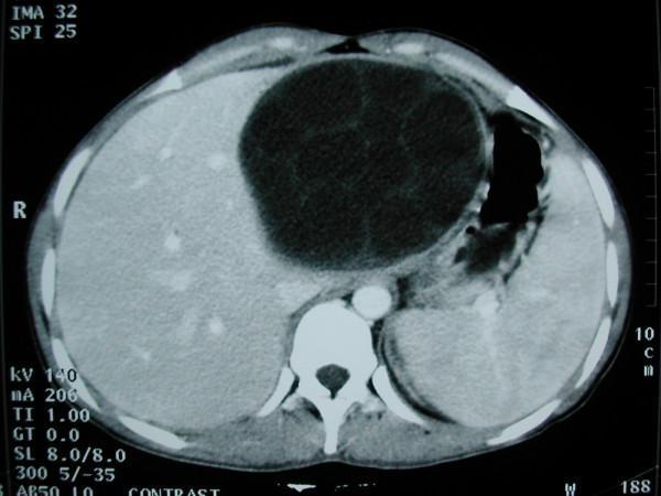
This diagnostic image is an axial contrast-enhanced computed tomography (CT) scan of the upper abdomen demonstrating a large, multilocular hepatic cyst. The primary lesion is a well-defined, hypo-attenuating (fluid-density) spherical mass located within the left lobe of the liver, specifically involving segments II, III, and IV. The cyst displays a classic 'multivesicular' appearance, characterized by multiple internal septations and smaller peripheral daughter cysts within the parent cyst, which is highly suggestive of a WHO Type CE2 or CE3a hepatic hydatid cyst caused by Echinococcus granulosus. The mass measures approximately 11 cm in diameter, resulting in a significant mass effect that displaces the surrounding liver parenchyma and causes mild compression of the adjacent stomach and splenic structures. Other visible anatomical landmarks include the right lobe of the liver, the spleen, the stomach containing air, and the vertebral body. This image is an essential educational example for parasitology and abdominal radiology, illustrating the classic diagnostic features used to differentiate hydatid disease from simple hepatic cysts or abscesses.
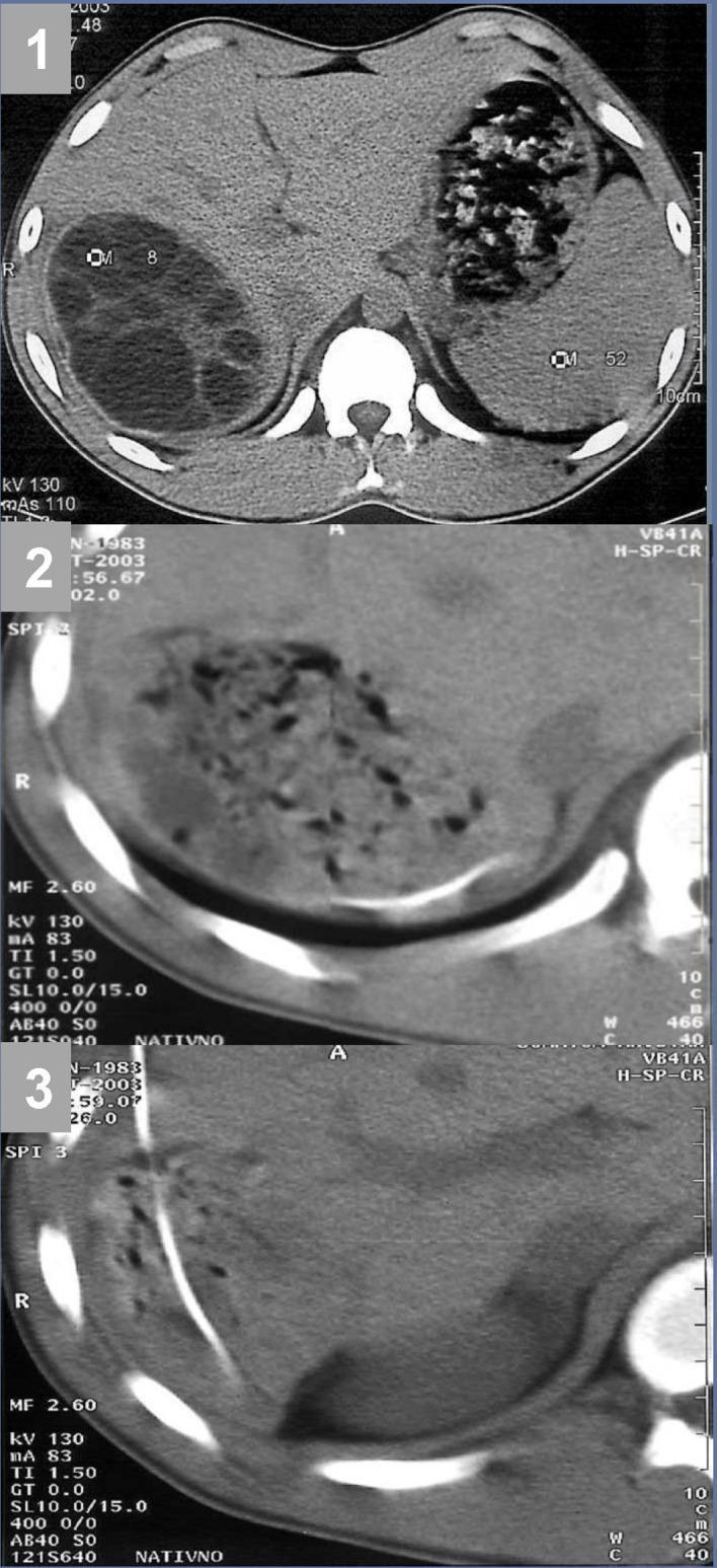
Diagnostic axial CT scans of the abdomen demonstrating a hepatic hydatid cyst (Echinococcus granulosus) and its management via the PAIR (Puncture, Aspiration, Injection, and Re-aspiration) technique. Frame (1) shows a large, well-circumscribed, multivesicular hydatid cyst in the right hepatic lobe, characterized by internal septations and multiple daughter cysts (Gharbi stage 3). Frame (2) displays the cyst cavity post-intervention, showing collapse and heterogeneous internal density as the vesicular fluid is removed and necrotic tissue remains. Frame (3) reveals a radiopaque percutaneous drainage catheter traversing the liver parenchyma with its tip positioned within the residual cyst cavity to facilitate evacuation of debris and disintegrated membranes. This series illustrates the interventional radiological approach to cystic echinococcosis, highlighting the transition from a tense multilocular lesion to a collapsed cavity undergoing drainage. Educational focus is on diagnostic imaging of parasitic liver disease and the visual confirmation of successful catheter placement during minimally invasive treatment.
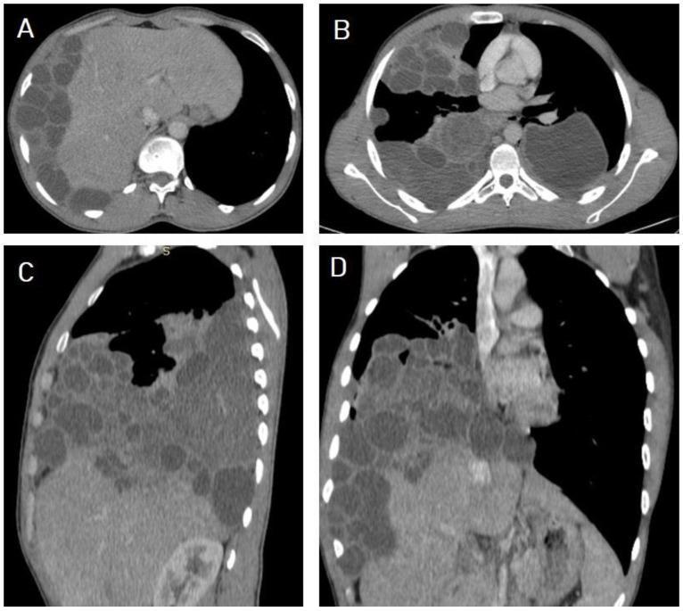
This set of non-contrast Computed Tomography (CT) images illustrates a case of disseminated echinococcosis (hydatid disease) in a 40-year-old male. Image A (axial section, abdomen) shows the liver with multiple well-circumscribed, hypodense cystic lesions consistent with WHO-IWGE CE2 type hydatid cysts containing internal daughter vesicles. There is evidence of hepatic cyst rupture with subsequent peritoneal dissemination. Images B (axial), C (sagittal), and D (coronal) of the chest reveal extensive bilateral pulmonary and pleural involvement. Multiple rounded, thin-walled cystic lesions of varying sizes occupy a significant portion of both lung fields, particularly on the right side. The distribution suggests secondary hematogenous or transdiaphragmatic seeding from the primary hepatic source. Key features include the multilocular cystic appearance, absence of significant perilesional edema, and widespread dissemination across the diaphragm into the thoracic cavity. This imaging demonstrates advanced complicated hydatid disease with catastrophic multisystemic involvement, useful for educational review of parasitic dissemination pathways and CT morphology of Echinococcus granulosus infections.
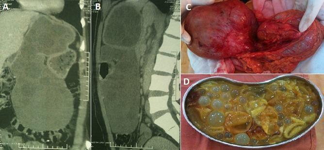
A four-panel composite image illustrating a large hepatic hydatid cyst through radiological and surgical views. Panels A and B are coronal and sagittal CT scans showing a massive, multilocular cystic lesion originating from the left hepatic lobe. The scans demonstrate significant mass effect, displacing the stomach anteriorly, the pancreas posteriorly, and the transverse colon inferiorly. Panel C is an intraoperative clinical photograph showing the surgical exposure of the large cystic mass and its anatomical relation to the surrounding viscera. Panel D displays the surgical specimen within a basin, revealing characteristic multivesicular contents consisting of numerous translucent daughter cysts of varying sizes suspended in yellow, bilious fluid. This educational visual documents the diagnostic imaging and macroscopic pathology of Echinococcus granulosus infection presenting as a voluminous liver cyst, emphasizing its impact on intra-abdominal anatomy.
Long case: hepatic hydatid disease with fever, jaundice and hematemesis
1. Case summary (presentation line)
- fever and vomiting
- one episode of altered (black, clotted) hematemesis
- dark yellow urine and watery stool
- reduced appetite
2. History (from your notes)
| Item | Finding |
|---|---|
| Name, age, sex | Bipul Sardar, 41 y, male |
| Occupation | Agricultural work |
| Admission / examination dates | DOA 16/10/26, DOE 29/10/26 |
| Chief complaints | Fever and vomiting, recorded as "x 2 days" |
| Fever | Intermittent, irregular, daily rise to a high temperature, with chills and rigor, body pain and headache. It settles with medicines. No cough or cold. |
| Vomiting | Sudden onset, non-projectile, non-bilious, 2-3 times per day, related to food or fever. One episode of acidic, blood-containing vomitus; the blood was altered (black, clotted). No abdominal or chest pain. |
| Urine and stool | Dark yellow urine; yellow, watery stool |
| Past history | Malaria 4-5 years ago. No significant surgical history. |
| Personal history | Sleep normal; appetite reduced (note says "Anorexia?"). Bowel and bladder entries are blank. |
| Addictions | Bidi, 20 pack-years; alcohol for 20 years |
| Family and allergy history | Nothing significant |
| Risk exposures | Blood transfusion, needle sharing and unprotected intercourse are listed, but the answers are unclear in the photo. Please confirm them. |
| Treatment so far | Antipyretics; imaging done |
- contact with dogs, sheep or cattle
- drinking untreated water and eating raw vegetables
- stool color (melena) and a dry-cough or hemoptysis history
- weight loss
- itching and pale stool
- paddy-field or flood-water exposure (leptospirosis)
- previous jaundice
- abdominal girth increase
- urticaria after a fall or trauma
3. General survey and examination (from your notes)
| Parameter | Finding |
|---|---|
| Consciousness | Alert, conscious, cooperative, well oriented to time, place and person |
| Pulse | 88/min |
| Respiratory rate | 18/min |
| BP, SpO2, temperature | Not recorded. Record all three. |
| Build and nutrition | Average build, normal nutrition |
| Facies | Normal |
| Pallor | Not clearly legible on the page; please verify |
| Icterus | Present |
| Cyanosis, clubbing | Absent (as I read the entries; please verify) |
| Lymphadenopathy | The entry is unclear; verify |
| Edema | Pitting edema present |
- Superficial: temperature normal, no tenderness.
- Deep: all 9 regions examined, no tenderness.
- Liver:
- palpable 5 cm below the right subcostal margin
- margin rounded
- consistency firm to hard
- surface smooth and non-nodular
- non-tender
- upper border of dullness at the 4th intercostal space
- The lower edge is down and the upper border is higher than the usual 5th space. This means a truly enlarged liver (a large span), not a ptotic one.
- A smooth, firm, non-tender, rounded liver fits space-occupying cysts or a chronic alcohol-related liver.
- A globular belly with an inverted umbilicus suggests ascites. The notes don't show the tests for it.
- Percussion (shifting dullness, fluid thrill) and auscultation (bruit, rub, bowel sounds).
- Spleen, kidneys and hernial orifices.
- External genitalia (your teacher's note, "10th quadrant, examine genitalia") and a digital rectal exam for melena.
- Signs of chronic liver disease: spider naevi, palmar erythema, gynecomastia, flapping tremor, caput medusae, testicular atrophy.
- Parotid enlargement and Dupuytren contracture (alcohol).
- Hydatid thrill (rare).
- JVP, CVS, chest (right basal dullness or effusion from a transdiaphragmatic cyst) and CNS (asterixis, orientation).
4. Provisional diagnosis
- biliary communication or cholangitis (fever with rigors plus jaundice); or
- secondary bacterial infection of a cyst.
- Upper GI bleed (altered hematemesis). Likely causes are portal hypertension with varices or portal hypertensive gastropathy (alcohol for 20 years, or cysts compressing portal or hepatic veins), or alcoholic gastritis or peptic ulcer.
- Pitting edema, probably from low albumin or chronic liver disease, to be confirmed.
5. Differential diagnosis
| Differential | For | Against |
|---|---|---|
| Amoebic liver abscess | Fever, hepatomegaly, alcohol, male | Usually tender, single cyst, brown "anchovy paste" fluid; CT shows an enhancing rim |
| Pyogenic liver abscess | Fever with rigors | Thick enhancing wall, tender liver, leukocytosis. It can also complicate an infected hydatid cyst. |
| Alcoholic hepatitis or cirrhosis with ascites | Alcohol, jaundice, edema, hematemesis | Cysts on CT; the liver is usually small in advanced cirrhosis |
| Polycystic liver disease or simple cysts | Multiple cysts | No septa or daughter cysts, no fever |
| Cystic neoplasm or necrotic metastasis | Hepatomegaly | Enhancing nodules or solid components |
| Viral hepatitis, typhoid | Listed in your notes | Do not explain the cysts |
| Malaria (relapse), leptospirosis, dengue | Past malaria; agricultural exposure; fever, jaundice, vomiting, edema | Need smear/RDT, leptospira IgM and dengue NS1 to exclude |
6. Investigations
- Blood:
- CBC with differential (eosinophilia, anemia, TLC, platelets)
- peripheral smear and malaria RDT
- blood culture ×2 (fever with rigors)
- Biochemistry:
- LFT (direct and indirect bilirubin, SGOT/SGPT, ALP and GGT, albumin, globulin)
- PT/INR
- RFT, electrolytes, blood sugar
- amylase or lipase if there is pain
- Serology:
- Echinococcus IgG (ELISA, indirect hemagglutination, immunoblot)
- HBsAg, anti-HCV, HIV
- Widal or typhoid culture
- leptospira IgM, dengue NS1
- amoebic serology
- Imaging:
- Ultrasound for Gharbi/WHO cyst staging.
- CECT whole abdomen (done); also look for peritoneal and splenic cysts, biliary dilatation and portal vein or IVC compression.
- MRCP if cholestasis or biliary dilatation is present.
- Chest X-ray or CT thorax (lung cysts; roughly 20% of hydatid disease is pulmonary).
- Endoscopy: upper GI endoscopy for varices, gastropathy or ulcer.
- Ascitic tap (if ascites is present): cell count, albumin (calculate SAAG), culture.
- Do not perform a diagnostic needle aspiration or biopsy without scolicidal cover. It risks spillage and anaphylaxis. The Casoni test is obsolete.
7. Treatment
A. Immediate and supportive
- Hematemesis:
- two large-bore IV lines, cross-match, restrictive transfusion
- IV PPI
- urgent endoscopy within 12-24 hours
- if varices are suspected, add terlipressin or octreotide, ceftriaxone prophylaxis and band ligation
- Fever and possible cholangitis: blood cultures first, then broad-spectrum IV antibiotics (for example ceftriaxone plus metronidazole, adjusted to culture). Urgent biliary drainage (ERCP) if there is cholangitis or obstruction.
- Alcohol: thiamine, folate, withdrawal monitoring. Advise stopping alcohol and bidi.
- Edema or ascites (if confirmed): salt restriction, albumin, spironolactone with or without furosemide.
- Avoid: NSAIDs and hepatotoxic drugs.
- Anaphylaxis preparedness: adrenaline, hydrocortisone and antihistamine ready for any cyst manipulation or rupture.
B. Definitive treatment of the hydatid cysts
- Albendazole:
- 10-15 mg/kg/day in 2 divided doses, taken with a fatty meal.
- Typically started 1-4 weeks before surgery.
- Continued for 1-3 months after surgery or intervention.
- Monitor LFT and CBC every 2 weeks (hepatotoxicity, leukopenia).
- Surgery (open or laparoscopic):
- Radical: pericystectomy or hepatic resection.
- Conservative: cystectomy or partial cystectomy with omentoplasty or capitonnage.
- In either, isolate the field with scolicidal-soaked packs.
- Avoid scolicidal agents (hypertonic saline, 20%) if there is a biliary fistula, because of the risk of sclerosing cholangitis.
- PAIR (Puncture, Aspiration, Injection, Re-aspiration): used for suitable large cysts, with albendazole cover. It is contraindicated with biliary communication, and daughter-cyst-filled cysts like this one are poor candidates. Modified catheterization is an alternative for CE2/3b if surgery isn't possible.
- Biliary fistula or cholangitis: ERCP with sphincterotomy and stent.
- Follow-up: ultrasound every 3-6 months for at least 2-3 years, to detect recurrence. Treat the hematemesis and complications first, then plan elective cyst management. Surgery in a bleeding, jaundiced alcoholic patient is high risk.
8. Related theory
Etiology and life cycle
- E. granulosus is a tapeworm (cestode). Its definitive hosts are dogs; sheep, cattle, goats and pigs are intermediate hosts. Humans are accidental (aberrant) intermediate hosts.
- Humans acquire it by swallowing eggs from dog feces on soil, vegetables, water or fur.
- The oncosphere penetrates the small intestine and travels via the portal vein. The liver (about 60-70%) is the first filter, then the lungs (about 20%), then spleen, kidney, brain and bone.
- Cyst growth is slow, roughly 1-5 cm per year. Many people are symptom-free for years.
- Humans are a dead end: the parasite does not mature to the adult tapeworm in us.
Cyst anatomy (three layers)
- Pericyst: host adventitia, which can calcify.
- Laminated layer: acellular, parasite-derived.
- Germinal layer: produces brood capsules, protoscolices ("hydatid sand") and daughter cysts.
Clinical features
- Most patients are asymptomatic.
- Growth causes low-grade fever, pain, tender hepatomegaly (right lobe) and eosinophilia.
- Rupture into the lungs causes dyspnea and hemoptysis.
- Rupture into the peritoneum or lungs can cause life-threatening anaphylaxis.
- Biliary rupture causes cholangitis, obstruction and jaundice.
- Superinfection causes pyogenic abscess in up to 20% of hepatic cases.
- Rare complications: pancreatitis, portal hypertension, Budd-Chiari syndrome, pericardial rupture.
Imaging
- Classic CT/USG: spherical cyst with a three-layer wall, septa and peripheral daughter cysts. Detached laminated membrane gives the "water lily" sign. Hydatid sand is seen as echoes.
- Calcification suggests an inactive or dead cyst (Current Surgical Therapy, 14e, p. 396).
| Gharbi | WHO-IWGE | Meaning | Usual management |
|---|---|---|---|
| I | CE1 | Unilocular, double wall | Albendazole + PAIR |
| II | CE3a | Detached membrane (water lily) | Albendazole + PAIR |
| III | CE2, CE3b | Multivesicular, daughter cysts, "honeycomb" | Surgery (or catheter technique) + albendazole |
| IV | CE4 | Heterogeneous, degenerating | Watch and wait |
| V | CE5 | Calcified | Watch and wait |
Differentiating liver cysts
Prevention
- Hand hygiene and washing vegetables.
- Regular deworming of dogs with praziquantel.
- Not feeding raw offal to dogs.
- Safe slaughter practices.
9. Recent evidence flag
Images
Questions for you
- What are the actual dates? Is the fever 2 days or 2 weeks old?
- Do you have labs (bilirubin, ALP, eosinophil count, albumin) and the CT report, such as cyst count, size and any biliary dilatation? I can then refine the diagnosis and the choice between surgery and PAIR.