
28 year old pregnant woman in her second trimester visited the antenatal clinic with complaints of mild fever and fatigue for the past few days. On examination, she had cervical lymphadenopathy. She mentioned cleaning her pet cat’s litter box regularly. Anomaly scan of the fetus showed signs of hydrocephalus. Blood was sent for serological testing. a) What is the probable clinical condition? b) Name the causative organism, its usual mode of transmission and life cycle of the organism. c) Briefly describe the risk factors and diagnostic methods. d) Write a note on preventive measures, especially in pregnancy.
Toxoplasma gondii life cycle cat oocyst diagram
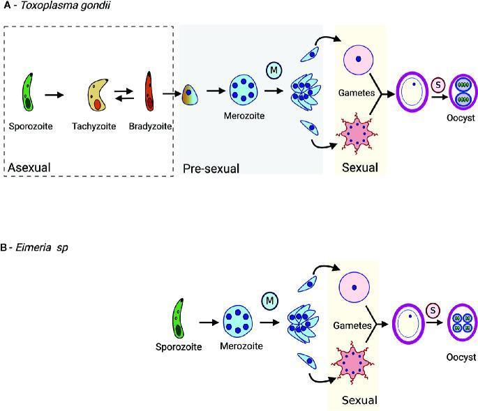
A comparative life cycle diagram of apicomplexan parasites Toxoplasma gondii (Panel A) and Eimeria sp. (Panel B), illustrating their developmental stages. The T. gondii cycle starts with asexual stages including elongated sporozoites, crescent-shaped tachyzoites, and cystic bradyzoites. It progresses to the pre-sexual phase where bradyzoites differentiate into merozoites. During merogony (indicated by a blue 'M' icon), multi-nucleated schizonts undergo cytoplasmic division to produce more merozoites. These enter the sexual phase, differentiating into macrogametes (large, round female cells) and flagellated microgametes (male cells). Fusion results in an oocyst, which undergoes sporulation (indicated by a pink 'S' icon) to form infectious sporozoites. Panel B illustrates the Eimeria cycle, which lacks the intermediate host tissue cyst phase (tachyzoite/bradyzoite) and proceeds directly from sporozoite to merozoite/pre-sexual development. This pathophysiology diagram highlights key morphological shifts and reproductive strategies critical for understanding parasitic transmission and pathogenesis in medical and veterinary microbiology.
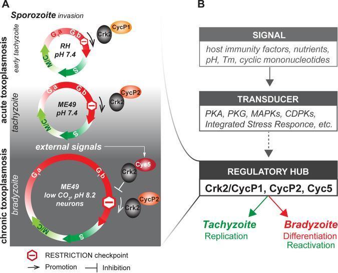
Educational medical diagram illustrating the cell cycle and signaling regulation of Toxoplasma gondii during stage conversion between tachyzoites and bradyzoites. Panel A depicts three cell cycle models: early tachyzoite (RH strain), acute tachyzoite (ME49 strain), and chronic bradyzoite (ME49 strain). The cycles show phases G1a/G1b (red), S (dark green), and Mitosis/Cytokinesis (M/C, light green). Key visual features include the progressive elongation of the G1 phase during bradyzoite development and the role of alternative cyclin-dependent kinase complexes (TgCrk2 with CycP1, CycP2, or Cyc5) at the G1 restriction point (stop sign icon). Panel B presents a vertical flow chart showing the hierarchy of development: external signals (immunity, nutrients, pH) are processed by transducers (PKA, PKG, MAPKs, Integrated Stress Response) which then act on a 'Regulatory Hub' containing the Crk2/Cyclin complexes. This hub determines the clinical outcome: Tachyzoite Replication (acute disease) or Bradyzoite Differentiation/Reactivation (chronic disease). The diagram explains the molecular basis of parasitological quiescence and pathogenesis in human toxoplasmosis.
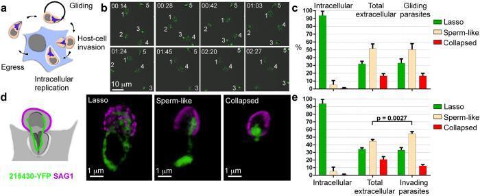
Educational panel illustrating the mitochondrial morphology and lytic cycle stages of the protozoan parasite Toxoplasma gondii. Panel (a) shows a pathophysiological diagram of the lytic cycle: intracellular replication, host-cell egress, gliding motility, and host-cell invasion. Panel (b) presents time-lapse fluorescence microscopy snapshots of gliding tachyzoites (labeled 1-5), tracking mitochondrial movement and shape changes over a 2:27 minute interval. Panel (d) displays high-resolution fluorescence images of invading parasites using two markers: TGME49_215430 (YFP, green) for the mitochondrial periphery and SAG1 (magenta) for the parasite surface. It visualizes three distinct mitochondrial phenotypes: 'Lasso' (circular/peripheral), 'Sperm-like' (rounded body with a tail-like extension), and 'Collapsed' (compact/contracted). Panels (c) and (e) are comparison bar charts showing the percentage distribution of these three morphologies across different physiological states: intracellular, total extracellular population, gliding parasites, and invading parasites. The data highlights that the lasso shape predominates in intracellular stages, while sperm-like and collapsed forms emerge significantly in extracellular and invading stages (p = 0.0027).
Congenital Toxoplasmosis - Complete Clinical Analysis
a) Probable Clinical Condition
- Maternal symptoms: mild fever, fatigue, cervical lymphadenopathy (a mononucleosis-like picture)
- Epidemiological exposure: regular cleaning of the cat's litter box (the single most significant risk factor in pregnancy)
- Fetal anomaly: hydrocephalus on anomaly scan - one of the classic signs of congenital toxoplasmosis
b) Causative Organism, Mode of Transmission, and Life Cycle
Causative Organism
Three Life Forms
| Form | Description | Location |
|---|---|---|
| Tachyzoites (trophozoites) | Rapidly dividing invasive form; responsible for acute infection and tissue damage | Blood and tissues during acute infection |
| Bradyzoites (tissue cysts) | Slowly dividing encysted form; persist in host tissues for years | Brain and muscle (latent/chronic phase) |
| Oocysts (sporozoites) | Sexual stage; environmentally resistant | Excreted in cat feces; infective in soil |
Life Cycle
- Cats (wild and domestic) are the only known host in which the sexual reproductive cycle occurs
- Cats become infected by eating raw meat containing tissue cysts or by ingesting oocysts from the environment
- In the cat's intestinal epithelium, the parasite undergoes sexual reproduction (gametogony), producing oocysts
- Oocysts are excreted in cat feces (a single infected cat can shed millions of oocysts per day)
- Oocysts sporulate in the environment within 1-5 days and become infectious, remaining viable in soil for months to years
- Intermediate hosts ingest oocysts via contaminated soil, food, or water
- Oocysts transform into tachyzoites in the gastrointestinal tract
- Tachyzoites disseminate hematogenously throughout the body (parasitemia)
- The immune system limits multiplication, leading to conversion to bradyzoites (tissue cysts) in brain and muscle
- Tissue cysts persist for the life of the host (latent infection)
- The cycle restarts when a cat eats an infected intermediate host
- Humans serve as an intermediate host only
- Vertical (transplacental) transmission can occur when a pregnant woman has primary infection
- Immunity is mediated primarily through T lymphocytes and is usually long-lasting
Modes of Human Transmission
- Ingestion of undercooked/raw meat (especially pork or lamb) containing tissue cysts - oocysts are completely destroyed by thorough cooking
- Ingestion of oocysts from material contaminated by cat feces (cat litter, soil, garden vegetables, contaminated water, unwashed fruits/vegetables)
- Transplacental transmission - from acutely infected mother to fetus (vertical transmission)
- Blood transfusion or organ transplantation (less common)
- Inhalation of contaminated dust (reported outbreaks in indoor riding stables)
- Drinking contaminated water or unpasteurized goat's milk
c) Risk Factors and Diagnostic Methods
Risk Factors
- Cleaning cat litter boxes (especially from outdoor/feral cats that eat raw meat) - the primary exposure in this case
- Consumption of raw or undercooked meat (pork, lamb, beef)
- Gardening without gloves (soil contaminated with oocysts)
- Immunocompromised state (HIV/AIDS, transplant recipients, corticosteroid therapy) - risk of reactivation
- Poor sanitation and crowded living conditions
- Areas with high seroprevalence (France, Brazil, tropical regions)
- Eating unwashed fruits and vegetables
- Drinking untreated or contaminated water
- Travel to or residence in Central/South America (more virulent genotypes)
- Timing of maternal infection is the critical determinant:
- First trimester infection: lowest risk of transmission (~10-15%) but greatest severity of fetal injury (intrauterine death, hydrocephalus, intracranial calcifications, microcephaly)
- Third trimester infection: highest risk of transmission (~~60-65%) but usually asymptomatic at birth
- Approximately 40% of neonates born to mothers with acute toxoplasmosis show evidence of infection
- Seronegative status in the mother (>90% of pregnant individuals have no past immunity)
- Chronic/latent maternal infection is unlikely to cause serious fetal injury
Diagnostic Methods
- IgG antibodies: Appear within 2-3 weeks of infection; peak at 6-8 weeks; persist for life. Low IgG titers have little significance as many have had asymptomatic prior infection. Tested by:
- Sabin-Feldman dye test (gold standard, available only in reference labs)
- Indirect Fluorescent Antibody (IFA) test
- Enzyme-Linked Immunosorbent Assay (ELISA)
- IgM antibodies: Appear early (1-2 weeks); useful for diagnosing acute/recent infection and congenital infection; however, can persist for >1 year, limiting its utility in timing acute infections - a critical limitation
- IgA antibodies: Presence favors acute infection
- Simultaneous IgG + IgM = acute infection
- If serologic testing is suspicious, confirmation at a reference laboratory (e.g., Palo Alto Medical Foundation's Toxoplasma Serology Laboratory, USA) is strongly recommended before concluding fetal risk
- PCR of amniotic fluid (amniocentesis after 18 weeks): Most valuable test to confirm congenital infection. Sensitivity ~64%, positive predictive value 100% (no false positives). Results available within 1 day
- PCR of blood, CSF, or bronchoalveolar lavage reflects active acute infection
- Isolation of tachyzoites from tissue/body fluids confirms acute infection
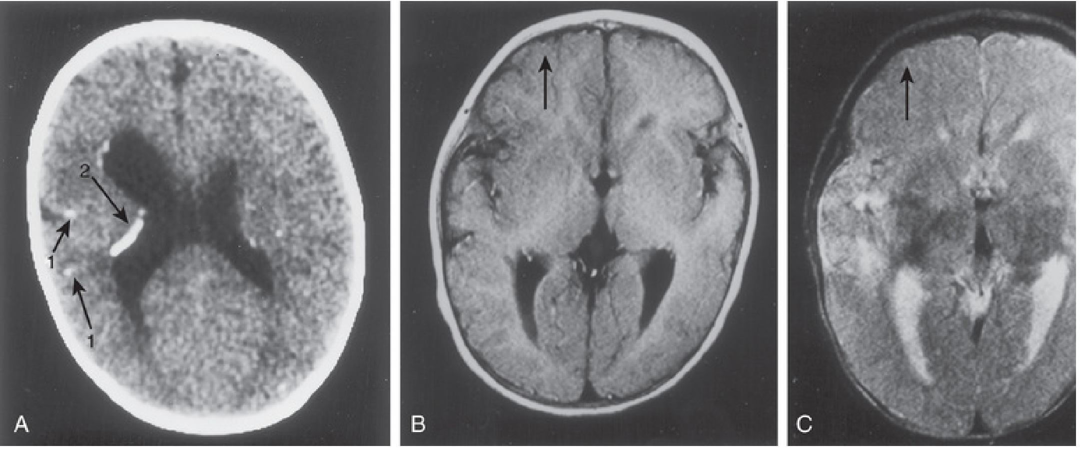
- Anomaly ultrasound (as in this case): Signs include ventriculomegaly/hydrocephalus, intracranial calcifications, microcephaly, hepatomegaly, liver calcifications, placentomegaly, growth restriction, ascites
- MRI can show cortical abnormalities
- Lymph node biopsy: shows reactive follicular hyperplasia with epithelioid histiocytes (characteristic pattern)
- Demonstration of tachyzoites in lymph nodes establishes acute diagnosis
- IgM dot-blot in neonates (sensitivity only 50-75% - may underestimate true incidence)
- IgA and IgM detection (since maternal IgG crosses the placenta, IgG alone is not diagnostic in the newborn)
d) Preventive Measures in Pregnancy
Regarding Cats and Litter Boxes (most important for this patient)
- Avoid cleaning the cat's litter box entirely if possible - delegate this task to someone else for the duration of pregnancy
- If unavoidable, change the litter daily (oocysts take 1-5 days to sporulate and become infectious, so daily cleaning before sporulation significantly reduces risk)
- Wear gloves when handling litter and wash hands thoroughly with soap and water afterward
- Keep cats indoors and feed them only commercially prepared (cooked) cat food - not raw meat or wild prey
- Consider testing the cat for T. gondii infection
Regarding Food Safety
- Never eat raw or rare meat (especially pork, lamb, or beef)
- Cook all meat thoroughly until juices run clear (internal temperature >67°C / 153°F); oocysts and tissue cysts are completely destroyed by proper cooking
- Wash hands after handling raw meat before preparing other foods
- Wash all fruits and vegetables carefully before consumption to remove possible oocyst contamination
- Avoid unpasteurized dairy products (goat's milk)
- Wash all kitchen surfaces and utensils that contact raw meat
Regarding Environmental Exposure
- Wear gloves while gardening (soil may be contaminated with cat feces/oocysts) and wash hands afterward
- Avoid contact with soil in areas frequented by cats
- Drink only treated/boiled water when traveling in areas with poor sanitation
- Wash hands before eating and after gardening
Regarding High-Risk Activities
- Avoid contact with stray or feral cats (more likely to be infected)
- Be aware of travel risks - strains in South/Central America are more virulent
Serological Monitoring
- Women who are seronegative at booking may benefit from repeat serologic testing each trimester in high-prevalence settings (standard practice in France)
- If seroconversion is detected, immediate referral to a specialist in maternal-fetal medicine is recommended
If Infection is Confirmed in Pregnancy - Treatment
- Spiramycin (1 g orally three times daily) is given for maternal infections confirmed before fetal infection is established - it concentrates in the placenta and reduces (but does not eliminate) vertical transmission risk; continued throughout gestation unless fetal PCR is positive
- If fetal infection is confirmed (positive amniotic fluid PCR or ultrasound findings): Pyrimethamine + Sulfadiazine + Folinic acid (leucovorin) - folinic acid protects against bone marrow suppression from pyrimethamine. Some experts now recommend switching to this regimen after the first trimester regardless of amniocentesis results
- Note: Pyrimethamine is contraindicated in the first trimester (teratogenic); Spiramycin is preferred in early pregnancy
- Spiramycin in the USA requires compassionate use authorization from the FDA
| Feature | Details |
|---|---|
| Diagnosis | Congenital Toxoplasmosis |
| Organism | Toxoplasma gondii (obligate intracellular protozoan) |
| Source in this case | Cat litter box (oocysts from cat feces) |
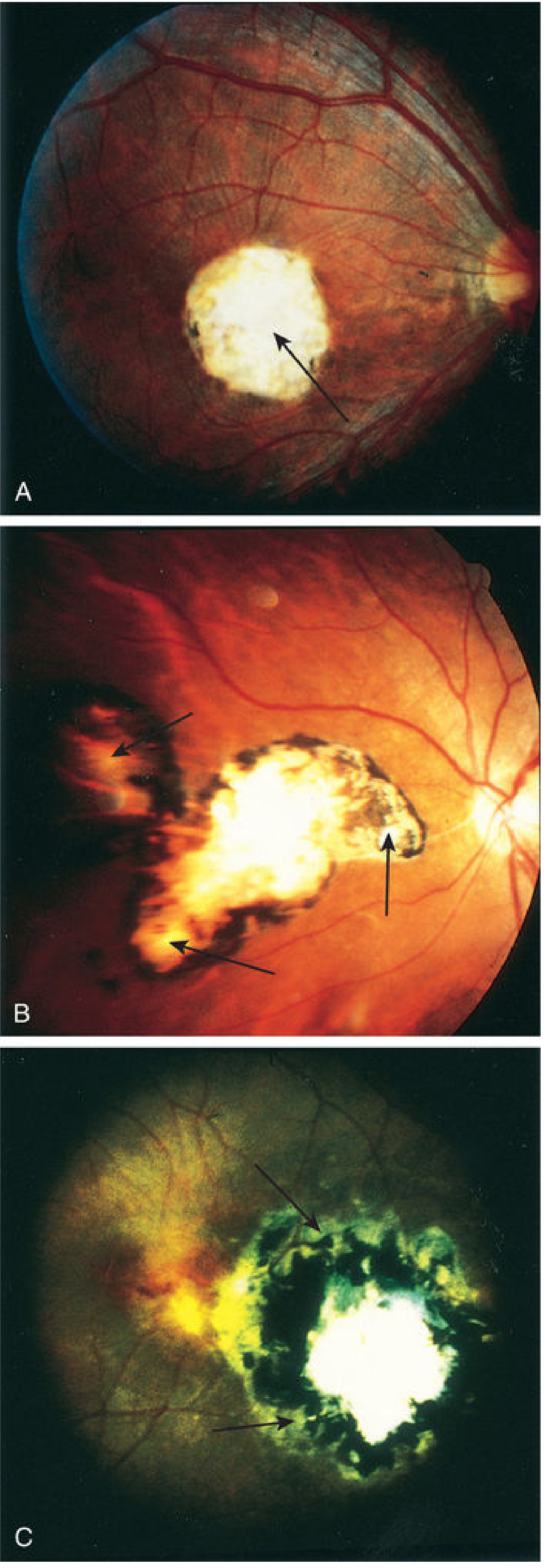
| Classic fetal triad | Chorioretinitis + Intracranial calcifications + Hydrocephalus |
| Key diagnostic test | Amniotic fluid PCR; Maternal IgG + IgM serology |
| Treatment (before fetal infection) | Spiramycin |
| Treatment (fetal infection confirmed) | Pyrimethamine + Sulfadiazine + Folinic acid |
| #1 preventive measure in pregnancy | Avoid cleaning cat litter box |
A 9-year-old girl brought to paediatric OPD with complaints of intermittent fever with chills, rigor, anorexia and tiredness for the past 10 days. On examination the patient had anaemia and hepatosplenomegaly. Her peripheral blood smear study showed accole forms and double dot forms inside the RBC. a) What is your probable diagnosis and the probable causative pathogenic species. b) Describe the life cycle of this parasite and laboratory diagnosis of this disease. c) Write a brief note on its prevention strategies
Plasmodium falciparum peripheral blood smear accolé ring forms double chromatin dot
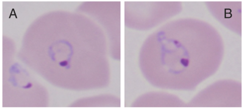
Summary : This figure shows two microscopic images (labeled A and B) of red blood cells infected with a parasite, likely Plasmodium species, stained to highlight intracellular structures. photo: Scene Overview : • Main subject: Red blood cells each containing a single intracellular parasite. • Setting: Microscopy, likely brightfield with Giemsa or similar stain. • Perspective: Close-up, high magnification, single-cell focus. • Composition: Two panels (A and B), each showing a single infected erythrocyte centrally placed. • Lighting: Even, typical of transmitted light microscopy. • Colour palette: Pale pink cytoplasm, purple-blue parasite structures. Technical Details : • No visible scale bar or magnification indicator. • Staining: Parasite nuclei and cytoplasm stained purple/blue, host cell cytoplasm pale pink. • On-image text: Labels “A” (left) and “B” (right) in black font at the upper left and right corners, respectively. Spatial Relationships : • Foreground: Infected erythrocytes with visible ring- or amoeboid-shaped parasites. • Background: Out-of-focus neighboring erythrocytes. • Placement: Each panel centers a single infected cell. Analysis : • Both panels show red blood cells with intracellular parasites at similar developmental stages, possibly ring or trophozoite forms. • The parasites are characterized by a purple chromatin dot and a lighter blue cytoplasmic ring or amoeboid structure. • The images may be used to compare morphological differences or diagnostic features between two conditions, stains, or parasite species.
Peripheral blood smear from a patient with suspected chronic lymphocytic leukemia (CLL) shows a predominance of small, mature-appearing lymphocytes with scant cytoplasm and clumped nuclear chromatin, accompanied by fragile cells that readily disrupt during smear preparation, producing numerous smudge or basket cells visible on the slide. The image depicts a single smudge cell in the upper right, with numerous small darkly staining lymphocytes occupying the field. The smear was prepared with Wright-Giemsa stain and examined under light microscopy at high magnification (approximately 1000x total magnification with oil immersion). Clinically, CLL is defined by absolute mature lymphocytosis of ≥5.0 × 10^9/L sustained for ≥3 months, often asymptomatic and detected incidentally on complete blood count. Morphology alone is insufficient for diagnosis; immunophenotyping showing co-expression of CD5, CD19, CD23 with weak surface immunoglobulin and light chain restriction supports CLL diagnosis. Smudge cells result from fragile leukemic cells and reflect underlying cytoskeletal fragility, though they are not specific. The image illustrates typical features: monomorphic lymphocytes, scant cytoplasm, condensed chromatin, and absent prominent nucleoli. This finding has diagnostic significance, guiding further workup, including flow cytometry, cytogenetics, and molecular studies, to distinguish CLL from other chronic lymphoproliferative disorders such as small lymphocytic lymphoma (SLL), mantle cell lymphoma, prolymphocytic leukemia, and reactive lymphocytosis.
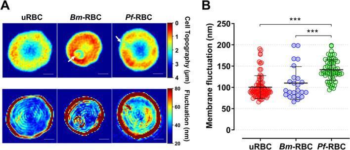
This medical visual consists of two parts: (A) qualitative 2D topographical and membrane fluctuation maps and (B) a quantitative dot plot, comparing uninfected red blood cells (uRBC) with those infected by Babesia microti (Bm-RBC) and Plasmodium falciparum (Pf-RBC). The top row of panel A shows topographical height maps (scale 0–4 µm), where uRBCs display a typical biconcave profile. Bm-RBCs and Pf-RBCs exhibit altered morphology, with arrows indicating the presence of single merozoites within parasitophorous vacuoles. The bottom row presents membrane fluctuation maps (scale 20–80 nm), illustrating variations in cellular biomechanics. uRBCs show relatively low, uniform fluctuation (mostly blue), while infected cells, particularly Pf-RBCs, display significantly increased and heterogeneous fluctuation patterns (yellow and red areas). Panel B is a statistical dot plot of mean membrane fluctuations in nanometers (nm). It confirms that Pf-RBCs have the highest membrane fluctuation (approximately 141.3 nm) compared to Bm-RBCs and uRBCs, with significant differences marked (***P < 0.001). This comparison highlights the biophysical changes induced by different intraerythrocytic parasites.
Falciparum Malaria - Complete Clinical Analysis
a) Probable Diagnosis and Causative Species
Diagnosis: Malaria - specifically Falciparum Malaria (Plasmodium falciparum)
| Finding | Significance |
|---|---|
| Intermittent fever with chills and rigors | Classic malarial paroxysm (erythrocytic schizogony) |
| Anorexia and fatigue | Systemic inflammatory response from cytokine release |
| Anaemia | Massive haemolysis of parasitized RBCs + immune-mediated destruction of uninfected RBCs |
| Hepatosplenomegaly | Reticuloendothelial hyperplasia, congestion, haemozoin pigment deposition |
| Accolé (appliqué) forms on smear | Pathognomonic for P. falciparum - ring trophozoites attached to the inner margin of the RBC membrane |
| Double chromatin dot (double dot/headphone form) | Pathognomonic for P. falciparum - ring-stage trophozoite with two chromatin masses |
Causative Species: Plasmodium falciparum
-
Accolé (appliqué/marginal) forms: Ring trophozoites positioned at the extreme periphery of the RBC, appearing "applied" or "pasted" to the inner surface of the red cell membrane. This occurs because of cytoadherence - P. falciparum infected RBCs sequester in deep capillaries, so only early ring forms circulate in peripheral blood.
-
Double chromatin dot forms (double dot/headphone forms): A single ring-stage trophozoite containing two chromatin dots (nuclei), giving a "headphone" or "double dot" appearance. Multiple rings per RBC can also be seen in heavy P. falciparum infections.
- Multiple infection of a single RBC (2-3 rings per cell) - unlike other species
- Infected RBCs are not enlarged (unlike P. vivax and P. ovale)
- No Schüffner's dots (these are found in P. vivax and P. ovale)
- Banana/crescent-shaped gametocytes (distinct to P. falciparum)
- Maurer's clefts (irregular blotches in the RBC cytoplasm)
b) Life Cycle and Laboratory Diagnosis
Life Cycle of Plasmodium falciparum
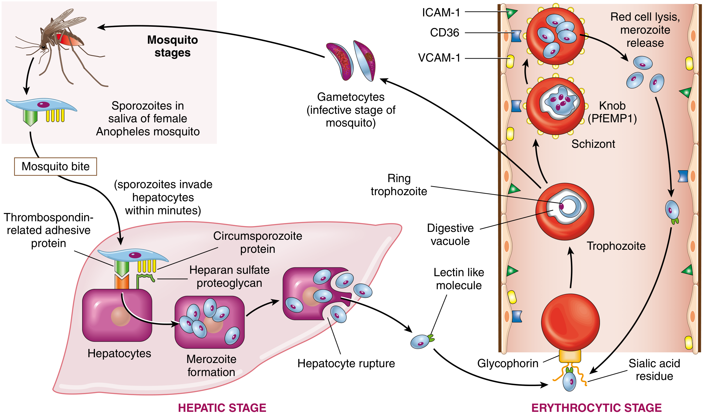
Phase 1: Sexual Cycle in the Mosquito (Sporogony)
- A female Anopheles mosquito takes a blood meal from an infected human and ingests gametocytes (male microgametocytes and female macrogametocytes)
- In the mosquito's midgut, male microgametes fertilize the female macrogamete to form a zygote
- The zygote elongates into a motile ookinete, which penetrates the gut wall and forms an oocyst on the outer gut surface
- The oocyst undergoes sporogony - repeated nuclear division - producing thousands of sporozoites
- The oocyst ruptures, releasing sporozoites that migrate to the mosquito's salivary glands
- The cycle in the mosquito takes approximately 10-14 days (extrinsic incubation period)
Phase 2: Pre-erythrocytic (Hepatic/Exoerythrocytic) Cycle in Humans
- When the infected mosquito takes a blood meal, sporozoites are injected into the human bloodstream
- Within minutes, sporozoites attach to hepatocytes using two surface proteins: thrombospondin-related adhesive protein and circumsporozoite protein, which bind to heparan sulfate proteoglycans on hepatocyte surfaces
- Sporozoites enter hepatocytes and undergo hepatic schizogony (merogony) - each sporozoite develops into a hepatic schizont containing up to 30,000 merozoites
- After 8-25 days (incubation period), infected hepatocytes rupture releasing merozoites into the bloodstream
- Important: Unlike P. vivax and P. ovale, P. falciparum does NOT form hypnozoites (dormant liver forms), so there is no true relapse in falciparum malaria - only recrudescence
Phase 3: Erythrocytic (Blood) Cycle - The Clinical Phase
- Released merozoites bind to glycophorin on RBC surfaces via a lectin-like molecule, invaginating into the cell within a "digestive" vacuole
- Inside the RBC, parasites develop sequentially: ring trophozoite → mature trophozoite → schizont
- The schizont expresses PfEMP1 (P. falciparum Erythrocyte Membrane Protein 1) - an adhesion molecule that concentrates in knob-like extensions on the RBC surface, causing parasitized RBCs to adhere to endothelial ICAM-1, VCAM-1, CD36, and E-selectin (sequestration in capillary beds)
- After ~48 hours (tertian periodicity), schizonts rupture infected RBCs, releasing 8-24 merozoites plus pyrogenic debris (malarial pigment/haemozoin) - this simultaneous rupture causes the characteristic malarial paroxysm (fever spike with chills and rigors)
- Released merozoites immediately invade new RBCs, continuing the cycle
- P. falciparum can infect RBCs of ANY age (unlike P. vivax which preferentially invades reticulocytes) - this is why it can cause extremely high parasitaemia
Gametogony (Transition to Mosquito Stage)
Laboratory Diagnosis
1. Peripheral Blood Smear Examination (Gold Standard)
- Used for screening/detection - concentrates the parasites up to 20-40 times
- More sensitive for low parasitaemia
- Used to detect and count parasites per microlitre
- Used for species identification - morphology of intraerythrocytic stages is well preserved
- Stained with Giemsa stain (or Wright-Giemsa)
- P. falciparum features on smear:
- Accolé/appliqué ring forms (marginal attachment)
- Double chromatin dot (headphone forms)
- Multiple rings per RBC
- No Schüffner's dots; no enlarged RBCs
- Crescent/banana-shaped gametocytes (diagnostic)
- Maurer's clefts
2. Rapid Diagnostic Tests (RDTs) - Immunochromatographic Tests
- Detect P. falciparum-specific antigens: HRP-2 (Histidine-Rich Protein 2) and pLDH (Plasmodium Lactate Dehydrogenase)
- Results in 15-20 minutes; useful at point-of-care where microscopy is unavailable
- FDA-approved kit: Alere BinaxNOW (qualitative, all 4 species)
- Sensitivity lower than microscopy - positive RDT must be confirmed by smear for species identification and parasitaemia quantification
3. PCR and Molecular Assays
- More sensitive than smear (detects very low parasitaemia)
- Useful for species confirmation (essential for appropriate therapy)
- Real-time PCR and LAMP (Loop-Mediated Isothermal Amplification) available
- Used when smear is negative but clinical suspicion is high, and for detecting mixed infections
- Limitation: Does not improve identification of symptomatic patients at low parasitaemia thresholds
4. Other Investigations
- Full blood count: Normocytic normochromic anaemia, thrombocytopaenia (characteristic), leucopenia or leucocytosis
- Serology (IFAT/ELISA): Not useful for acute diagnosis (IgG persists for years); used for epidemiological surveys and blood bank screening
- Antigen detection: Detects HRP-2 (persists in blood for weeks after treatment - can give false-positive post-treatment)
- Malarial pigment (haemozoin) in peripheral blood monocytes on smear
- Liver function tests: Elevated bilirubin (haemolytic), elevated transaminases
- Renal function tests: Rule out blackwater fever (haemoglobinuria) and acute renal failure
- Blood glucose: Hypoglycaemia common in children with severe falciparum malaria
- CSF examination if cerebral malaria is suspected
c) Prevention Strategies
1. Personal Protection Measures (Anti-Mosquito)
- Insecticide-Treated Bed Nets (ITNs) and Long-Lasting Insecticidal Nets (LLINs): The single most effective intervention for reducing malaria transmission, especially for children and pregnant women. LLINs maintain insecticidal effect for 3-5 years
- Protective clothing: Wearing long-sleeved shirts, long trousers, socks, and shoes especially during peak biting hours (dusk to dawn, as Anopheles mosquitoes are night-biters)
- Insect repellents: DEET (N,N-diethyl-meta-toluamide)-containing repellents applied to exposed skin; picaridin and IR3535 are alternatives
- Window/door screens: Screening doors and windows with fine mesh
- Mosquito coils and vaporizers: Burning insecticide coils or using electric vaporizers indoors during night hours
2. Vector Control - Killing Adult Mosquitoes
- Indoor Residual Spraying (IRS): Spraying insecticides (DDT, pyrethroids, malathion) on the interior walls of houses where Anopheles mosquitoes rest after feeding. One of the most powerful vector control tools
- Space spraying: Aerial or ground-level fogging of outdoor areas during outbreaks
3. Antilarval Measures - Source Reduction
- Environmental management: Drainage or filling of stagnant water bodies, clearing weeds from water surfaces, desilting irrigation channels
- Biological control: Introducing larvivorous fish (Gambusia affinis, Lebistes reticulatus) in water bodies; use of Bacillus thuringiensis israelensis (Bti), a biological larvicide
- Chemical larviciding: Application of larvicidal oils, temephos (Abate), or other larvicides to breeding sites
- Source reduction: Eliminating artificial water collection sites (discarded tyres, open containers, flower vases, blocked drains)
4. Chemoprophylaxis (for travellers and high-risk groups)
- Chloroquine: For travel to chloroquine-sensitive areas (Haiti, Dominican Republic, Central America north of Panama Canal, parts of Middle East)
- Atovaquone-proguanil (Malarone): Daily; started 1-2 days before travel, continued 7 days after return
- Mefloquine: Weekly; started 2-3 weeks before travel, continued 4 weeks after
- Doxycycline: Daily; started 1-2 days before travel, continued 4 weeks after
- Primaquine: For terminal prophylaxis against P. vivax and P. ovale hypnozoites (contraindicated in G6PD deficiency and pregnancy)
5. Surveillance and Case Management (Public Health Level)
- Passive case detection: Diagnosis and treatment of all fever cases at health facilities
- Active case detection: Screening of population in endemic areas, especially high-risk groups (children under 5, pregnant women)
- Early diagnosis and complete treatment: The India National Vector Borne Disease Control Programme (NVBDCP) mandates prompt treatment of all microscopy/RDT-confirmed cases
- Sentinel surveillance: Monitoring drug resistance and epidemic trends
- Epidemic preparedness and response: Rapid deployment of control measures during outbreaks
6. Vaccine
- RTS,S/AS01 (Mosquirix): WHO-recommended malaria vaccine (2021) for children in sub-Saharan Africa. Targets P. falciparum circumsporozoite protein. Provides ~30-40% protection against clinical malaria in young children; recommended in 4-dose schedule (3 doses from 5 months, 4th dose ~18 months later)
- R21/Matrix-M: Newer vaccine showing ~75-80% efficacy in clinical trials; WHO approved in 2023
7. Treatment as Prevention
- Prompt and effective treatment of confirmed cases (using ACT - Artemisinin Combination Therapy) interrupts transmission by eliminating gametocytes
- Artemisinin Combination Therapy (ACT) for P. falciparum:
- Artesunate + Sulfadoxine-Pyrimethamine (ACT-SP): standard in most of India
- Artemether + Lumefantrine (ACT-AL): used in North-Eastern India (SP resistance)
- Single dose Primaquine (0.75 mg/kg) on Day 2 to kill gametocytes and block transmission
- Banning artemisinin monotherapy to prevent drug resistance
| Feature | P. falciparum | P. vivax | P. ovale | P. malariae |
|---|---|---|---|---|
| Fever periodicity | 48 hrs (tertian) | 48 hrs (tertian) | 48 hrs | 72 hrs (quartan) |
| RBC preference | All ages | Reticulocytes | Reticulocytes | Older cells |
| RBC enlargement | No | Yes | Yes | No |
| Schüffner's dots | No (Maurer's clefts) | Yes | Yes | No |
| Hypnozoites | No | Yes (relapses) | Yes (relapses) | No |
| Gametocytes | Crescent/banana | Round | Round | Round |
| Distinctive smear findings | Accolé forms, double dot, multiple rings per RBC | Amoeboid trophozoites, Schüffner's dots | Fimbriated/oval RBCs | Band/ribbon trophozoites |
| Severity | Most severe, fatal | Moderate | Mild | Mild (nephrotic syndrome) |
What is full and reasonable disclosure? Explain about contrast autonomy and Paternalism.
Full and Reasonable Disclosure, Autonomy vs. Paternalism
I. Informed Consent: The Foundation
- Informed - based on adequate disclosure of relevant facts
- Intelligent - given after understanding what is being consented to, and the risks involved
- Voluntary - free from coercion, manipulation, or undue influence
- Competent - given by a person with legal and mental capacity
II. Full Disclosure
Definition
- Fully informed of every risk and factual material necessary for the making of a proper consent
- That the consent itself be based upon such material disclosure
What Must Be Disclosed
| Category | What It Covers |
|---|---|
| (a) Diagnosis | What the condition is, including uncertainty if present |
| (b) Nature of treatment/procedure | What will be done, how, and why |
| (c) Risks involved | All material risks, both common minor ones and rare serious ones |
| (d) Prospects of success | Expected outcome and prognosis |
| (e) Prognosis without treatment | What will happen if the procedure is declined |
| (f) Alternative methods | Other available treatment options |
Guidelines for Disclosure of Risks
"If the risk of untoward result is statistically high, the patient should be informed regardless of the effect on his morale. If the risk is statistically low, but the consequences of a rare untoward occurrence may be severe, the patient should likewise be informed. On the other hand, if the statistical risk is low and the severity of the risk is not great, the physician may safely tailor his warning so as not to excite the patient's fears."
- High probability + any severity → must disclose
- Low probability + high severity → must disclose
- Low probability + low severity → discretion permitted
Limits of Full Disclosure: Therapeutic Privilege
- Discarding a needed procedure that carries minimal risk, or
- Suffering psychological harm from such disclosure, thereby becoming an increased surgical risk
- Obtain informed consent from a responsible relative
- In the relative's absence, obtain medical consultation
- Chart the intentional omission and the therapeutic exception basis in regard to it
- Medical emergency (implied consent for life-saving treatment)
- Patient's waiver of the physician's disclosure requirement
- Lack of capacity/competence (consent from a substitute decision-maker)
III. Reasonable Disclosure
The Two Standards
Standard 1: The Professional Practice Standard (Doctor/Physician-Based)
- The physician is required to provide information that a reasonable physician in the same specialty would customarily disclose in similar circumstances
- This is the older, traditional standard
- Also known as the Bolam test in UK law (Bolam v. Friern Hospital Management Committee, 1957)
- Under this standard, disclosure is measured against what doctors do in practice, not against what patients need
- Criticism: The medical profession itself set the benchmark, which could protect physicians even when patients were given insufficient information
Standard 2: The Reasonable Patient Standard (Patient-Based)
- Developed in the landmark 1972 US case Canterbury v. Spence, which rejected the professional practice standard
- The physician must disclose all information regarding diagnosis, treatment options, and risks that a "reasonable patient" would want to know in a similar situation
- Rather than relying on the practices of the medical community, the reasonable person standard empowers patients to determine how much information should be disclosed
- The court recognized practical limits: "what a prudent person in the patient's position would have decided if suitably informed of all perils bearing significance"
| Variant | Standard Applied |
|---|---|
| Objective patient standard | What a reasonable patient in similar circumstances would want to know - the more common approach |
| Subjective patient standard | What this particular patient would want to know given their specific circumstances |
Evolution of Disclosure Standards in Case Law
| Case | Year | Jurisdiction | Key Principle |
|---|---|---|---|
| Schloendorff v. Society of New York Hospital | 1914 | USA | "Every human being of adult years and sound mind has a right to determine what shall be done with his own body" (Justice Cardozo) - foundational autonomy principle |
| Salgo v. Leland Stanford Jr. University Board | 1957 | USA | Formally established informed consent doctrine; physicians must provide facts "necessary to form the basis of an intelligent consent" |
| Canterbury v. Spence | 1972 | USA | Rejected professional standard; adopted reasonable patient standard |
| Rogers v. Whittaker | 1992 | Australia | Extended to what risks affect that one patient, not just the generality of reasonable patients |
| Montgomery v. Lanarkshire Health Board | 2016 | UK (Supreme Court) | Effectively ended Bolam applying to consent; enshrined "what a reasonable person would expect to be told" - patient autonomy fully endorsed |
IV. Autonomy vs. Paternalism
Medical Paternalism
Definition
Historical Roots
- The doctor-patient relationship initially resembled an archaic priest-supplicant relationship - a parental figure with the power to manipulate nature on behalf of a helpless ward
- The Hippocratic tradition and the principle of beneficence (acting for the patient's medical benefit) were used to justify paternalism
- For centuries, patients were expected to passively accept medical care directed by their physicians
- The attitude was: "Doctor knows best"
Justification for Paternalism
- Beneficence: The physician's primary duty is to act for the patient's medical benefit
- Non-maleficence (Primum non nocere): Preventing harm may sometimes require withholding distressing information
- Patients may lack medical knowledge to make truly informed decisions
- Illness itself may compromise a patient's rational decision-making
- Therapeutic privilege is a preserved form of "soft paternalism" in modern ethics
Problems with Paternalism
- Fails to respect the patient as a person with individual values, beliefs, and preferences
- May lead to decisions that are medically correct but personally unacceptable to the patient
- Removes the patient's right to self-determination
- Historically led to inadequate disclosure, "blanket consent forms," and uninformed patients
- Can result in patients refusing needed care or being subjected to unwanted procedures
Patient Autonomy
Definition
Historical Development
- Growing sentiment from the late 19th and early 20th centuries that patients had the right to actively influence their care
- Schloendorff v. Society of New York Hospital (1914): Justice Cardozo's ruling was the "canary in the coalmine" for medical paternalism
- Autonomy gradually gained ground and ultimately displaced the long-accepted institution of medical paternalism
- Canterbury v. Spence (1972) and subsequent cases enshrined autonomy in law
Core Elements of Autonomy in Practice
- Right to information: The patient must receive complete and comprehensible information about their diagnosis, treatment options, risks, benefits, and prognosis
- Right to decide: The patient makes the ultimate decision about their own care
- Right to refuse treatment: A competent adult may refuse any treatment, even life-saving treatment - courts have consistently upheld this right
- Voluntariness: Decisions must be free from coercion, manipulation, or deception
- Informed refusal: Patients can decline procedures after being informed of the risks of refusal
Legal Basis for Autonomy
- Competent adults enjoy a legal presumption of competence in medical decision-making
- Courts have upheld the right of competent adults to refuse treatment even when this decision may result in death
- Even patients with psychiatric illness may be found competent if they can communicate a choice, understand their situation, and rationally manipulate relevant information (In re Maida Yetter, 1973)
The Contrast: Autonomy vs. Paternalism
| Feature | Paternalism | Autonomy |
|---|---|---|
| Driving principle | Beneficence / Non-maleficence | Respect for person / Self-determination |
| Who decides? | Physician decides for the patient | Patient decides, guided by physician |
| Role of the patient | Passive recipient of care | Active participant in decision-making |
| Information sharing | Selective - physician decides what to tell | Full - patient receives all material information |
| Patient refusal | May be overridden "for the patient's good" | Must be respected if patient is competent |
| Doctor-patient dynamic | Hierarchical (physician dominant) | Collaborative / Partnership |
| Ethical foundation | Beneficence as the overriding principle | Autonomy as the overriding principle |
| Historical era | Traditional/classical medicine | Modern medicine (20th century onwards) |
| Typical phrase | "I know what's best for you" | "Here is the information - what do you want?" |
| Legal position today | Disfavoured; only therapeutic privilege preserved | Fully endorsed in law (Schloendorff, Canterbury, Montgomery) |
The Balance: Shared Decision-Making
- The physician contributes expertise, medical knowledge, and clinical judgment
- The patient contributes personal values, preferences, and ultimate authority
- Together, they reach a mutually acceptable decision that is both medically appropriate and consistent with the patient's autonomy
- Emergencies where consent cannot be obtained and delay would cause serious harm
- Incompetent patients (children, those with severe cognitive impairment) where substitute decision-makers act in the patient's best interest
- Therapeutic privilege - limited and carefully documented situations where full disclosure would cause disproportionate psychological harm
- The patient's decision poses serious risk to third parties or public health (e.g., quarantine of infectious disease)
- The patient is legally incompetent or lacks decision-making capacity
- The patient is a minor (parental/guardian consent required, though a child's assent is increasingly sought)
Summary
Full disclosure means providing the patient with all information a reasonable patient would need to make a truly informed decision - including diagnosis, risks, benefits, alternatives, and prognosis with and without treatment.
Reasonable disclosure is a legal standard - either what a reasonable doctor would disclose (professional standard) or what a reasonable patient would want to know (patient-based standard) - with modern law favouring the latter.
Paternalism is the traditional physician-centred model where the doctor acts in the patient's presumed best interest, overriding the patient's own wishes.
Autonomy is the modern patient-centred model where the patient's right to self-determination is paramount, and the doctor's role is to inform and advise rather than to decide.