
What is intususseption
Reading File
Now let me search the medical library for intussusception content.
Finding Sources
Searching PubMed
intussusception
Good results. Now let me read the most detailed textbook sections in parallel.
Reading File
Reading File
Reading File
Let me also fetch the image mentioned and get the Rosen's Emergency Medicine detailed section.
Reading File
Loading Image
I now have comprehensive information from multiple authoritative sources. Here is a complete answer:
Intussusception
Definition
Intussusception is the telescoping of a proximal segment of bowel (the intussusceptum) into the lumen of a more distal, adjacent segment (the intussuscipiens). This creates a "bowel within bowel" configuration that obstructs the intestinal lumen and compresses the mesenteric blood supply, risking ischemia, gangrene, and perforation.
- Grainger & Allison's Diagnostic Radiology, p. 1805
- Bailey and Love's Short Practice of Surgery, 28th ed.
Epidemiology
- The second most common cause of acute abdomen in children after appendicitis.
- Most cases occur between 2 months and 2 years of age, with peak incidence at 5-9 months.
- Can occur up to school age; cases beyond 6-7 years are more likely to have a pathological lead point.
- In adults, intussusception is uncommon and frequently associated with neoplasm or malignancy.
Types (by location)
| Type | Frequency |
|---|---|
| Ileocolic (ileum telescopes into colon) | Most common, >80% |
| Ileoileocolic | Less common |
| Ileo-ileal (small bowel only) | Less common; often reduces spontaneously |
| Colocolic | Rare |
Causes and Lead Points
In children (>90% have NO lead point):
The most common cause is lymphoid hypertrophy (enlarged Peyer's patches), typically following a viral infection.
Pathological lead points (5-10%):
- Meckel's diverticulum
- Intestinal polyp
- Duplication cyst
- Small bowel lymphoma
- Nasojejunal tubes
Pathological lead points are more common in children >2 years, in recurrences, and in adults.
Clinical Presentation
The classic triad in infants:
- Colicky abdominal pain - the infant draws up their legs during spasms
- Vomiting
- "Redcurrant jelly" stools - blood-stained mucus passed rectally (a late sign)
Other features:
- Between pain episodes, infant initially appears well
- Dehydration and abdominal distension develop over time
- Palpable right upper quadrant mass - present in less than 50% of patients
- Can rapidly deteriorate to hypovolemia, shock, and become life-threatening
Diagnosis
Ultrasound (first-line, gold standard)
- Sensitivity and specificity approaching 100%
- Characteristic signs:
- "Target" or "doughnut" sign - concentric rings in the transverse plane
- "Sandwich" appearance - in the longitudinal plane
- "Crescent in doughnut" sign - hyperechoic semilunar structure from mesenteric fat pulled into the intussusceptum
- Helpful to identify lead points and assess bowel viability
Plain radiograph
- Small bowel obstruction pattern (dilated loops, fluid levels)
- Paucity of gas in the right iliac fossa
- "Meniscus sign" - soft-tissue mass contrasting an air-filled bowel loop
CT scan
- Reserved for adults, complex cases, or when ultrasound is insufficient
Intraoperative Appearance
Below is a surgical photo showing intussusception - the proximal bowel (intussusceptum) has telescoped into the distal segment (intussuscipiens), creating the classic "bowel within bowel" appearance:
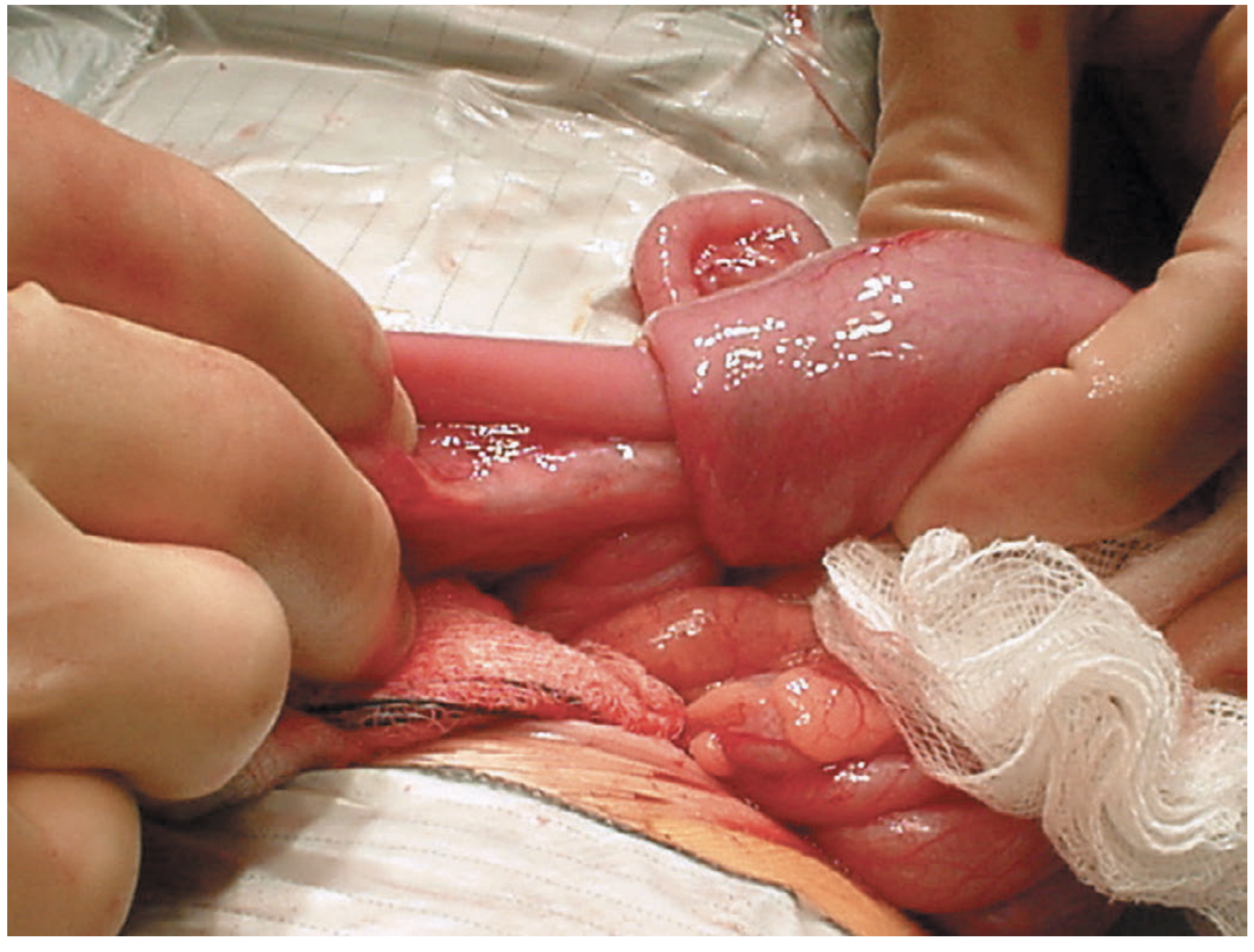
Bailey and Love's Short Practice of Surgery, 28th ed.
Treatment
Resuscitation first:
- IV fluids
- Nasogastric drainage
- Broad-spectrum antibiotics
Non-operative reduction (first-line):
- Pneumatic (air) enema or hydrostatic enema - successful in >70% of cases
- Success confirmed by air flowing into the small bowel and resolution of symptoms
- Contraindications: peritonitis, bowel perforation, shock, or free intraperitoneal air
Surgical reduction:
Indicated when:
- Enema reduction fails
- Peritonitis or perforation present
- Strangulation suspected
- Pathological lead point identified
- Can be open or laparoscopic
Bowel resection required when:
- Irreducible intussusception
- Infarction/gangrene
- Pathological lead point requiring excision
Recurrence
- Occurs in approximately 5% after non-operative reduction
- Recurrences are more likely to have a pathological lead point
Adults vs. Children
| Feature | Children | Adults |
|---|---|---|
| Lead point | Usually absent (viral/idiopathic) | Usually present (neoplasm) |
| Most common type | Ileocolic | Variable |
| Treatment | Enema reduction first | Typically surgical |
| Malignancy association | Rare | Common |
Recent systematic reviews (2024-2025) confirm that adult intussusception requires careful evaluation for underlying malignancy, and surgical resection is the standard management in most adult cases - PMID 39085533, PMID 40831154.
Sources: Grainger & Allison's Diagnostic Radiology; Bailey and Love's Short Practice of Surgery, 28th ed.; Rosen's Emergency Medicine; Goldman-Cecil Medicine.
This is a shared conversation. Sign in to Orris to start your own chat.