


Patient presented with this swelling in the inner aspect of left thigh ! What it could be
cellulitis erythema thigh swelling skin
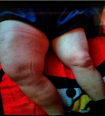
This clinical photograph shows a side-by-side comparison of a patient's lower extremities, specifically highlighting the anterior thighs and knees. The right thigh displays significant gross asymmetry characterized by massive edema and swelling, appearing substantially larger in circumference than the left. The skin over the right thigh is taut, shiny, and demonstrates diffuse erythema, suggesting underlying inflammation or cellulitis. A localized, linear indentation or superficial skin change is visible on the distal anterolateral aspect of the right thigh. In contrast, the left thigh appears relatively normal with visible anatomical contours of the musculature and knee joint. The visual findings are consistent with acute inflammatory conditions such as diabetic muscle infarction, severe myositis, or complicated cellulitis. This image serves as an educational tool for identifying lower extremity edema, inflammatory skin changes, and limb asymmetry during a physical examination in the context of systemic diseases like diabetes mellitus.
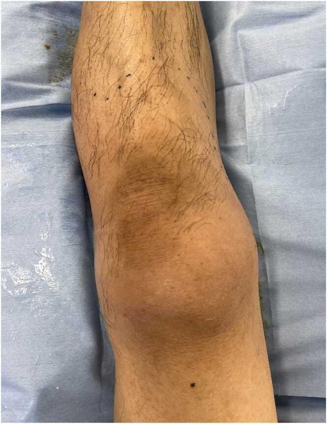
This clinical photograph shows the anterior view of a patient's left leg, including the distal thigh, knee, and proximal lower leg, resting on a blue medical drape. The image demonstrates a primary finding of diffuse, mild erythema (redness) extending from the thigh down to the shin. The erythema is most concentrated and visible over the patellar region (knee), presenting as a dusky, pinkish-tan discoloration. There is no visible evidence of joint effusion, gross swelling, bullae, or skin necrosis at this stage. Tiny black markings are visible on the skin, likely surgical or clinical landmarks used to track the progression of the redness. This visual finding is significant in the context of early-stage necrotizing fasciitis or cellulitis, particularly highlighting cases where clinical pain and tenderness out of proportion to visible skin changes (erythema) are key diagnostic indicators of deep tissue infection.
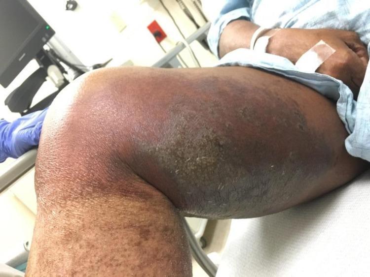
This clinical photograph shows the medial and anterior aspects of the right thigh and knee of a patient in a clinical setting. The image demonstrates a large, confluent area of skin pathology characterized by centrally located hyperkeratotic plaques and evidence of bullous lesions. The central plaques exhibit a thickened, scaly texture with significant hyperpigmentation. Surrounding the central lesions is a broad zone of peripheral erythema, indicating secondary inflammation or cellulitis. The affected area extends from the mid-thigh down toward the knee, showing significant swelling and skin textural changes. This image serves as a clinical example of severe inflammatory dermatosis, potentially lichen planus or pyoderma gangrenosum, with superimposed infection. The presentation highlights the morphological features of hyperkeratosis, plaque formation, and inflammatory erythema in a patient with complex medical comorbidities.
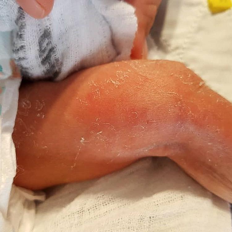
Clinical photograph of a neonatal left distal medial thigh showing an localized inflammatory skin reaction. The central area exhibits prominent erythema (redness) and visible surface desquamation (skin peeling), consistent with an extravasation injury or localized cellulitis. The affected region is described as indurated, though the borders are diffuse and lack sharp demarcation, fading into the surrounding normal skin tone. This visual presentation is characteristic of tissue irritation following intravenous (IV) infusion, where vesicant or irritant substances (such as human albumin or dextrose) have leaked into the subcutaneous space. The educational focus is on identifying early signs of extravasation, including erythema, swelling, and induration, which require prompt clinical intervention to prevent progression to necrosis or ulceration.
Clinical Assessment: Inner Thigh Swelling (Left)
What the Image Shows
- A large, oval-to-circular, dome-shaped erythematous swelling approximately 8-12 cm in diameter on the inner/medial aspect of the left thigh
- Erythema that is most intense centrally and fades peripherally (central hyperemia)
- Skin is tense, smooth, intact - no ulceration, blistering, or necrosis
- Green marker lines traced around the perimeter - a classic clinical technique to monitor erythema spread (strongly suggesting cellulitis is already the working diagnosis)
- No visible overlying fluctuance described, though this cannot be excluded clinically without palpation
Differential Diagnosis
1. Cellulitis (Most Likely)
- Defined as a diffuse spreading infection of the deep dermis and subcutaneous tissue, most often caused by Group A β-hemolytic Streptococcus (more diffuse spread) or Staphylococcus aureus (more localized) - Fitzpatrick's Dermatology
- Classic signs: erythema, swelling, warmth, and pain (rubor, tumor, calor, dolor)
- Unilateral lower extremity involvement is typical
- The green marker demarcation is the standard bedside method to monitor spread or regression - this is already in use here
- Diagnosis is primarily clinical; labs and imaging are often equivocal
- Risk factors: skin barrier compromise (insect bite, minor wound, tinea pedis, lymphedema)
2. Erysipelas (Close Variant)
- Superficial lymphatic involvement; characteristically sharply demarcated bright-red plaque
- This lesion has somewhat diffuse edges, making classic erysipelas slightly less likely, but the two overlap clinically
- Treatment is the same as cellulitis
3. Insect Bite Reaction / Hypersensitivity Reaction
- A large local reaction to an insect bite (bee, hornet, tick, spider) can produce an erythematous, warm, circular plaque of this size
- The smooth surface, circular outline, and absence of central punctum visible here makes this worth considering
- Tick bite reactions can mimic Lyme disease early target lesion (erythema migrans)
4. Erythema Migrans (Lyme Disease)
- If the patient has had outdoor exposure, a bull's-eye or expanding erythematous ring on the thigh is the classic early lesion of Borrelia burgdorferi infection
- This case: the erythema appears more of a solid blush (not a clearing center ring), making this less likely, but cannot be excluded without history
- Serology (ELISA + Western blot) warranted if clinical suspicion
5. Deep Vein Thrombosis (DVT) of Femoral/Iliac Vein
- Proximal (thigh-level) DVT can present with marked thigh swelling, tenderness, and erythema - Harrison's Principles of Internal Medicine 22E
- The erythema in DVT is usually more diffuse/streaky; there is often pitting edema of the whole limb
- This lesion is well-localized rather than involving the whole thigh, making DVT less likely - but the imaging study ordered (MRI thigh) will help exclude this
- Wells score and D-dimer/Doppler ultrasound should be considered
6. Abscess (Early/Pre-fluctuant)
- Would present similarly to cellulitis but with a central area of fluctuance
- If there is tenderness disproportionate to the surface area or central softening on palpation, an underlying abscess must be excluded
- Ultrasound is the investigation of choice to differentiate
7. Liposarcoma / Soft Tissue Sarcoma (Important red flag - less likely acutely)
- A soft tissue mass >10 cm in the thigh should raise concern for liposarcoma - the upper thigh is the most common site - Andrews' Diseases of the Skin
- However, this is a much more gradual process and would not present with acute erythema and warmth unless complicated by hemorrhage or inflammation
- The MRI study already ordered is appropriate to exclude this
Key Investigations Suggested
| Test | Purpose |
|---|---|
| MRI Thigh (already ordered) | Characterize depth, rule out soft tissue mass, necrotizing fasciitis, DVT |
| CBC + CRP/ESR + blood culture | Severity of infection, bacteremia |
| D-dimer + Doppler US | If DVT suspected |
| Wound/skin culture | If pustule, break, or drainage present |
| Lyme serology | If outdoor exposure history |
Red Flags Requiring Urgent Escalation
- Rapid spread beyond marker lines within 12-24 hrs
- High fever, rigors, hypotension (sepsis)
- Severe pain out of proportion to appearance (necrotizing fasciitis)
- Skin discoloration turning gray/blue/black (fascial involvement)
- Failure to improve after 48 hrs on appropriate antibiotics
Sources: Fitzpatrick's Dermatology Vol 1-2; Schwartz's Principles of Surgery 11th Ed; Harrison's Principles of Internal Medicine 22E (2025); Andrews' Diseases of the Skin


Patients gives a history that the lesion is painful even on rest and has a high crp of 58 but she has this lesion from a very long time but now suddenly this is painful and growing in size
Revised Assessment: Critical Update
| Feature | Significance |
|---|---|
| Lesion present for a long time | Pre-existing benign soft tissue lesion (most likely lipoma) |
| Suddenly painful at rest | Active inflammatory/infective or malignant transformation |
| Growing in size | Expansion - concerning for infection, hemorrhage, or malignancy |
| CRP 58 mg/L | Elevated - confirms active systemic inflammation |
| Medial thigh location | Classic location for lipoma / atypical lipomatous tumor |
| Young female, 29 yrs | Age less typical for liposarcoma, but not impossible |
MRI Analysis (5 Sequences - Aug 7, 2026)
Image 1 - SPR / Coronal Scout (pd_tse_fs_dixon_cor)
- Coronal scout view of both thighs
- Bilateral thighs visible; the left medial thigh shows a well-defined subcutaneous/deep lesion with distinct signal characteristics compared to the right
- The lesion appears superficial to the adductor muscle compartment
Image 2 - Axial PD fat-sat (pd_tse_fs_tra)
- Left medial thigh: A well-circumscribed ovoid lesion visible in the subcutaneous/superficial fascial layer of the medial thigh
- The lesion appears hyperintense on PD fat-sat (consistent with fluid/edematous content)
- There is surrounding soft tissue signal change - edema/reactive inflammation extending around the lesion
- The right thigh appears normal at the same level
- The lesion has a bright signal with surrounding reactive changes on this fat-suppressed sequence
Image 3 - Axial T1 (t1_tse_tra)
- Left medial thigh: The lesion shows mixed T1 signal - areas of fat signal (bright on T1) alongside areas of intermediate-to-low signal
- This T1 fat bright component strongly suggests fat-containing lesion (lipoma)
- The mixed signal (not uniformly fat-bright) raises concern for non-fatty components - septations, nodular areas, or areas of dedifferentiation
- Surrounding musculature appears preserved
Image 4 - Sagittal T2 (t2_tse_sag)
- Sagittal view clearly shows the well-defined round/oval lesion with a bright T2 signal center (could be fluid, myxoid, or necrotic content)
- The mass appears to have an outer rim/capsule with internal heterogeneity
- Size appears consistent with clinical estimate (~8-10 cm)
- No obvious bony involvement
Image 5 - Axial T1 fat-sat post-contrast (t1_tse_fs_tra KM = Kontrastmittel = gadolinium)
- This is the most diagnostically important image
- The left medial lesion shows avid contrast enhancement - notably peripheral and internal enhancement
- The enhancement is heterogeneous - not the uniform thin-rim of a simple cyst
- There is surrounding inflammatory enhancement in the subcutaneous fat
- The right thigh is normal with no corresponding lesion
- This enhancement pattern is not consistent with simple lipoma (which should show no enhancement)
Leading Diagnoses - Revised
🔴 1. INFECTED LIPOMA / SUPPURATING LIPOMA (Most Likely Immediate Cause)
- Explains the chronicity (lesion present for a long time)
- Explains the sudden onset of rest pain and rapid growth (from inflammation/pus accumulation)
- Explains elevated CRP
- Explains the overlying skin erythema and clinical marker demarcation
- MRI findings: Post-contrast enhancement + surrounding edema is consistent with infected/inflamed lipoma rather than a simple lipoma
- Treatment: IV antibiotics + likely surgical drainage/excision
🔴 2. ATYPICAL LIPOMATOUS TUMOR (ALT) / WELL-DIFFERENTIATED LIPOSARCOMA - Must Be Excluded
- "When a fatty tumor becomes larger than 10 cm, liposarcoma should be seriously considered" - Andrews' Diseases of the Skin
- ALT is the most common form of liposarcoma - occurs in extremities (especially proximal thigh) - Campbell's Operative Orthopaedics 15th Ed
- MRI: Well-differentiated liposarcoma shows fat signal but also thick septations (>2 mm), nodular non-fatty components, and enhancement - exactly what is suspected here
- Dedifferentiated liposarcoma: when a WDL/ALT "suddenly grows" and becomes painful, dedifferentiation (high-grade transformation) must be excluded
- This is a young patient (~29 yrs) - myxoid liposarcoma, the most common subtype in younger patients, must be considered
- CRP elevation and rapid growth are red flags for dedifferentiation
🟡 3. INTRAMUSCULAR LIPOMA WITH SECONDARY INFLAMMATION
- A lipoma extending into or between muscle fibers, prone to trauma-related inflammation
- Can become painful with exercise or direct pressure
🟡 4. HEMATOMA INTO PRE-EXISTING LIPOMA
- Traumatic hemorrhage into a lipoma (e.g., sports injury - note the foam mat/gym setting in the clinical photo)
- Would cause sudden pain and enlargement
- T1-bright areas could also represent methemoglobin if subacute blood
What the MRI is Telling You - Key Warning Signs
- Fat signal on T1 (pre-existing lipomatous tissue)
- Non-fatty components / mixed signal
- Post-contrast enhancement (lipomas should NOT enhance)
- Surrounding soft tissue edema/reactive changes
- Rapid growth + rest pain + elevated CRP
Urgent Recommended Action Plan
Immediate
- Do NOT assume this is cellulitis of a simple lipoma and treat with antibiotics alone
- Urgent surgical/orthopedic oncology referral - this cannot wait
- Do NOT perform incision and drainage without specialist input - if this is a sarcoma, biopsy tract contamination will complicate definitive resection
Investigations
| Test | Priority | Reason |
|---|---|---|
| Full radiologist report on this MRI | Urgent | Need formal characterization of lesion |
| CBC, CRP, ESR, LDH | Today | Baseline inflammatory markers |
| MRI with gadolinium (full sequences) | Already done - report needed | Enhancement pattern is key |
| CT Chest/Abdomen/Pelvis | Urgent if sarcoma suspected | Staging |
| Core needle biopsy (by specialist) | After radiology review | Tissue diagnosis - must be done along planned resection tract |
| PET-CT | If staging required | High-grade transformation |
Biopsy Approach
Summary
- Long-standing subcutaneous lesion (pre-existing lipoma)
- Sudden painful growth
- Elevated CRP + post-contrast MRI enhancement
Sources: Campbell's Operative Orthopaedics 15th Ed (2026); Andrews' Diseases of the Skin; Fitzpatrick's Dermatology; Schwartz's Principles of Surgery 11th Ed