
Charqcters of pa thorax x ray
normal PA chest X-ray labeled anatomy
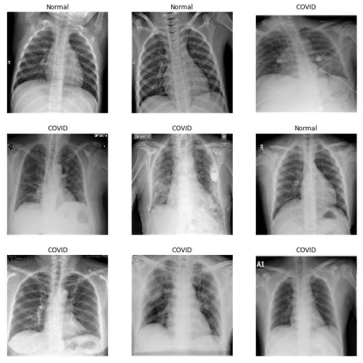
A comparison grid of nine chest X-ray (CXR) images in the posterior-anterior (PA) or anterior-posterior (AP) view, categorized into 'Normal' and 'COVID' groups for medical educational purposes. The images labeled 'Normal' demonstrate healthy pulmonary anatomy, characterized by clear lung fields, sharp costophrenic angles, and well-defined diaphragmatic borders. In contrast, the 'COVID' images illustrate various stages of COVID-19 pneumonia. Key visible findings in the COVID-positive cases include bilateral and unilateral ground-glass opacities (GGOs), peripheral patchy consolidations, and increased interstitial markings indicating pulmonary inflammation. Some clinical images show advanced pathology with diffuse lung involvement, while others exhibit medical equipment such as electrocardiogram leads or cardiovascular devices. This comparative layout is designed to help students and clinicians distinguish between normal thoracic structures and the characteristic radiologic manifestations of viral pneumonia, specifically focusing on the distribution and density of pulmonary infiltrates.
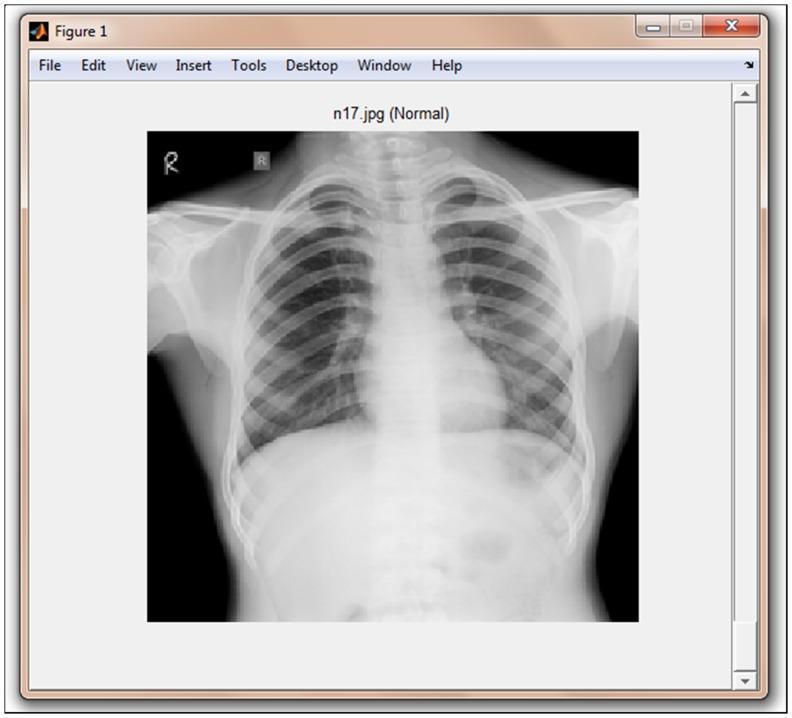
This diagnostic image shows a posteroanterior (PA) or anteroposterior (AP) chest X-ray (CXR) displayed within a MATLAB graphical user interface titled 'TB-Xpredict'. The radiograph depicts a normal adult thorax. Key anatomical structures include the radiopaque bony framework—specifically the clavicles, ribs, and vertebral column—and the radiolucent lung fields. The cardiac silhouette is visible within the central mediastinum, with clear costophrenic and cardiophrenic angles. The pulmonary vasculature exhibits normal branching patterns without evidence of consolidation, infiltrates, or pleural effusion. The image is labeled 'n17.jpg (Normal)', serving as a baseline reference for computer-aided detection (CAD) systems designed to screen for pulmonary tuberculosis (TB). This content is relevant for medical students and radiology residents focusing on normal thoracic anatomy and the application of machine learning algorithms (such as Gist or PHOG features) in automated diagnostic screening.
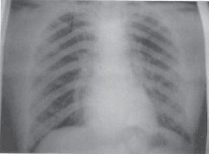
A posterior-anterior (PA) view chest X-ray illustrating normal thoracic anatomy. The radiographic image shows clear and well-aerated bilateral lung fields with no visible pulmonary opacities, infiltrates, or masses. The cardiac silhouette is of normal size and position within the mediastinum, and the trachea is observed at the midline. Both costophrenic angles are sharp, and the diaphragmatic domes are clearly defined and appropriately contoured, indicating the absence of pleural effusion. The skeletal structures, including the ribs and clavicles, appear intact without evidence of acute fracture or deformity. This diagnostic image serves as a baseline for a normal chest examination, illustrating healthy pulmonary vasculature radiating from the hilum toward the lung periphery.
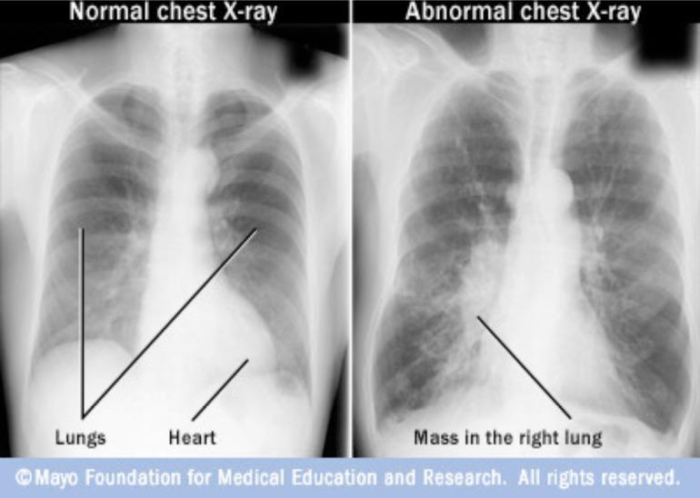
This Comparison Chart features two side-by-side posterior-anterior (PA) chest radiographs labeled 'Normal chest X-ray' and 'Abnormal chest X-ray'. The 'Normal' image depicts clear, radiolucent lung fields, sharp costophrenic angles, and a distinct cardiac silhouette. In contrast, the 'Abnormal' image demonstrates a significant focal opacity in the lower-to-middle zone of the right lung, labeled as a 'Mass in the right lung'. This finding represents a radiopaque lesion that obscures the normal pulmonary vascular markings and silhouettes the right heart border. The comparison serves as a diagnostic example for identifying primary lung neoplasms or large pulmonary masses. Key educational concepts include recognizing abnormal densities, understanding radiographic silhouettes, and differentiating healthy pulmonary parenchyma from pathological mass lesions. This material is suitable for undergraduate and intermediate medical education within the fields of radiology, pulmonology, and oncology.
PA Chest X-Ray - Schematic Anatomy
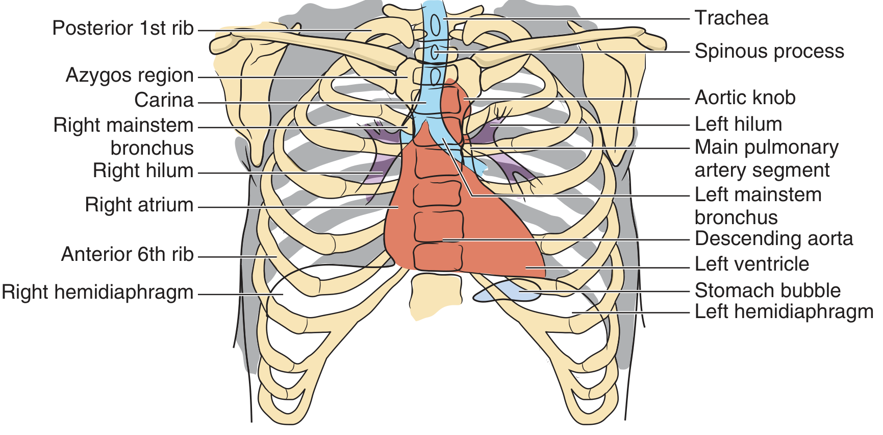
What is a PA Chest X-Ray?
Step 1 - Assess Technical Quality (RIP)
| Parameter | How to Check | Normal Standard |
|---|---|---|
| R - Rotation | Measure distance from vertebral spinous processes to medial clavicular heads on each side | Distances should be equal (up to 2-3 mm difference is acceptable) |
| I - Inspiration | Count posterior ribs as they join the spine | At least 9 posterior ribs should be visible |
| P - Penetration (Exposure) | Check visibility of vertebral interspaces | Should be visible but lung parenchyma should not be "burned out"; vessels should fade 3-4 mm from chest wall |
Step 2 - Systematic Reading (Structures to Assess)
1. Trachea
- Midline structure, visible as a lucent column
- Should be central - deviation indicates tension pneumothorax (away from affected side), collapse, or mediastinal mass
- Carina angle (tracheal bifurcation): normally <70°; widening suggests left atrial enlargement
2. Mediastinum
- Normal width: <8 cm in adults; or <25% of the thoracic diameter at the level of the carina
- Widening suggests: aortic dissection, pericardial tamponade, lymphoma, thymoma, germ cell tumours
- A "thin" mediastinum has no clinical significance
- Note: children <5 years normally have a wider mediastinum
3. Cardiac Silhouette
- Cardiothoracic ratio (CTR): transverse cardiac diameter divided by transthoracic diameter at the same level
- Normal CTR on PA: ≤0.5 (>0.5 = cardiomegaly)
- Right heart border: formed by the right atrium
- Left heart border (top to bottom): aortic knob, main pulmonary artery segment, left atrial appendage, left ventricle
- An extra hump on the left = enlarged pulmonary artery
- Pneumopericardium creates a black line around the heart border
4. Hila (Pulmonary Roots)
- Formed by pulmonary arteries and veins, with bronchi and lymph nodes
- Normal: left hilum is higher than right in ~70% of people; equal level in ~30%; right hilum is never normally higher than left
- Hilar enlargement/adenopathy suggests: sarcoidosis, tuberculosis, lymphoma, malignancy
5. Lung Fields
- Vascular markings should be visible and taper as they extend toward the periphery, stopping 3-5 mm short of the chest wall
- "Cephalization of flow": upper lobe vessels become enlarged - a sign of congestive heart failure (CHF)
- Silhouette sign: when a parenchymal opacity erases the border of an adjacent structure (e.g., right middle lobe pneumonia erases the right heart border)
- Nodules: 5-30 mm; masses: >30 mm
- Rapid "ping-pong" comparison of left vs. right lung fields helps detect flagrant asymmetries
6. Diaphragm
- Visualized as a smooth, curved line convex upward, where air-containing lung lies adjacent above
- Right hemidiaphragm is normally higher than the left by 15-30 mm (up to 20 mm is cited as normal)
- Normal level: mid-lung field at 5th-6th anterior rib interspace
- Sharp costophrenic angles (lateral): any blunting suggests pleural fluid (200-500 mL needed to blunt the lateral angle)
- Sharp cardiophrenic angle (medial): fat pads here can mimic cardiomegaly if the film is underexposed
- Air under the diaphragm (pneumoperitoneum) = surgical emergency - bowel perforation until proven otherwise
- Depression/flattening: seen in emphysema; Elevation: lung atelectasis, phrenic nerve palsy, subphrenic abscess
7. Pleura
- Normally invisible on chest X-ray
- No visible pleural line - absence of vascular markings reaching the inner edge of the chest = pneumothorax until proven otherwise
- Pleural effusion signs:
- Small: blunting of costophrenic angles (200-500 mL)
- Moderate: homogeneous opacity with concave superior margin higher laterally (meniscus sign)
- Massive: dense hemithoracic opacification with contralateral mediastinal shift (if no shift, suspect ipsilateral collapse or mesothelioma)
8. Bones and Soft Tissues
- Ribs, clavicles, scapulae, thoracic spine, sternum
- Look for fractures, lytic lesions, exostoses, or destructive lesions
- Soft tissue: subcutaneous emphysema, pacemaker leads, foreign bodies, mastectomy
Key Normal Measurements Summary
| Feature | Normal Value |
|---|---|
| Cardiothoracic ratio (CTR) | ≤0.5 on PA |
| Mediastinal width | <8 cm (or <25% of thoracic diameter) |
| Posterior ribs visible on inspiration | ≥9 |
| Right vs. left hemidiaphragm | Right higher by 15-30 mm |
| Left vs. right hilum | Left normally higher or equal (right never higher) |
| Tracheal carina angle | <70° |
| Lung vessels from chest wall | Fade 3-5 mm before chest wall |
A Useful Mnemonic for Systematic Reading
- A - Airway (trachea, carina, bronchi)
- B - Bones and soft tissues
- C - Cardiac silhouette and mediastinum
- D - Diaphragm and costophrenic angles
- E - Everything else (lung fields, pleura, hila, extra-thoracic structures)