
Extracellular matrix reference from biochemistry Dr. Dinesh puri
Reading File
The extracellular matrix (ECM) is a network of proteins and carbohydrates surrounding cells. Key components include collagens, proteoglycans, glycoproteins (fibronectin, laminin), and elastin.
Searching sources...
Finding Sources
Finding Sources
The library doesn't have a book specifically by "Dr. Dinesh Puri." Let me search the biochemistry books available and look for ECM content there, as well as search for the specific book.
Finding Sources
Finding Sources
I can see "Basic Medical Biochemistry - A Clinical Approach - 6e" has a dedicated chapter on ECM. Let me also check the Lippincott and Harper's biochemistry books. The user is asking for a "Dr. Dinesh Puri" reference - this is likely referring to the style/teaching of that author but the library has the standard biochemistry texts. Let me read the ECM content from the biochemistry books.
Reading File
Finding Sources
Reading File
Finding Sources
Reading File
Reading File
I now have comprehensive ECM content from the biochemistry textbook. Let me fetch the key diagrams and compile the full answer.
Loading Image
Loading Image
Loading Image
I now have all the content needed. Note: The library does not contain a book specifically by "Dr. Dinesh Puri" - this is a popular Indian biochemistry author whose book is commonly used in Indian medical schools but is not in the library. The content below is drawn from the closest equivalent standard biochemistry reference available - Basic Medical Biochemistry: A Clinical Approach, 6e - covering the same ECM topic.
Extracellular Matrix (ECM) - Biochemistry Reference
Note: The library does not contain a book specifically authored by Dr. Dinesh Puri. The following is compiled from Basic Medical Biochemistry - A Clinical Approach, 6e (the most detailed biochemistry ECM chapter available), which covers the same content Dr. Puri's book addresses.
Overview
The extracellular matrix fills the spaces between cells and binds cells and tissue together, determining the shape of tissues and the nature of partitioning between tissue types.
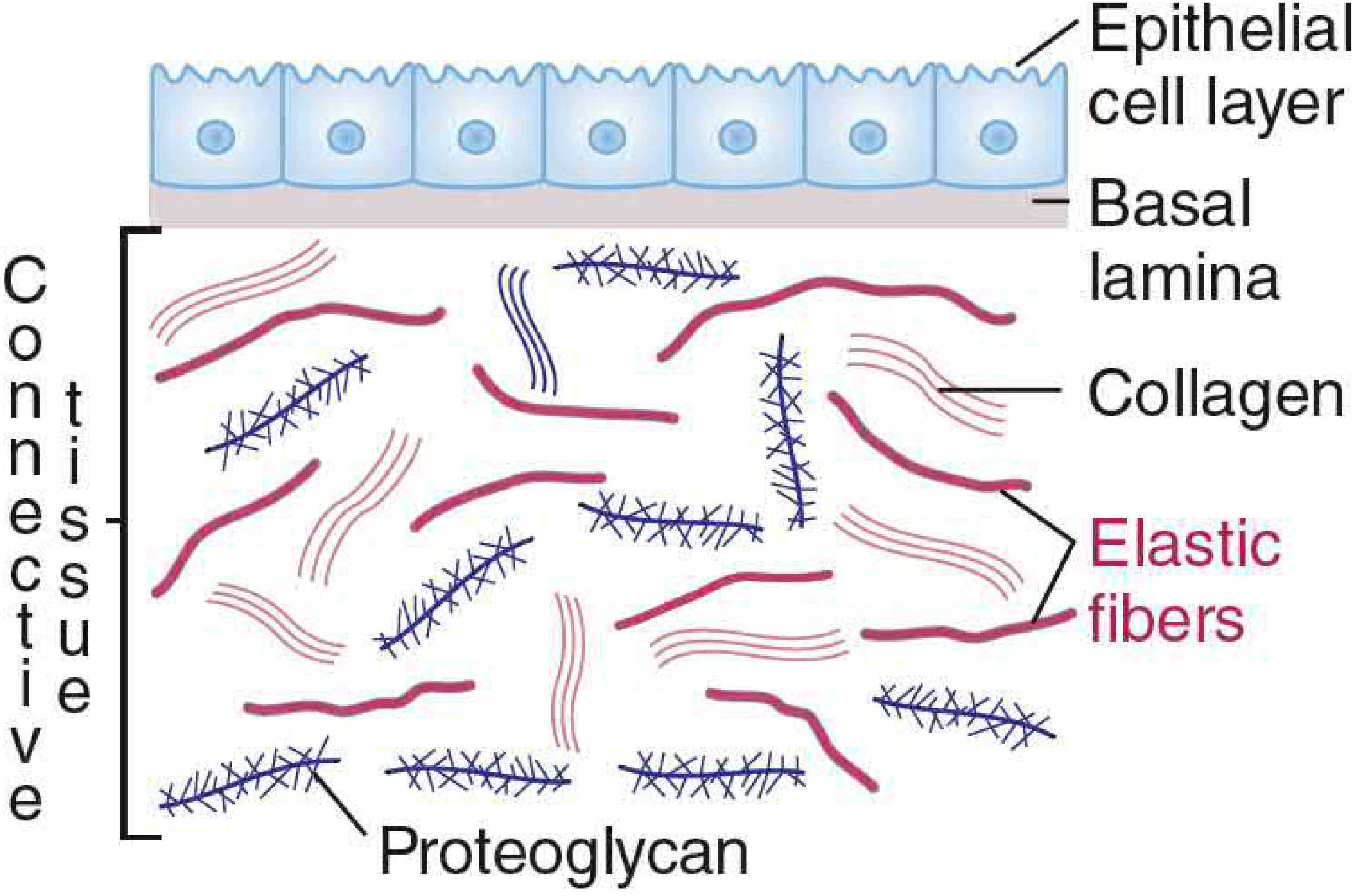
Three basic components of the ECM:
- Fibrous structural proteins - collagens, elastin, laminin
- Proteoglycans - long glycosaminoglycan (GAG) chains on a protein backbone
- Adhesion proteins - fibronectin and laminin, linking ECM components to cells
I. Fibrous Proteins
1. Collagen
Collagen is the most abundant protein in mammals (Type I collagen). It is produced principally by fibroblasts, muscle cells, and epithelial cells.
Composition:
- ~33% glycine, ~21% proline and hydroxyproline
- Hydroxyproline is formed by posttranslational modification of peptidyl proline residues (requires Vitamin C)
Structure:
- Three pro-alpha chains wound into a triple helix (ropelike)
- Every 3rd amino acid is glycine (Gly-X-Y repeats) - only Gly fits the center
- X = often proline; Y = often hydroxyproline or hydroxylysine
- Individual chains ~1,000 amino acid residues
- Chains linked by interchain hydrogen bonds
Key collagen types:
| Type | Location |
|---|---|
| Type I | Loose connective tissue, bone, tendons, skin, blood vessels, cornea |
| Type II | Cartilage |
| Type III | Blood vessels, fetal skin |
| Type IV | Basement membrane (basal lamina) - forms lattice, not fibrils |
Synthesis of Collagen (Steps):
- Synthesized in RER as preprocollagen (signal sequence present)
- Signal sequence cleaved → procollagen formed in ER
- Three procollagen chains associate via disulfide bonds at carboxy terminus
- Triple helix forms from C-terminus → N-terminus
- Procollagen (triple helix + globular ends) secreted from cell
- Extracellular proteases remove N- and C-terminal extensions → tropocollagen
- Tropocollagen assembles in ordered fashion → collagen fibrils
- Fibrils strengthened by covalent cross-links between lysine residues
Types based on function:
- Fibril-forming collagens (I, II, III) - form large insoluble fibers; give tensile strength (e.g., tendons have fibrils aligned parallel to long axis)
- Fibril-associated collagens - bind fibril surfaces; link to other matrix components
- Transmembrane collagens - form anchoring fibrils linking ECM to connective tissue
- Network-forming collagens (Type IV) - form flexible sheets for basement membranes
2. Elastin
Elastin is the major protein in elastic fibers found in smooth muscle, endothelial cells, chondrocytes, and fibroblasts. Allows tissues to expand and contract - important for blood vessels and lungs.
Composition: Also contains microfibrils made of glycoproteins fibrillin-1 and fibrillin-2
Precursor: Tropoelastin
- Highly soluble
- Synthesized on RER for eventual secretion
- Has a highly cross-linked, insoluble, amorphous structure once secreted
II. Proteoglycans
Proteoglycans = core protein + many long, linear glycosaminoglycan (GAG) chains covalently attached.
Structure: Resembles a "bottlebrush" - GAG chains extending from the core protein
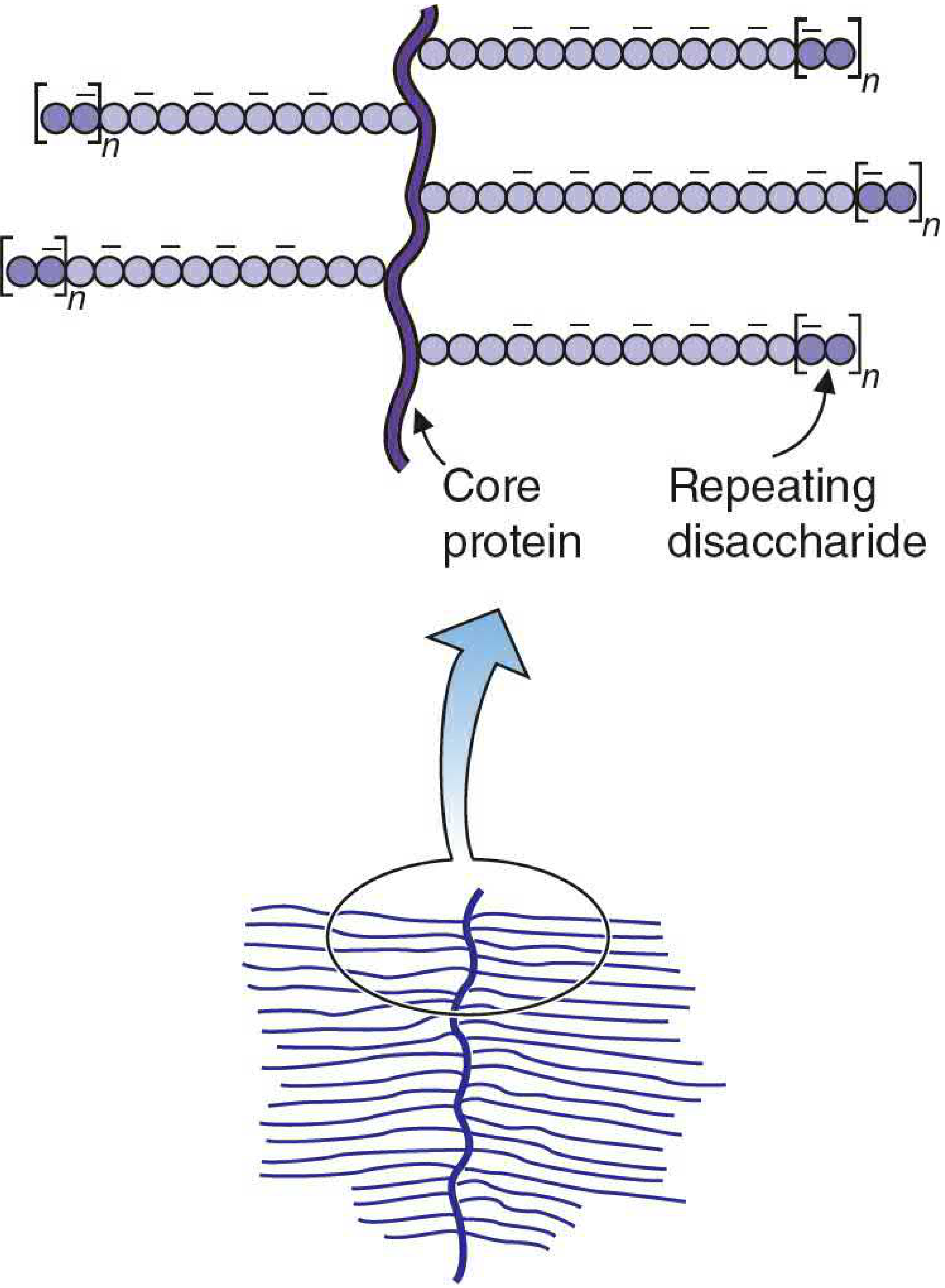
Glycosaminoglycans (GAGs):
- Repeating disaccharide units containing a hexosamine + a uronic acid
- Frequently sulfated (adding negative charge)
- Common GAGs: chondroitin sulfate, keratan sulfate, heparan sulfate, heparin, hyaluronic acid
Synthesis:
- Starts with attachment of a sugar to Ser, Thr, or Asn residue of core protein
- Additional sugars donated by UDP-sugar precursors, added sequentially to the non-reducing end
- Synthesized in ER and Golgi complex
Proteoglycan Aggregates:
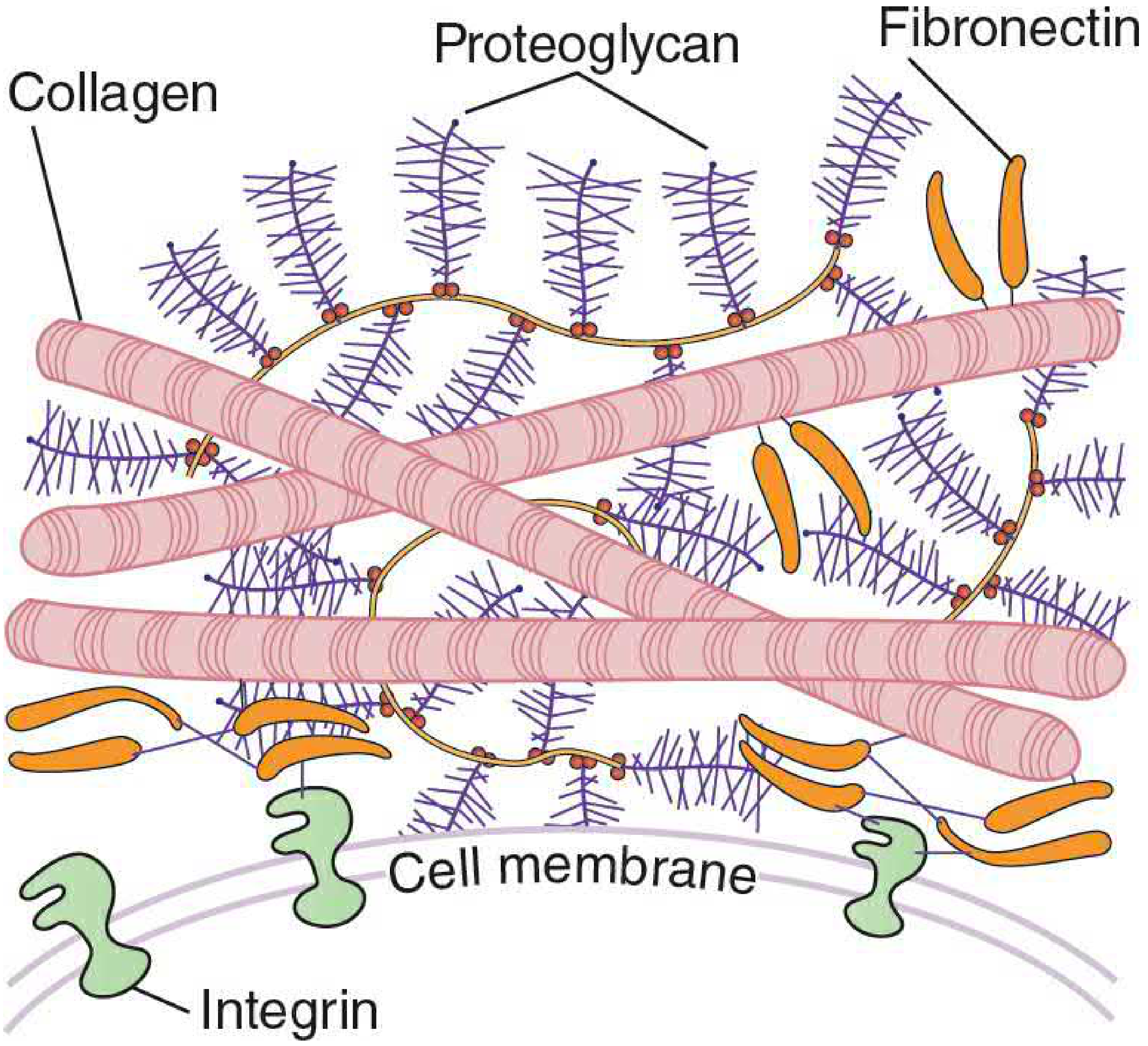
- Large aggregates form by noncovalent attachment to hyaluronic acid via a "link" protein
- These aggregates then interact with fibronectin (attached to integrin on cell membrane)
Role in cartilage:
- Long polysaccharide chains carry many anionic groups
- High concentration of negative charges attracts cations → high osmotic pressure → draws water into cartilage
- Resulting tension balances swelling pressure → gives cartilage resilience (withstands compressive load and re-expands)
III. Adhesion Proteins
Fibronectin and Laminin are extracellular glycoproteins with distinct binding domains for:
- Proteoglycans
- Collagen
- Fibrin
- Cell surface integrins
Integrins:
- Span the plasma membrane
- Bind fibronectin on the external side
- Adhere to proteins that bind intracellular actin filaments of the cytoskeleton
- Provide signaling mechanisms between cells and via the ECM
IV. Degradation of ECM
Matrix Metalloproteinases (MMPs):
- Degrade ECM components to allow cell movement and remodeling
TIMPs (Tissue Inhibitors of MMPs):
- Regulate MMP activity
Dysregulation of this balance allows cancer cells to metastasize - spreading to other body parts and locally to adjacent tissues.
Proteoglycan Degradation:
- Lysosomal enzymes degrade proteoglycans brought in by endocytosis
- Lysosomal proteases digest the protein core
- Glycosidases (endo- and exo-) degrade the carbohydrate chains
- Enzymes are specific - cleave one sugar at a time from the non-reducing end
- Deficiency of these enzymes → Mucopolysaccharidoses (accumulation of partially degraded proteoglycans)
V. Clinical Correlations
| Disease | ECM Defect |
|---|---|
| Scurvy | Vitamin C deficiency → impaired proline/lysine hydroxylation → defective collagen cross-linking |
| Osteogenesis imperfecta | Mutations in Type I collagen genes → brittle bones |
| Ehlers-Danlos syndrome | Defective collagen synthesis or cross-linking → hyperextensible skin/joints |
| Marfan syndrome | Fibrillin-1 mutation → defective elastic fibers in aorta, lens |
| Mucopolysaccharidoses | Lysosomal enzyme deficiency → GAG accumulation (e.g., Hurler syndrome - iduronidase deficiency) |
| Cancer metastasis | MMP/TIMP imbalance → ECM degradation facilitating invasion |
Source: Basic Medical Biochemistry - A Clinical Approach, 6e, Chapter 47
This is a shared conversation. Sign in to Orris to start your own chat.