
UV prolapse in absolute details gyn
Reading File
Now let me run comprehensive parallel searches on uterovaginal prolapse.
Finding Sources
Finding Sources
Finding Sources
Excellent finds. Now let me read the main uterovaginal prolapse sections from gynecology textbooks in parallel.
Finding Sources
Reading File
Finding Sources
Good. Now let me read the detailed gynecology content from Berek & Novak and Campbell Walsh, and also continue reading the Bailey & Love section.
Reading File
Reading File
Reading File
Excellent. Now let me read the deeper Berek & Novak content (pathophysiology, POP-Q, treatment) and the rest of the Campbell Walsh section.
Reading File
Reading File
Now let me read the Berek & Novak detailed pathophysiology and POP-Q sections, and the Campbell Walsh pathophysiology section.
Reading File
Reading File
I have enough comprehensive content now. Let me also fetch the key anatomical diagrams from Bailey & Love and Berek & Novak.
Reading File
Loading Image
Loading Image
Now let me fetch a few more key images and read the surgical treatment section in more detail.
Loading Image
Reading File
I now have very comprehensive content. Let me compile the full, detailed response.
Uterovaginal (UV) Prolapse - Complete Gynaecological Overview
Definition
Pelvic organ prolapse (POP) - of which uterovaginal prolapse is the central type - is defined as the descent of one or more of the anterior vaginal wall, posterior vaginal wall, the uterus (cervix), or the apex of the vagina (vaginal vault or cuff scar after hysterectomy), correlated with symptoms. It represents the progressive herniation of pelvic organs through the urogenital diaphragm, most commonly leading to vaginal bulge symptoms. - Berek & Novak's Gynecology, p. 1593
Epidemiology
- Affects up to 40% of women at some point in their lifetime. - Bailey & Love's Short Practice of Surgery, 28th Ed., p. 1611
- The prevalence of symptomatic prolapse ranges from 3-12%; asymptomatic prolapse on examination is present in the majority of adult women.
- Prevalence of at least one pelvic floor disorder is 23.7% in U.S. women; more than doubles in women aged 80+.
- Lifetime probability of undergoing surgical correction by age 80: 1 in 5 women; cumulative risk for POP surgery is 12.6%.
- Annual risk of POP surgery increases progressively until age 73 (4.3 per 1,000 women).
- POP is more common in Caucasian and Hispanic women compared to African-American women. - Campbell-Walsh-Wein Urology, p. 3404
Pelvic Support Anatomy - Three Levels (DeLancey's Classification)
Understanding the three levels is essential for identifying and repairing specific defects.
Level I (Apical Support)
- Provided by the cardinal and uterosacral ligaments - condensations of areolar connective tissue.
- Origin: lateral borders of sacral vertebrae S2-S4; travel retroperitoneally to insert at upper vagina and cervix.
- Also includes paracervical fascia and upper paravaginal fibromuscular connective tissue.
- The ureter is closest to the uterosacral ligament at or just posterior to its insertion on the posterolateral cervix.
Level II (Lateral Support)
- Provided by the arcus tendineus fasciae pelvis (ATFP) - the "white line."
- The anterior vaginal wall is suspended laterally by paravaginal attachments from the ATFP.
- Lateral detachment from the ATFP results in paravaginal defects causing anterior compartment prolapse.
Level III (Distal Support)
- Provided by the perineal body, perineal membrane, and external anal sphincter complex.
- Loss leads to perineal descent and distal posterior vaginal wall defects.
Types of Uterovaginal Prolapse
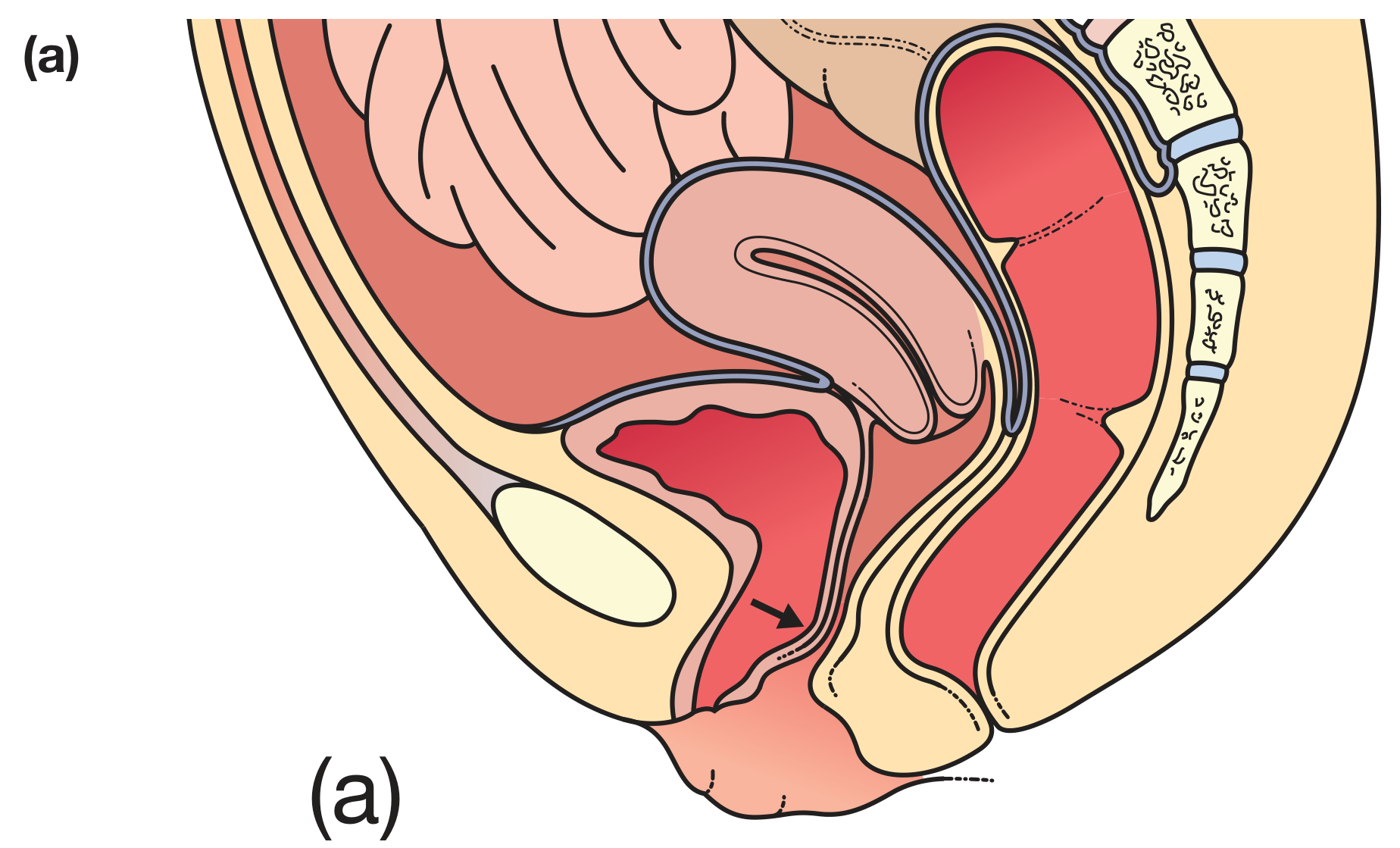
1. Anterior Compartment (Cystocele / Urethrocele / Cystourethrocele)
- Most common segment to prolapse.
- Represents descent of the anterior vaginal wall, most commonly with the bladder (cystocele), but can also represent an anterior enterocele (especially after prior surgery).
- Cystourethrocele = combined prolapse of bladder + urethra.
- Symptoms: sensation of vaginal lump, urinary urgency (OAB symptoms), recurrent UTIs, voiding difficulty.
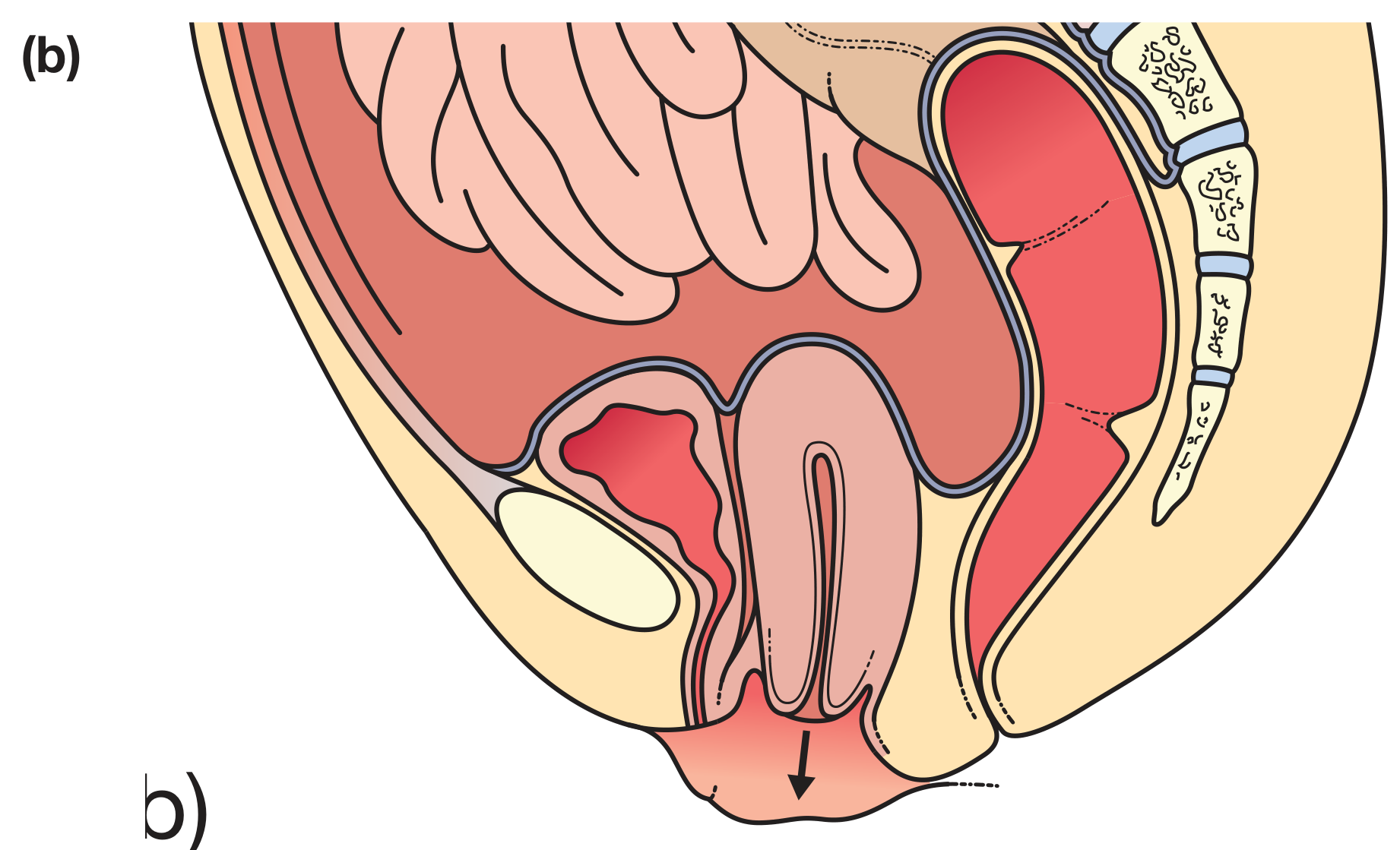
2. Apical Compartment (Uterine Prolapse / Vaginal Vault Prolapse)
- Descent of the uterus and cervix (or vaginal cuff post-hysterectomy).
- Involves failure of Level I support (cardinal/uterosacral ligament complex).
- Procidentia = complete prolapse of the uterus through the introitus (stage IV); associated with vaginal discharge, ulceration of vaginal mucosa, and bleeding.
- Enterocele: a peritoneal sac containing small intestine ± omentum descending in the rectovaginal space. Can be asymptomatic or cause defecatory dysfunction and pelvic pain.
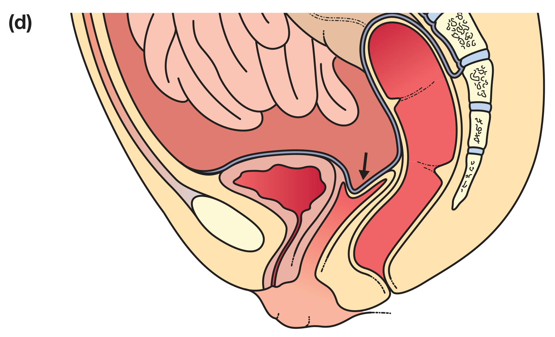
3. Posterior Compartment (Rectocele)
- Weakness of the posterior vaginal wall, usually involving the rectum.
- Symptoms: difficulty defecating, sensation of incomplete rectal emptying, need to "splint" (digitally reduce the posterior vaginal wall) to defecate.
- Can also include small bowel or colon even in the presence of an intact uterus.
4. Vaginal Vault Prolapse
- Occurs after hysterectomy when the vaginal cuff descends.
- Can include enterocele component.
Risk Factors
| Category | Specific Factors |
|---|---|
| Obstetric | Vaginal delivery, increased parity, prolonged labour, forceps delivery, large birth weight |
| Age | Post-menopausal; risk increases progressively to age 73 |
| Hormonal | Oestrogen deficiency |
| Mechanical | Obesity (major risk factor), chronic constipation, heavy lifting occupations |
| Iatrogenic | Hysterectomy (especially for prolapse) - strong predictor of secondary pelvic floor surgery |
| Genetic | Family history, connective tissue disorders (Ehlers-Danlos syndrome, Marfan syndrome) |
| Racial | More common in Caucasian and Hispanic women |
| Protective | Elective or unlaboured caesarean section |
- Campbell-Walsh-Wein Urology, p. 3404; Bailey & Love, p. 1611
Pathophysiology
Causes are multifactorial, contributing to weakening of pelvic support connective tissue, muscles, and nerve damage:
- Connective tissue defects: Cardinal and uterosacral ligament laxity; changes in collagen composition and quantity (types I and III collagen ratio altered).
- Levator ani muscle damage: Vaginal delivery can cause levator ani avulsion injury or nerve damage, leading to pelvic floor descent under loading.
- Neurological injury: Pudendal nerve stretch/injury during labour impairs pelvic floor muscle tone.
- Oestrogen deficiency: Reduces tissue strength and elasticity in postmenopausal women.
- Increased intra-abdominal pressure: Chronic constipation, heavy lifting, obesity - all force pelvic organs downward against weakened supports.
- Apical detachment: Uterine prolapse involves detachment from the uterosacral ligament complex and lateral wall detachment from endopelvic connective tissue.
Clinical Features / Symptoms
The most specific symptom for POP is a vaginal bulge that can be seen or felt by the patient. - Campbell-Walsh-Wein, p. 3405
| Symptom | Mechanism |
|---|---|
| "Something coming down" / bulge | Prolapsed organ reaching/passing introitus |
| Pelvic pressure or dragging discomfort | Organ descent, ligament stretch |
| Urinary urgency / OAB symptoms | Bladder stretch, detrusor overactivity |
| Recurrent UTIs | Incomplete bladder emptying due to cystocele |
| Voiding difficulty / retention | Urethral kinking with large cystocele |
| Stress urinary incontinence (SUI) | Associated urethral support defect (note: SUI prevalence paradoxically declines as prolapse extends beyond hymen due to urethral kinking - "occult SUI") |
| Difficulty defecating / incomplete emptying | Rectocele trapping stool |
| Need to splint to defecate | Rectocele defect |
| Low backache | Pelvic floor and ligament strain |
| Vaginal discharge, ulceration, bleeding | Advanced/complete prolapse (procidentia) with mucosal exposure |
| Dyspareunia | Anatomic distortion |
Important clinical note: There is a poor correlation between bowel/urinary symptoms and the severity and site of prolapse. Many prolapse-related symptoms are not caused by the prolapsing vagina itself but by co-existing bladder, anorectal, or pelvic floor dysfunction. Symptoms such as vaginal bulge and need to splint correlate most strongly when the leading edge of prolapse is beyond the vaginal hymen. - Campbell-Walsh-Wein, p. 3405
Grading / Staging: POP-Q System
The Pelvic Organ Prolapse Quantification (POP-Q) System is the current standard. It uses 9 measurements (six vaginal site measurements + total vaginal length, genital hiatus, perineal body), all relative to the hymen (negative = above, positive = below).
POP-Q Reference Points
| Point | Description | Range |
|---|---|---|
| Aa | Anterior wall 3 cm from hymen (corresponds to urethrovesical junction) | -3 to +3 cm |
| Ba | Most dependent portion of the remaining anterior wall | -3 cm to +TVL |
| C | Most distal edge of cervix or vaginal cuff | ±TVL |
| D | Posterior fornix (omitted after hysterectomy) - marks uterosacral ligament attachment | ±TVL |
| Ap | Posterior wall 3 cm from hymen | -3 to +3 cm |
| Bp | Most dependent portion of the remaining posterior wall | -3 cm to +TVL |
| TVL | Total vaginal length | - |
| GH | Genital hiatus (urethral meatus to posterior fourchette) | - |
| PB | Perineal body | - |
TVL = total vaginal length. Negative values = above hymen (normal). Positive values = beyond hymen (prolapsed).
Point D differentiates suspensory failure from cervical elongation (elongated cervix: C well below D; true apical descent: C and D both descend).
POP-Q Stages
| Stage | Definition |
|---|---|
| 0 | No prolapse demonstrated |
| 1 | Most distal portion >1 cm above the level of the hymen |
| 2 | Most distal portion ≤1 cm above or below the level of the hymen |
| 3 | Most distal portion >1 cm below the hymen but less than (TVL - 2 cm) |
| 4 | Maximal descent (complete eversion / procidentia) |
Stage assigned according to the most dependent portion of the prolapse - Berek & Novak's Gynecology, p. 1611-1612
Example normal POP-Q: -3, -3, -8, -10, -3, -3, 11, 4, 3 (Aa, Ba, C, D, Ap, Bp, TVL, GH, PB)
Evaluation
History
- Nature of the bulge, onset, duration, impact on quality of life.
- Urinary symptoms (frequency, urgency, incontinence, voiding difficulty).
- Bowel symptoms (constipation, fecal incontinence, straining, splinting).
- Sexual function.
- Obstetric history, prior pelvic surgery.
Physical Examination
- Patient in lithotomy position; examination ideally at maximum Valsalva to reproduce maximal descent.
- Evaluate all three compartments (anterior, apical, posterior) systematically.
- Assess levator ani muscle tone and integrity.
- Assess anal sphincter complex.
- Half-speculum exam to isolate each compartment individually.
- POP-Q measurement using a marked ring forceps or cotton-tipped applicator.
Bladder Function Evaluation
- Post-void residual (PVR) measurement - large cystocele may cause significant urinary retention.
- Urodynamic studies when urinary symptoms are prominent or before surgical planning.
- Assess for occult SUI: reduce prolapse (with ring forceps or pessary) and re-test for stress incontinence.
Bowel Function Evaluation
- Defecating proctogram (evacuation proctography) for suspected significant rectocele or enterocele.
- Anorectal manometry for suspected sphincter dysfunction.
Imaging
- Dynamic (defecation) MRI is increasingly useful for all three compartments - provides detailed multicompartment assessment of pelvic floor anatomy.
- Pelvic floor electromyography (EMG): growing utility for assessing neuromuscular integrity.
Management
Non-Surgical / Conservative Management
1. Lifestyle Modifications
- Avoidance of constipation (high-fibre diet, adequate hydration, stool softeners).
- Weight reduction in obese patients.
- Avoidance of heavy lifting.
2. Pelvic Floor Muscle Training (PFMT)
- Recommended for at least 16 weeks for grade 1-2 prolapse.
- Supervised physiotherapy is more effective than unsupervised.
- Strengthens levator ani, narrows the genital hiatus, and reduces prolapse symptoms.
3. Topical Oestrogen (Vaginal)
- Indicated for oestrogen deficiency.
- Increases tissue strength and elasticity in vaginal and pelvic floor tissues.
- Also used prior to surgical repair to optimise tissue quality.
4. Vaginal Pessaries
- Mechanical devices that support the prolapsed organs.
- Replaced every 3-6 months.
- Main complications: vaginal ulceration, infection, discharge, bleeding.
Types of Pessaries:
| Type | Use | Notes |
|---|---|---|
| Ring pessary | Most commonly used for all degrees of prolapse | Inserted between posterior fornix and pubic bone; easiest to self-manage |
| Ring with support | Anterior compartment / cystocele | Adds a membrane for additional support |
| Gellhorn pessary | More significant prolapse | Held in place by suction/space occupation; harder to self-insert/remove |
| Cube pessary | Severe prolapse / vault prolapse | Must be removed daily; strong suction retention |
| Donut pessary | Large vault prolapse, complete procidentia with decreased perineal support | Space-filling; popular |
| Continence ring/dish | Co-existent SUI | Provides bladder neck support |
- Berek & Novak's Gynecology, p. 1618-1619
Surgical Management
Surgical management aims to restore uterovaginal anatomy and position, with an individualised, multicompartmental approach based on: prior treatments, activity level, health status, desire for uterine preservation, and approach (vaginal vs. abdominal). - Berek & Novak's, p. 1594
Surgical Treatments by Compartment (Table 87.12, Bailey & Love)
| Condition | Procedure | Key Points / Complications |
|---|---|---|
| Urethrocele/Cystocele | Anterior colporrhaphy (anterior vaginal wall repair) | Plicates pubocervical fascia in the midline; no mesh required for primary repair; risk of recurrence ~30-40% |
| Cystocele (paravaginal defect) | Paravaginal repair (vaginal or abdominal) | Reattaches lateral vaginal wall to ATFP; can be done laparoscopically |
| Uterine prolapse | Vaginal hysterectomy + vault suspension | Standard approach; vault suspended to uterosacral/sacrospinous ligaments |
| Uterine prolapse (uterine preservation) | Sacrospinous hysteropexy or Manchester (Fothergill) repair | Increasingly preferred; avoids hysterectomy; sacrospinous ligament fixation of cervix |
| Enterocele | Enterocele repair (McCall culdoplasty or similar) | Peritoneal sac excised; pouch of Douglas obliterated with purse-string sutures |
| Vaginal vault prolapse | Sacrospinous ligament fixation (SSLF) | Vaginal approach; attaches vaginal vault to sacrospinous ligament (usually right side); risk of buttock pain, pudendal nerve injury |
| Vaginal vault prolapse | Uterosacral ligament suspension (USLS) | Vaginal or abdominal; re-attaches vault to uterosacral ligaments; ureteral injury risk (~1-2%) |
| Vaginal vault prolapse | Abdominal/laparoscopic sacrocolpopexy | Gold standard for vault prolapse; mesh bridge from vaginal apex to sacral promontory (S1/S2); low recurrence rate (~5-15%); risk of mesh erosion, sacral bleeding |
| Rectocele | Posterior colporrhaphy | Plicates rectovaginal fascia in the midline; can be enhanced with levatorplasty (caution: dyspareunia) |
| Posterior enterocele | Posterior enterocele repair ± posterior colporrhaphy | Peritoneal sac excised + posterior wall reinforced |
| Obliterative procedure | Colpocleisis (Le Fort's colpocleisis) | For elderly/unfit patients who do not wish to be sexually active; low morbidity, high success rate; closes the vagina permanently |
Abdominal Approaches
- Abdominal sacrocolpopexy: Considered the "gold standard" for vault prolapse; synthetic mesh attached from vaginal apex to anterior longitudinal ligament at S1/S2.
- Laparoscopic/Robotic sacrocolpopexy: Equivalent outcomes to open; less blood loss, faster recovery.
- Abdominal uterosacral suspension: Re-suspends vaginal apex to uterosacral ligaments; can be combined with hysterectomy.
Vaginal Obliterative Procedures (Colpocleisis)
- Suitable for elderly, sexually inactive women with good life expectancy but poor surgical risk.
- Very high success rates (95-98%), low morbidity.
- Permanent loss of vaginal function.
Transvaginal Mesh (TVM)
- Use is now highly restricted in many countries (FDA warnings 2019) due to serious complications (mesh erosion/extrusion through vaginal epithelium, dyspareunia, chronic pelvic pain, organ perforation, infection).
- Native tissue repair is now preferred; mesh reserved for specific recurrent prolapse cases.
Uterine Preservation vs. Hysterectomy
- Hysterectomy has historically been performed routinely at UV prolapse repair, but high-level evidence does not uniformly support this.
- Uterine-preserving procedures (sacrospinous hysteropexy, uterosacral ligament suspension with uterus in situ, Manchester repair) have comparable outcomes and are increasingly preferred in younger women.
- However, hysterectomy at the time of repair has been performed to facilitate access to uterosacral ligaments for apical suspension. - Berek & Novak's, p. 1654
Management of Co-existing Urinary Symptoms with POP Repair
- Prolapse repair alone can resolve OAB symptoms in >50% of women (anterior compartment repair reduces frequency by 33%, urge incontinence by 49%, voiding difficulty by 74%).
- Occult SUI: Urethral kinking by large prolapse can mask genuine SUI. Reduce prolapse with pessary/ring forceps and stress test; if SUI unmasked, consider concurrent anti-incontinence procedure.
- Failure to recognise POP at the time of UI surgery significantly increases the need for subsequent prolapse surgery.
Summary of Treatment Approach
UV Prolapse Confirmed
│
├── Symptomatic? ──No──→ Reassurance + PFMT
│
└── Yes
    │
    ├── Mild (Stage 1-2)
    │   → PFMT ≥16 weeks + lifestyle + topical oestrogen ± pessary
    │
    ├── Moderate-Severe (Stage 2-4) + Wishes Non-Surgical
    │   → Pessary (ring / Gellhorn / donut) + follow-up q3-6mo
    │
    └── Surgical Candidate
        ├── Wants uterine preservation → Hysteropexy / Manchester repair
        ├── Consents to hysterectomy → Vaginal hysterectomy + vault suspension
        │   (SSLF or USLS vaginal) or sacrocolpopexy (abdominal/laparoscopic)
        ├── Rectocele → Posterior colporrhaphy
        ├── Cystocele → Anterior colporrhaphy ± paravaginal repair
        └── Elderly / not sexually active → Colpocleisis
Key Points Summary
- UV prolapse affects up to 40% of women; anterior compartment is the most common site.
- The POP-Q system is the validated standard for staging; stage is determined by the most dependent point.
- Point D (posterior fornix) differentiates true apical descent from cervical elongation.
- Vaginal bulge is the most specific symptom; urinary/bowel symptom correlation with prolapse severity is poor.
- Pelvic floor muscle training for ≥16 weeks is the first-line treatment for stage 1-2.
- Ring pessary is the most commonly used mechanical device; replaced every 3-6 months.
- Sacrocolpopexy (abdominal/laparoscopic) is the gold standard for vault prolapse.
- Transvaginal mesh is now heavily restricted due to serious complications.
- Occult SUI must be screened for before surgical repair by reducing the prolapse and stress-testing.
- Colpocleisis is the preferred procedure in elderly, sexually inactive women with high surgical risk.
Sources: Bailey & Love's Short Practice of Surgery 28th Ed., p. 1611-1612; Berek & Novak's Gynecology, pp. 1593-1654; Campbell-Walsh-Wein Urology, pp. 3404-3411
This is a shared conversation. Sign in to Orris to start your own chat.