
Can you teach me all clinical examinations used in general medicine
Reading File
Clinical examinations in general medicine is a very broad topic covering multiple body systems. Let me clarify what would work best for you before diving in.
Asking for Preferences
Which systems or areas would you like to cover? · What is your learning level?
Great - a complete systems overview at junior doctor level. Let me pull from the medical library across all major examination areas simultaneously.
Searching sources...
Finding Sources
Finding Sources
Finding Sources
Finding Sources
Good - found key sources. Now reading the actual content from Harrison's (CVS), Goldman-Cecil (abdominal), Daroff's Neurology, and searching for general/respiratory exam content simultaneously.
Reading File
Reading File
Reading File
Finding Sources
Excellent content retrieved. Now let me get the respiratory exam content and the rest of the CVS exam (heart sounds, murmurs) simultaneously.
Finding Sources
Reading File
Finding Sources
Reading File
Finding Sources
Reading File
I now have rich content from Harrison's and Goldman-Cecil. Let me get the respiratory/chest exam and the rest of the heart sounds sections.
Reading File
Finding Sources
Reading File
Reading File
I now have comprehensive data from Harrison's on CVS, and from Goldman-Cecil on abdominal examination, and from Daroff's on neurology. Let me read the respiratory chest exam section from Harrison's and get key images.
Reading File
Searching Images
clinical examination techniques cardiovascular respiratory abdominal
I now have all the key content needed. Let me update the todos and compile the comprehensive teaching document.Here is a complete structured guide to clinical examinations in general medicine, compiled from Harrison's Principles of Internal Medicine 22E, Goldman-Cecil Medicine, and Daroff's Neurology in Clinical Practice.
Clinical Examinations in General Medicine
The Universal Sequence (for any system)
Every clinical examination follows: Inspection → Palpation → Percussion → Auscultation
(Exception: the abdomen - always auscultate before palpating/percussing to avoid altering bowel sounds.)
1. GENERAL / SYSTEMIC EXAMINATION
Before going system-by-system, every patient gets a general survey.
1a. Overall Inspection
- Build: thin/obese, nutritional status, hydration
- Conscious level: alert, drowsy, confused (GCS if needed)
- Appearance: distressed, comfortable, jaundiced, pale, cyanosed
- Posture and gait (briefly, walking into the room)
- Speech: dysarthria, aphasia, hoarseness
1b. Vital Signs
| Parameter | Technique | Key Notes |
|---|---|---|
| Temperature | Oral/axillary/tympanic | >38°C = fever; <36°C = hypothermia |
| Pulse | Radial - rate, rhythm, volume, character | Normal 60-100 bpm; regular/irregular |
| Blood Pressure | Seated, arm at heart level, 5-10 min rest, correct cuff size | Both arms (>10 mmHg diff is abnormal). Korotkoff I = systolic, V = diastolic |
| Respiratory Rate | Count breaths for 60 seconds | Normal 12-20/min; most underrecorded vital sign |
| SpO2 | Pulse oximetry | Normal ≥95%; finger vs toe gap >2% = peripheral arterial disease |
| BMI / Weight | Scale + height | Document at every admission |
Blood pressure pearls (Harrison's):
- Systolic leg pressure is normally up to 20 mmHg higher than arm - reversed = coarctation or aortic dissection
- "White coat hypertension": BP >130/80 in clinic but <130/80 out of clinic, no end-organ damage
- Ankle-Brachial Index (ABI): ankle systolic ÷ higher brachial pressure. Low ABI = peripheral arterial disease; powerful predictor of cardiovascular mortality
1c. Hands
- Clubbing: loss of nail-fold angle; 5 causes per system (lung cancer, fibrosis, bronchiectasis, cyanotic heart disease, liver cirrhosis, IBD)
- Peripheral cyanosis: blue/cold fingers (low flow)
- Koilonychia: spoon-shaped nails - iron deficiency anaemia
- Leukonychia: white nails - hypoalbuminaemia
- Pallor: palmar crease pallor = Hb <9 g/dL roughly
- Asterixis (flap): outstretched hands, wrists extended - hepatic, uraemic, or CO2 retention encephalopathy
- Dupuytren's contracture: palmar fibrosis - liver disease, diabetes, familial
- Splinter haemorrhages: infective endocarditis, vasculitis
- Osler's nodes / Janeway lesions: endocarditis
- Tremor (resting, postural, intention): see Neurology section
1d. Face
- Eyes: jaundice (sclerae), anaemia (conjunctivae), Horner's (ptosis + miosis + anhidrosis), exophthalmos (thyroid), xanthelasma, arcus cornealis
- Mouth: angular stomatitis (B12/iron def), central cyanosis (tongue and lips), leukoplakia, oral candidiasis, gum hypertrophy (phenytoin, CCBs, leukaemia)
- Parotid enlargement: alcoholic liver disease
- Malar flush: mitral stenosis
- Facies: myxoedema, Cushingoid, acromegaly, Parkinsonism (hypomimia), progressive supranuclear palsy (worried expression)
1e. Neck
- JVP: see CVS section (very important bedside venous pressure measure)
- Lymph nodes: anterior/posterior cervical, supraclavicular (Virchow's node = gastric/lung cancer), axillary, inguinal
- Thyroid: palpate from behind, swallows on swallowing; auscultate for bruit
- Carotid bruit: auscultate at angle of mandible
1f. Oedema
- Peripheral: pitting oedema at ankles/sacrum - cardiac, hepatic, nephrotic, nutritional
- Non-pitting: lymphoedema, myxoedema
- Press firmly for 10-15 seconds - depth of pit correlates roughly with severity
2. CARDIOVASCULAR SYSTEM (CVS) EXAMINATION
(Based on Harrison's Principles of Internal Medicine 22E, Chapter 246)
Sequence: Position patient at 45°, adequately exposed from waist up.
2a. Jugular Venous Pressure (JVP)
The JVP is the single most important bedside measurement of volume status.
- Use the internal jugular vein (preferred - direct line to RA; external JV is valved)
- Measure the vertical height of venous pulsation above the sternal angle (angle of Louis) at 45°
- Normal: <4.5 cm above sternal angle at 45°
- Tip: pulsations above the clavicle in the sitting position are always abnormal (RA is ≥10 cm below clavicle)
Distinguishing JVP from carotid pulse:
| Feature | JVP | Carotid |
|---|---|---|
| Obliterable by pressure | Yes | No |
| Waveform | Biphasic (a and v waves) | Monophasic |
| Changes with posture | Yes | No |
| Changes with inspiration | Yes | No |
JVP waveform components:
- a wave - RA presystolic contraction (after P wave on ECG); absent in AF; cannon a wave = AV dissociation (VT!)
- x descent - RA pressure fall after tricuspid opening
- c wave - tricuspid bulging into RA during early systole
- v wave - atrial filling during ventricular systole; giant v wave = tricuspid regurgitation
- y descent - tricuspid opens, RA empties
- Prominent y descent + pericardial knock = constrictive pericarditis
Clinical meanings:
- Elevated JVP: fluid overload, right heart failure, tamponade, SVC obstruction
- Elevated JVP with elevated JVP rising on inspiration (Kussmaul's sign): constrictive pericarditis, restrictive cardiomyopathy
- Elevated JVP (>10 mmHg) predicts pulmonary wedge pressure >22 mmHg (PPV 88%)
2b. Arterial Pulse
- Best assessed at the carotid for character; radial for rate and rhythm
- Rate, rhythm, volume, character, radio-radial/radio-femoral delay, vessel wall
Pulse character:
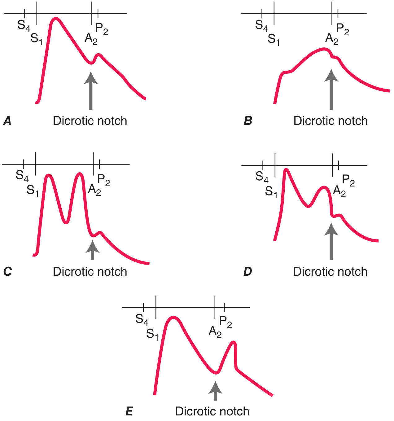
| Pulse Type | Character | Cause |
|---|---|---|
| Pulsus parvus et tardus | Weak, slow upstroke | Severe aortic stenosis |
| Corrigan's/water-hammer | Sharp rise, rapid collapse | Severe aortic regurgitation |
| Bisferiens | Two systolic peaks | HOCM, AR |
| Pulsus paradoxus | >10 mmHg fall in systolic on inspiration | Cardiac tamponade, massive PE, severe obstructive lung disease, tension pneumothorax |
| Pulsus alternans | Beat-to-beat amplitude variation (regular rhythm) | Severe LV systolic dysfunction |
| Anacrotic | Slow notched upstroke | Severe AS |
Pulsus paradoxus measurement: inflate BP cuff above systolic; slowly deflate; note the pressure at which Korotkoff sounds are heard only in expiration, then the pressure at which heard in every beat. Difference >10 mmHg = positive.
2c. Blood Pressure
- Both arms; >10 mmHg difference = subclavian/atherosclerotic disease, coarctation, dissection
- Systolic legs > arms normally; reversed = coarctation
- Measure ABI in peripheral arterial disease
2d. Precordial Examination
Inspection:
- Visible apex beat: midclavicular line, 5th ICS in thin adults
- Visible pulsations elsewhere = abnormal
- Right parasternal pulsation = ascending aortic aneurysm
Palpation:
- Patient supine 30°, enhanced in left lateral decubitus
- Apex beat: location (normally ≤5th ICS, midclavicular line), size (<2 cm), character
- Heaving/sustained = LV hypertrophy (pressure overload: AS, hypertension)
- Hyperdynamic/thrusting = LV volume overload (MR, AR)
- Displaced/diffuse = dilated cardiomyopathy
- Parasternal heave: RV enlargement (pressure or volume overload)
- Thrills (palpable murmurs): grade 4+ murmurs; location identifies origin
- Loud P2 palpable at left upper sternal border = pulmonary hypertension
2e. Cardiac Auscultation
Areas:
- Aortic: 2nd right ICS
- Pulmonary: 2nd left ICS
- Tricuspid: left lower sternal border (4th/5th ICS)
- Mitral: apex (5th ICS, midclavicular line) - listen with bell for low-pitched sounds
First Heart Sound (S1):
- Mitral + tricuspid closure
- Loud S1: early rheumatic mitral stenosis, short PR interval, hyperkinetic states
- Soft S1: late rheumatic MS (calcified leaflets), long PR interval, LV systolic dysfunction, beta-blockers
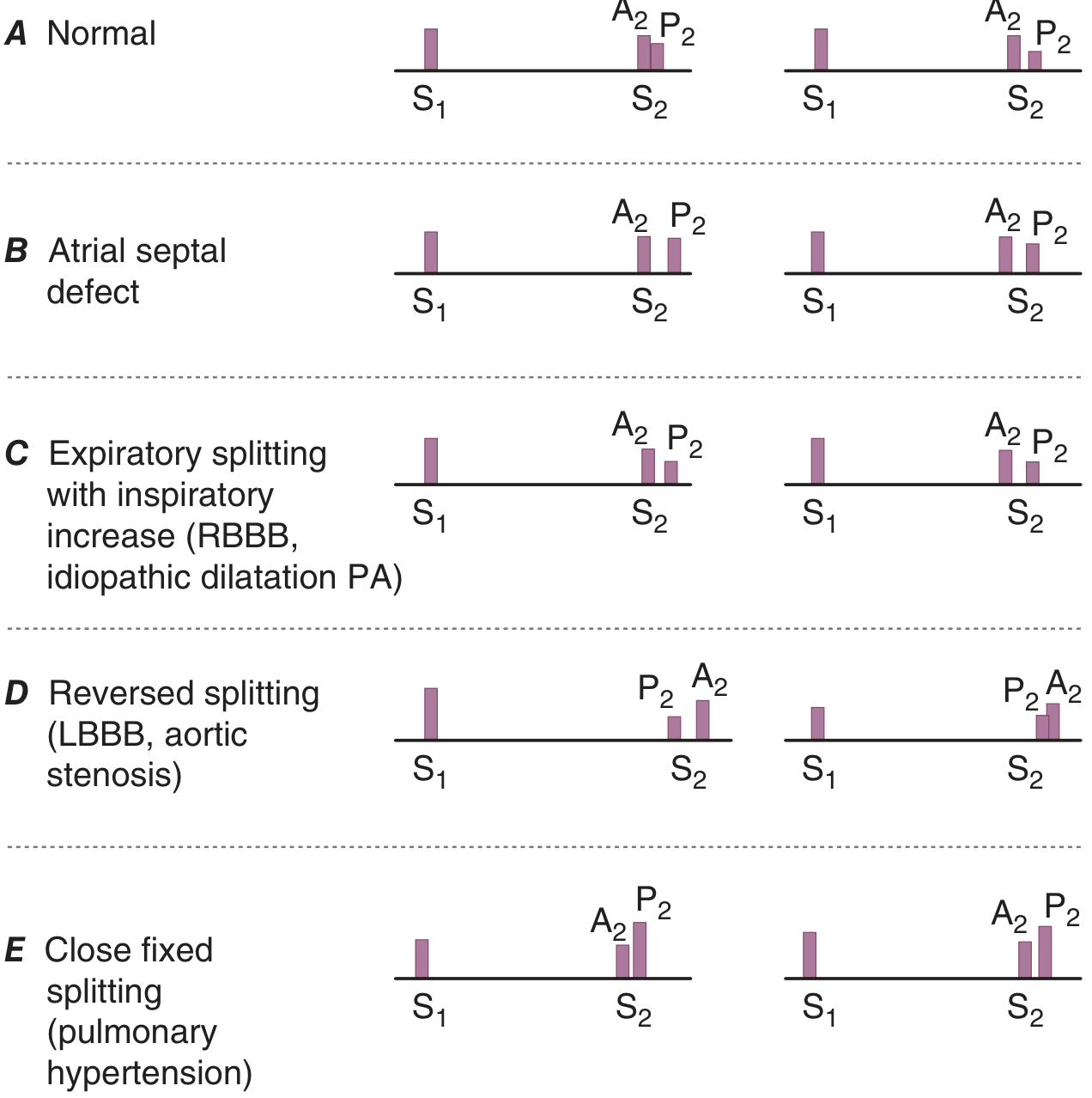
- Splitting: heard in young patients and RBBB (delayed tricuspid closure)
Second Heart Sound (S2):
- Aortic (A2) + pulmonary (P2) closure
- Physiologic splitting: widens on inspiration (more right-sided filling), narrows on expiration
- Wide splitting: RBBB, severe MR (early aortic closure)
- Fixed splitting: ASD (no respiratory variation) - classic sign
- Reversed/paradoxical splitting: components audible at end-expiration, narrow on inspiration - LBBB, RV pacing, severe AS, HOCM, acute MI
- Loud P2: pulmonary hypertension (may be palpable)
- Narrow single S2: pulmonary hypertension
S3 (Third Heart Sound):
- Low-pitched, early diastolic; heard at apex with bell
- In adults: always pathological - LV failure, dilated cardiomyopathy
- In children/young adults: can be normal ("physiologic S3")
- Sounds like "Ken-TUCK-y"
S4 (Fourth Heart Sound):
- Low-pitched, late diastolic (pre-systolic); heard at apex
- Reflects stiff ventricle (LVH, ischaemia, hypertrophic cardiomyopathy)
- Sounds like "TEN-nes-see"
- Never normal in adults
Heart Murmur Grading:
| Grade | Description |
|---|---|
| 1 | Barely audible (only heard in ideal conditions) |
| 2 | Faint but clearly audible |
| 3 | Moderate, no thrill |
| 4 | Loud + palpable thrill |
| 5 | Very loud, heard with stethoscope partially off chest |
| 6 | Heard without stethoscope |
Systolic Murmurs:
| Murmur | Timing | Best Heard | Radiation | Quality |
|---|---|---|---|---|
| MR (chronic) | Holosystolic | Apex | Axilla | Blowing |
| AS | Midsystolic (ejection) | Aortic area | Carotids | Harsh, crescendo-decrescendo |
| TR | Holosystolic | Left lower sternal border | Increases with inspiration (Carvallo's sign) | Blowing |
| VSD | Holosystolic | Left sternal border | Radiates all over | Harsh |
| HOCM | Midsystolic | Left lower sternal border | Increases with Valsalva/standing, decreases with squatting | Harsh |
| Pulmonary stenosis | Midsystolic | Pulmonary area | Ejection click | Harsh |
Severe AS signs: parvus et tardus carotid pulse, late-peaking grade ≥3 murmur, soft or absent A2, S4 at apex, sustained LV impulse
Diastolic Murmurs (always pathological):
| Murmur | Timing | Best Heard | Radiation |
|---|---|---|---|
| AR | Early diastolic decrescendo | Left sternal border (lean forward, expiration) | - |
| MS | Mid-diastolic, low-pitched rumble | Apex (bell, left lateral decubitus, expiration) | - |
Manoeuvres:
- Valsalva: decreases preload - most murmurs softer; HOCM and MVP murmur louder
- Squatting: increases preload and afterload - HOCM murmur softer
- Standing: decreases preload - HOCM murmur louder
- Handgrip: increases afterload - MR/AR louder, AS softer
2f. Peripheral Vascular Examination
- Palpate all peripheral pulses: radial, brachial, carotid, femoral, popliteal, dorsalis pedis, posterior tibial
- Auscultate for bruits: carotid, subclavian, renal (epigastric), femoral
- Bruit extending into diastole or with thrill = severe obstruction
- Check for lower limb oedema, skin changes (lipodermatosclerosis, venous ulcers), temperature asymmetry
3. RESPIRATORY SYSTEM EXAMINATION
(Harrison's Principles of Internal Medicine 22E, Chapter 262)
Position: Patient sitting, examine posterior chest first, then anterior.
3a. Inspection
- Rate and pattern: tachypnoea (>20/min), bradypnoea, Cheyne-Stokes (central apnoeic cycles - heart failure, CNS disease), Kussmaul (deep, sighing - metabolic acidosis), Biot's (irregular - brainstem damage)
- Accessory muscle use: sternocleidomastoid, scalenes - respiratory distress
- Intercostal recession: severe obstruction in children
- Barrel chest: AP diameter equal to transverse; COPD/emphysema
- Kyphoscoliosis: can cause restrictive defect
- Asymmetric expansion: collapse, pneumothorax, effusion on affected side
- Tracheal position: midline? Deviation away from effusion/tension pneumothorax; towards collapse
3b. Palpation
- Tracheal position (finger in sternal notch)
- Chest expansion: thumbs meet at midline over lower posterior chest; should expand symmetrically ≥5 cm
- Tactile (vocal) fremitus: place ulnar aspect of hand or palm on chest; say "99"
- Increased = consolidation (fluid-filled alveoli transmit better)
- Decreased/absent = effusion, pneumothorax, thick chest wall
3c. Percussion
- Middle finger of non-dominant hand as pleximeter; tap with middle finger of dominant hand
- Percuss comparing side to side symmetrically
- Normal: resonant
- Dull: consolidation, collapse, pleural effusion, mass
- Stony dull: pleural effusion (classically)
- Hyper-resonant: pneumothorax, emphysema
- Diaphragm excursion: mark dullness at end-expiration and end-inspiration (normal excursion ~5 cm)
3d. Auscultation
- Use diaphragm; compare sides methodically
- Breath sound character:
| Sound | Normal? | Cause when abnormal |
|---|---|---|
| Vesicular | Normal peripheral lung | - |
| Bronchial | Normal only over trachea | Consolidation (pneumonia) when heard peripherally |
| Bronchovesicular | Normal over main bronchi | Mild consolidation when elsewhere |
| Diminished/absent | Abnormal | Emphysema (diffuse), pneumothorax, effusion (unilateral) |
- Added sounds:
| Sound | Description | Cause |
|---|---|---|
| Crackles (rales) | Discontinuous, popping sounds | Fine: pulmonary oedema (bases), IPF (Velcro-like, bilateral bases); Coarse: pneumonia, bronchiectasis |
| Wheezes | Continuous, musical, usually expiratory | Asthma (polyphonic), COPD, tumour (monophonic, fixed) |
| Rhonchi | Low-pitched continuous, like snoring | Secretions in medium airways; bronchitis, COPD |
| Stridor | High-pitched inspiratory wheeze over neck | Upper airway obstruction (epiglottitis, foreign body, tumour) |
| Pleural rub | Creaking, leathery; both phases | Pleuritis (pneumonia, PE, malignancy) |
Vocal resonance tests:
- Whispered pectoriloquy: ask patient to whisper "1-2-3"; if clearly heard = consolidation
- Bronchophony: say "99"; louder than normal = consolidation
- Egophony: say "EEE"; if you hear "AHH" = consolidation (pneumonia); not heard in fibrosis
Summary of clinical signs by diagnosis:
| Condition | Expansion | Percussion | Breath sounds | Vocal fremitus | Added |
|---|---|---|---|---|---|
| Consolidation | Reduced (affected) | Dull | Bronchial | Increased | Crackles, egophony |
| Effusion | Reduced (affected) | Stony dull | Absent/reduced | Absent | Pleural rub (edge) |
| Pneumothorax | Reduced (affected) | Hyper-resonant | Absent | Absent | - |
| Collapse | Reduced (affected) | Dull | Reduced/absent | Reduced | - |
| Emphysema | Reduced (both, barrel) | Hyper-resonant | Reduced (both) | Reduced | Prolonged expiration |
| Fibrosis | Reduced (both) | Normal | Vesicular reduced | Normal | Fine basal crackles (Velcro) |
| Asthma (acute) | Reduced (both) | Normal/hyper | Reduced | Normal | Polyphonic wheeze |
4. ABDOMINAL EXAMINATION
(Goldman-Cecil Medicine, International Edition)
Position: Patient supine, one pillow, adequately exposed from xiphisternum to pubic symphysis. Ask them to keep arms at sides and legs flat.
4a. Inspection
Survey for:
- Scars: location suggests prior surgery (RIF appendicectomy, midline laparotomy, RUQ cholecystectomy)
- Distension: generalised (the 5 Fs: Fat, Fluid/ascites, Faeces, Flatus, Fetus/mass) vs localised
- Prominent veins: periumbilical - portal hypertension (caput medusae); inferior epigastric collaterals - IVC obstruction
- Visible peristalsis: small bowel obstruction, pyloric stenosis (epigastric, left to right)
- Hernias: umbilical, epigastric, inguinal (ask patient to cough/stand)
- Skin: striae (purple = Cushing's; white = pregnancy/obesity), spider naevi (>5 = liver disease), jaundice, Grey-Turner's/Cullen's sign (bruising in flanks/periumbilical = retroperitoneal haemorrhage, severe pancreatitis)
- Asymmetry: organomegaly, mass
4b. Auscultation (before palpation/percussion in the abdomen)
- Bowel sounds: listen in one spot (e.g., right iliac fossa) for 15-30 seconds
- Normal: intermittent gurgles
- Increased/hyperactive: early obstruction, gastroenteritis, after eating
- Absent (>3 min silence): paralytic ileus, peritonitis, late obstruction
- Tinkling/high-pitched: mechanical obstruction
- Bruits: renal arteries (lateral to umbilicus - renovascular hypertension); aortic (epigastric - aneurysm/stenosis); hepatic bruit (hepatocellular carcinoma)
- Note: routine auscultation for bowel sounds has limited value in patients without abdominal pain
4c. Percussion
- Cursory light percussion across all 9 regions first - dullness, tympany, unexpected tenderness
- Liver size: upper border of dullness in midclavicular line (MCL) on full expiration, lower border dullness in MCL on full inspiration; normal span 6-12 cm
- Splenomegaly: percussion in left upper quadrant near 10th rib posterior to midaxillary line - dullness distinct from gastric tympany suggests spleen
- Ascites: percuss for flank dullness (flanks dull, central tympany); if dull, test for shifting dullness: percuss flank to mark dullness, keep finger there, roll patient away - if line of dullness shifts upward = positive (most sensitive bedside sign for ascites)
- Fluid thrill (for massive ascites): flick one flank while pressing midline with other hand's edge, second hand on opposite flank detects impulse
4d. Palpation
Always start away from the site of pain. Begin with light palpation, then deep.
Light palpation:
- Watch the patient's face, not the abdomen
- Detect guarding (voluntary or involuntary muscle spasm), tenderness, superficial masses
- Involuntary guarding + rigidity = peritonism
Deep palpation - organ assessment:
Liver: start in RIF, hand flat, ask patient to breathe in and out; edge moves down on inspiration; palpate upward on each inspiration, feel for the edge. Assess: size (cm below costal margin), consistency (hard = malignancy, fibrosis; soft = congestive), tenderness, surface (smooth vs nodular), pulsatility (TR)
Spleen: start in RIF, move toward LUQ; use bimanual technique (left hand posteriorly supporting left costal margin, right hand palpating below); asks patient to breathe in. Cannot get above it (contrast with enlarged left kidney). Tip of enlarged spleen = splenomegaly:
- Grade I: just palpable
- Grade II: to umbilicus
- Grade III: into RIF (massive)
Kidneys: bimanual palpation (ballot technique); right kidney easier to feel than left; kidneys ballottable (unlike spleen). Polycystic kidneys can be massive bilateral.
Aorta: palpate in epigastrium, expansile pulsation (width >3 cm = aneurysm). In thin patients normal aorta felt easily.
Bladder: dull to percussion, arises from pelvis, cannot get below it.
Renal angle tenderness: fist percussion over costovertebral angle (CVA) - positive in pyelonephritis, renal stones.
Special signs:
- Murphy's sign: 2 fingers in RUQ, patient takes deep breath; arrest of inspiration due to pain = positive = acute cholecystitis (false positive: hepatitis)
- McBurney's sign: tenderness at McBurney's point (1/3 from ASIS to umbilicus) = appendicitis
- Rebound tenderness (Blumberg's): press slowly, release rapidly - pain worse on release = peritoneal irritation
- Rovsing's sign: pressure in LIF causes pain in RIF = appendicitis
- Obturator test: flex and internally rotate right hip - pain in RIF = inflamed appendix/pelvic abscess
- Psoas sign: extend right hip (patient left lateral) - pain in RIF = retrocaecal appendicitis
- Carnett's test: patient tenses abdominal wall (head lift) - if tenderness worse = abdominal wall source; if less = intra-abdominal source
4e. Rectal Examination
Indicated in: rectal/perianal symptoms, suspected GI bleeding, incontinence, acute abdomen.
- Inspect perianal area: fissures, fistulas, haemorrhoids, skin tags
- Digital: assess anal tone, rectal wall circumferentially for masses, prostate in males (size, consistency, tenderness), cervix in females
5. NEUROLOGICAL EXAMINATION
(Bradley and Daroff's Neurology in Clinical Practice)
5a. Philosophy: Start During the Interview
- Hypomimia (expressionless face) = Parkinsonism or depression
- Worried/astonished expression = progressive supranuclear palsy
- Ptosis = myasthenia gravis or brainstem lesion
- Speech = dysarthria, aphasia, spasmodic dysphonia
- Involuntary movements during history-taking
5b. Mental Status
- Orientation: time, place, person
- Attention: months backwards, serial 7s
- Memory: 3-word recall (immediate and 5-minute)
- Language: naming, repetition, comprehension, reading, writing
- Calculation, visuospatial function, executive function (frontal lobe)
- Validated tools: MMSE (30 points), MoCA (for mild cognitive impairment, 30 points)
5c. Cranial Nerves (CNs)
| CN | Test | Key Findings |
|---|---|---|
| I - Olfactory | Test each nostril (coffee, vanilla - not pungent) | Anosmia: head injury, Parkinson's, olfactory groove meningioma |
| II - Optic | Visual acuity (Snellen), visual fields (confrontation), pupil reactions (RAPD = swinging flashlight test), fundoscopy | Papilloedema = raised ICP; optic atrophy; RAPD = optic nerve lesion |
| III, IV, VI | Eye movements (horizontal and vertical), nystagmus, pupil shape/size/reactivity, ptosis | CN III palsy: ptosis, "down and out", dilated pupil; CN VI: failure to abduct |
| V - Trigeminal | Facial sensation (all 3 divisions), corneal reflex, jaw opening/deviation, masseter/temporalis power | Corneal reflex absent = V or early VII lesion |
| VII - Facial | Raise eyebrows, close eyes tight, show teeth, puff cheeks | UMN VII (stroke): forehead spared (bilateral cortical supply); LMN VII (Bell's): forehead involved, complete weakness |
| VIII - Vestibulocochlear | Whisper test, tuning fork (Rinne and Weber) | Rinne positive = normal or SNHL; Weber lateralises to better ear in SNHL, worse ear in conductive |
| IX, X | Palatal movement (say "ahhh"), gag reflex, voice quality | Unilateral palate deviation away from lesion |
| XI - Accessory | Trapezius (shrug against resistance), sternocleidomastoid (turn head against resistance) | Lesion: ipsilateral trapezius + contralateral SCM weakness |
| XII - Hypoglossal | Protrude tongue | Deviation toward side of LMN lesion |
5d. Motor System
Inspection:
- Wasting, fasciculations (LMN)
- Involuntary movements: tremor, chorea, dystonia, myoclonus, hemiballismus
Tone:
- Spasticity (velocity-dependent resistance, clasp-knife): UMN - stroke, MS, cord lesion
- Rigidity (equal resistance throughout range): extrapyramidal - Parkinson's (lead-pipe or cog-wheel with tremor)
- Hypotonia/flaccidity: LMN, cerebellar, acute UMN shock
- Test: flex/extend limbs passively at each joint
Power (MRC Grading):
| Grade | Description |
|---|---|
| 0 | No movement |
| 1 | Flicker of movement |
| 2 | Movement with gravity eliminated |
| 3 | Movement against gravity only |
| 4 | Movement against some resistance (4-, 4, 4+) |
| 5 | Normal full power |
Upper vs Lower Motor Neuron:
| Feature | UMN | LMN |
|---|---|---|
| Tone | Increased (spastic) | Decreased (flaccid) |
| Power | Weakness (pyramidal distribution) | Weakness (focal/radicular) |
| Reflexes | Hyperreflexia | Hyporeflexia/absent |
| Plantars | Extensor (Babinski) | Flexor |
| Fasciculations | Absent | Present |
| Wasting | Late/mild | Early/prominent |
Practical tests (save time):
- Pushup from floor, rising from chair without arms = proximal power
- Walk on toes (S1/gastrocnemius), walk on heels (L4-5/tibialis anterior)
- Pronator drift: arms outstretched, palms up, eyes closed; weak arm drifts down and pronates
5e. Reflexes
| Reflex | Test | Root Level |
|---|---|---|
| Biceps jerk | Tap biceps tendon | C5-C6 |
| Supinator/brachioradialis jerk | Tap distal radius | C5-C6 |
| Triceps jerk | Tap triceps tendon | C7-C8 |
| Finger jerk | Tap flexed fingers | C8 |
| Knee jerk (patellar) | Tap patellar tendon | L3-L4 |
| Ankle jerk (Achilles) | Tap Achilles tendon | S1-S2 |
| Plantar response | Stroke lateral sole with key | L5-S1 |
- Grading: 0 (absent), 1+ (reduced), 2+ (normal), 3+ (brisk), 4+ (clonus)
- Clonus: >5 beats = UMN lesion
- Hoffman's reflex: flick distal middle finger = thumb flexion; UMN sign in upper limb
- Plantar (Babinski): extensor (big toe extends, toes fan) = UMN lesion
Superficial reflexes (absent in UMN lesion):
- Abdominal reflexes: stroke toward umbilicus in each quadrant (T8-T12)
- Cremasteric reflex: stroke inner thigh = testis rises (L1-L2)
5f. Coordination
Cerebellar signs (mnemonic: DANISH):
- Dysdiadochokinesis - rapidly alternating movements (forearm pronation/supination)
- Ataxia - gait (broad-based, staggering), Romberg's negative (cerebellar)
- Nystagmus - fast phase to side of lesion (horizontal)
- Intention tremor - finger-nose test (tremor increases approaching target), heel-shin test
- Scanning speech - dysarthric, monotone, staccato
- Hypotonia - reduced tone
Romberg's test: stand feet together, arms outstretched, eyes open then closed. Falls with eyes closed but not open = proprioceptive ataxia (posterior column/dorsal root lesion). Cerebellar ataxia = falls with eyes open AND closed.
5g. Sensory Examination
Test in a dermatomal pattern; compare left vs right; distal vs proximal.
Primary modalities:
- Pain/temperature (spinothalamic tract): pinprick; temperature
- Light touch: cotton wool
- Vibration sense (posterior column): 128 Hz tuning fork on bony prominences (great toe, malleolus, knee, anterior iliac spine, sternum - go distal to proximal)
- Proprioception (joint position sense, JPS) (posterior column): grip the toe/finger at the sides, move up or down; patient identifies direction with eyes closed
Higher cortical sensory tests (parietal lobe):
- Graphesthesia: write number on palm
- Stereognosis: identify object in hand
- Sensory extinction: stimulate both sides simultaneously; parietal lesion extinguishes contralateral stimulus
5h. Gait Assessment
Examine every neurological patient walking:
| Gait Type | Description | Cause |
|---|---|---|
| Spastic (hemiplegic) | Circumduction of extended leg, arm flexed | Stroke, UMN lesion |
| Scissor gait | Both legs spastic, adducted, cross over | Bilateral UMN (cerebral palsy, bilateral stroke) |
| Parkinsonian | Stooped, shuffling, small steps, reduced arm swing, festination, en bloc turns | Parkinson's disease |
| Cerebellar (ataxic) | Wide-based, staggering, titubation | Cerebellar lesion (alcohol, MS, tumor) |
| High-stepping (foot drop) | Exaggerated hip and knee flexion, steppage | L4-5 lesion, peroneal nerve palsy |
| Waddling | Side-to-side swaying | Proximal myopathy, hip disease |
| Sensory ataxia | Wide-based, stamps feet, worse in dark, Romberg positive | Posterior column lesion (B12 def, tabes dorsalis) |
| Apraxic | Feet appear glued to floor, shuffling, normal in bed | Normal-pressure hydrocephalus, frontal lesion |
6. THYROID EXAMINATION
- Inspection: neck swelling? Moves on swallowing (thyroid tissue) vs protrusion of tongue (thyroglossal cyst)
- Palpation: stand behind patient; use 2-3 fingers bilaterally to palpate lobes and isthmus; ask patient to swallow sip of water; feel for size, consistency, tenderness, nodules, tracheal deviation
- Percussion: retrosternal extension? Percuss over sternum
- Auscultation: thyroid bruit = hyperthyroidism (increased vascularity)
- Signs of hyper/hypothyroidism: pulse rate, AF, tremor, eye signs (exophthalmos, lid lag, lid retraction, ophthalmoplegia in Graves'), proximal myopathy, pretibial myxoedema, onycholysis, reflexes (brisk in hyper, delayed relaxation in hypo)
- Pemberton's sign: raise both arms above head; facial plethora/congestion = SVC obstruction from retrosternal goitre
7. MUSCULOSKELETAL EXAMINATION
"GALS" Screening (Gait, Arms, Legs, Spine)
Quick screen; then go system/joint-specific if abnormal.
| Component | What to assess |
|---|---|
| Gait | Smooth, symmetric, arm swing, turns |
| Arms | Hands (squeeze MCP joints), wrists, elbows, shoulders (behind head, behind back), cervical spine |
| Legs | Hip (passive flexion/rotation), knees (effusion - ballottement, bulge sign), ankles, subtalar joint |
| Spine | Cervical (rotation, flexion), lumbar (forward flexion - Schober's test), SI joints (sacral pressure) |
Joint Examination Sequence (any joint):
- Look (swelling, deformity, erythema, wasting, rash)
- Feel (temperature, tenderness, swelling type: bony/synovial/effusion, crepitus)
- Move (active then passive range of motion; end-feel)
- Special tests (joint-specific)
- Function (grip strength, gait, ADLs)
8. QUICK REFERENCE: BEDSIDE DIAGNOSTIC TESTS ACROSS SYSTEMS
| Test | System | Positive Finding | Meaning |
|---|---|---|---|
| Shifting dullness | Abdomen | Dullness shifts | Ascites |
| Murphy's sign | Abdomen | Inspiratory arrest in RUQ | Acute cholecystitis |
| Babinski sign | Neurology | Extensor plantar | UMN lesion |
| Romberg's test | Neurology | Falls with eyes closed | Posterior column/sensory ataxia |
| Kernig's sign | Neurology | Pain/resistance to knee extension with hip flexed 90° | Meningism |
| Brudzinski's sign | Neurology | Hips flex on neck flexion | Meningism |
| Pulsus paradoxus | CVS | >10 mmHg fall in systolic on inspiration | Cardiac tamponade, severe asthma, PE |
| Kussmaul's sign | CVS | JVP rises on inspiration | Constrictive pericarditis |
| Hepatojugular reflux | CVS | JVP rises with sustained RUQ pressure | RV failure |
| Egophony | Respiratory | "EEE" heard as "AHH" | Consolidation |
| Whispered pectoriloquy | Respiratory | Whisper clearly audible | Consolidation |
| Tactile fremitus | Respiratory | Increased vs decreased | Consolidation vs effusion/pneumothorax |
9. ORDER OF A COMPLETE GENERAL MEDICINE WARD ROUND EXAMINATION
A structured approach at the bedside:
- End of bed inspection: general appearance, IV lines, O2, monitoring
- Hands: nails, palms, asterixis, temperature, pulses
- Face/neck: eyes, mouth, JVP, lymph nodes, thyroid
- Vital signs: BP (both arms if needed), pulse, RR, SpO2, temperature
- CVS: precordium, heart sounds, peripheral pulses, oedema
- Respiratory: trachea, chest expansion, percussion, auscultation
- Abdomen: inspection, auscultation, percussion, palpation; offer PR
- Neurology (if indicated): conscious level, cranial nerves, motor, reflexes, sensation, cerebellar, gait
- Lymph nodes, skin: full body skin check if relevant
- Genitalia/hernias: if clinically indicated
- Fundoscopy: HTN, diabetes, raised ICP, endocarditis
- Urine dipstick: routine in most medical patients
Sources:
- Harrison's Principles of Internal Medicine 22E (2025, McGraw Hill Medical) - Chapter 246 (Cardiovascular Examination), Chapter 262 (Respiratory Examination)
- Goldman-Cecil Medicine, International Edition - Chapter on Abdominal Examination
- Bradley and Daroff's Neurology in Clinical Practice - Chapter 1 (Neurological Examination)
Specifically on musculoskeletal examination with methods and interpretation
Searching sources...
Finding Sources
Finding Sources
Excellent - Rheumatology textbook has the most comprehensive content. Reading all key sections in parallel.
Reading File
Reading File
Reading File
Reading File
Reading File
Reading File
Reading File
Reading File
Reading File
Reading File
Reading File
Reading File
Finding Sources
Reading File
Reading File
Reading File
Finding Sources
Reading File
I now have comprehensive content across all MSK regions. Let me compile the full structured guide.
Musculoskeletal (MSK) Examination - Complete Guide
Source: Rheumatology 2-Volume Set (2022, Elsevier), Harrison's Principles of Internal Medicine 22E
CORE FRAMEWORK: LOOK - FEEL - MOVE - STRESS
Every joint, every time, in this order:
| Step | What you are assessing |
|---|---|
| LOOK | Swelling, deformity, wasting, skin, attitude at rest, during movement |
| FEEL | Warmth, tenderness (location), swelling type (synovial/bony/fluid), crepitus |
| MOVE | Active, then passive, then resisted (against resistance) |
| STRESS | Ligamentous stability, special tests |
STEP 1: GENERAL SURVEY (Before Touching the Patient)
Posture (patient standing, in underwear)
- Compare heights of: shoulders, scapulae, pelvic brim, gluteal creases
- Inspect the spine: normal cervical lordosis, thoracic kyphosis, lumbar lordosis
- Look at feet in neutral standing position
- Any asymmetry = scoliosis, leg length discrepancy, joint deformity
Gait (ask them to walk across the room and back)
| Gait Pattern | Description | Cause |
|---|---|---|
| Antalgic | Shortened stance phase on affected side, leaning to that side | Pain in any lower limb joint or bone |
| Trendelenburg | Pelvis dips to opposite side during stance on affected leg; body sways over the hip | Weak hip abductors (gluteus medius), hip OA, hip fracture |
| High-stepping | Exaggerated hip and knee flexion during swing phase | Foot drop (L4/5, peroneal nerve palsy) |
| Leg length discrepancy | Tilting pelvis, knee flexion of longer leg | Leg length inequality |
Trendelenburg test: ask patient to stand on one leg. Normally the pelvis stays level or rises slightly. If the pelvis drops to the opposite side = positive = weak hip abductors on the standing leg.
Functional MSK Screen (GALS equivalent)
Before detailed regional exam, these rapid manoeuvres screen all major regions:
| Action | Structures assessed |
|---|---|
| Sit cross-legged on floor | Hip abduction, external rotation, flexion; knee flexion |
| Rise to standing (no hands) | Leg extension, back extension, proximal muscle power |
| Bend forward to touch toes | Lumbar/thoracic flexion, scoliosis |
| Remove shoes and socks | Hip + knee flexion, hand + wrist function |
| Remove top/shirt | Shoulder + elbow range |
| Walk normally | Full gait cycle |
| Walk on tiptoes | Ankle plantar flexion, toe extension, calf power |
| Walk on heels | Ankle dorsiflexion, knee extension, enthesitis |
| Arms extended, hands pronated | Elbow extension, shoulder power |
| Make a fist | Finger flexion |
| "Hands praying" position | Wrist extension, elbow flexion, finger extension |
| Arms above head reaching | Shoulder flexion |
| Look over each shoulder | Cervical spine rotation |
STEP 2: INTERPRETATION PRINCIPLES
Before going joint by joint, know how to interpret what you find.
Types of Swelling - How to Distinguish
| Swelling Type | Feel | Interpretation |
|---|---|---|
| Synovial thickening | Boggy, rubbery, not fluctuant, non-tender in early RA | Synovitis (RA, psoriatic arthritis, reactive) |
| Joint effusion | Fluctuant, ballottable, may be warm | Inflammation, trauma, haemarthrosis, OA |
| Bony swelling | Hard, non-tender, irregular joint line | OA (osteophytes), Heberden's/Bouchard's nodes |
| Bursal swelling | Fluctuant, outside joint line, overlies bony prominence | Bursitis (olecranon, prepatellar, infrapatellar) |
Inflammation vs Damage vs Mechanical
| Feature | Inflammatory (e.g., RA) | Damage/OA | Mechanical/Periarticular |
|---|---|---|---|
| Swelling | Boggy synovial or effusion | Bony enlargement | Bursa or tendon sheath |
| Warmth | Present (esp. medium joints) | Mild or absent | Localised to structure |
| Redness | Rare (acute gout, septic) | Absent | Absent (unless gout) |
| Stiffness | Morning stiffness >30-60 min | Short morning stiffness <30 min | None at rest |
| Pain with movement | All directions | Throughout range (mechanical) | One plane of movement |
| Passive vs active | Both restricted equally in synovitis | Passive > active usually in OA | Active much more painful than passive |
Key rule: Pain in ALL directions of movement = synovitis. Pain in ONE plane = localised articular or periarticular problem. Pain THROUGHOUT range = OA.
Active vs Passive vs Resisted Movement
| Method | Use |
|---|---|
| Active | Patient moves joint themselves; tests integrated function; pain/restriction reflects all structures |
| Passive | Examiner moves the joint; isolates joint from muscle/tendon; more range than active = pain/weakness in muscle-tendon |
| Resisted (isometric) | Joint in neutral; patient resists movement; tests tendon and enthesis specifically; pain = tendon or enthesis problem |
Tenderness Location
| Location of tenderness | Meaning |
|---|---|
| Joint line | Articular disease (OA, inflammatory arthritis) |
| Periarticular (away from joint line) | Bursitis, tendinitis, enthesitis |
| Muscle belly | Myositis, fibromyalgia |
| Bone | Fracture, osteomyelitis, metastases, Paget's disease |
| Vertebral body (percussion) | Infection, fracture, myeloma |
STEP 3: REGIONAL EXAMINATION
3.1 HANDS AND WRISTS
Position: patient seated, hands rested on a pillow, palms up then down.
LOOK
Dorsal surface:
- Small muscle wasting: interossei wasting (guttering between tendons) = RA, ulnar nerve, T1 lesion
- Tendon nodules: over extensor tendons = RA
- Deformities (critical to recognise):
| Deformity | Description | Cause |
|---|---|---|
| Ulnar deviation | Fingers deviate ulnar-ward at MCPs | RA (MCP synovitis + ulnar drift) |
| Swan-neck deformity | PIP hyperextension + DIP flexion | RA (volar plate laxity + extensor imbalance) |
| Boutonnière deformity | PIP flexion + DIP hyperextension | RA (central slip rupture) |
| Z-deformity of thumb | MCP hyperextension + IP flexion | RA, SLE |
| Heberden's nodes | Bony swelling at DIP joints | OA |
| Bouchard's nodes | Bony swelling at PIP joints | OA |
| Telescoping fingers | Digits shorten and elongate (opera glass deformity) | Psoriatic arthritis (arthritis mutilans) |
| Spindle-shaped PIP swelling | Fusiform synovial swelling at PIPs | Early RA |
| Dactylitis ("sausage digit") | Entire digit swollen | Psoriatic arthritis, reactive arthritis |
Palmar surface:
- Palmar erythema (liver disease, pregnancy, RA)
- Dupuytren's contracture (flexion contracture of ring/little finger)
- Thenar wasting (median nerve/carpal tunnel syndrome, T1)
- Hypothenar wasting (ulnar nerve)
Skin and nails:
- Psoriatic plaques
- Nail pitting (>20 pits = psoriasis), onycholysis, ridging
- Splinter haemorrhages (endocarditis, vasculitis)
- Periungual erythema (dermatomyositis, SLE)
- Raynaud's changes (colour, ulcers)
- Calcinosis (systemic sclerosis)
- Gottron's papules over MCPs/PIPs (dermatomyositis)
- Tight, thickened, bound-down skin (systemic sclerosis - scleroderma)
FEEL
- Warmth: use dorsum of YOUR hand, compare both sides
- Squeeze across all MCPs together (composite tenderness test) - positive if tender = MCP synovitis
- Palpate each MCP individually: index finger on dorsal joint line, thumb beneath - feel for boggy synovial thickening
- Palpate PIPs and DIPs: using "interlocking C" technique - index and thumb of both hands around joint; feel for warmth, synovitis, bony swelling
- Palpate tendon sheaths during movement: crepitus = tenosynovitis
MOVE
- Active: make a tight fist (all fingers should fully flex to touch palm); observe palmar blanching on release (grip strength)
- Pinch grip: thumb to each finger individually (tip-to-tip, lateral)
- Grip strength: patient squeezes your two fingers; grade or use dynamometer
- Wrist: active flexion/extension; note if full extension achievable ("hands-praying" position tests bilateral wrist extension)
- Passive wrist: flexion (normal ~80°), extension (normal ~70°), radial deviation (15°), ulnar deviation (30°)
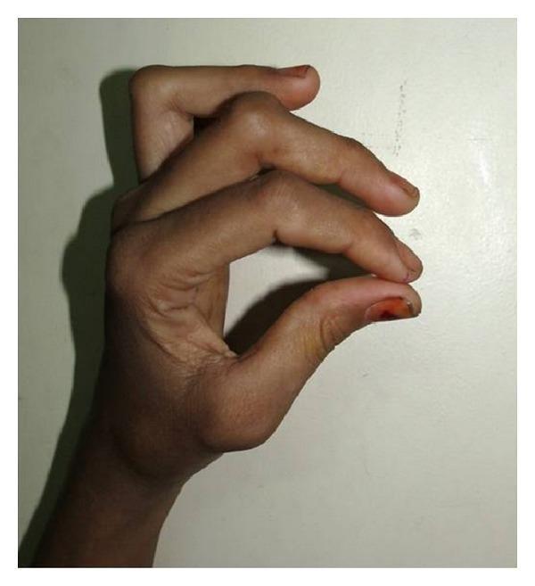
- Hypermobility test: passively extend 5th finger to >90° at MCP = one criterion of Beighton score
RESISTED TESTS (Tendon/Enthesis)
- Resisted wrist extension (elbow extended) → pain at lateral epicondyle = lateral epicondylitis (tennis elbow)
- Resisted wrist flexion → pain at medial epicondyle = medial epicondylitis (golfer's elbow)
3.2 ELBOW
Position: patient seated, examine from front and behind.
LOOK
- Para-olecranon groove: first place to see elbow joint effusion (fills the groove, making it convex)
- Olecranon bursa: discrete fluctuant swelling directly over olecranon = bursitis (gout, septic, trauma, RA)
- Rheumatoid nodules: firm, non-tender, subcutaneous, over olecranon and extensor surface
- Carrying angle: normal valgus ~10-15° women, ~5° men; increased = cubitus valgus (ulnar nerve risk); decreased = cubitus varus
FEEL
- Lateral epicondyle: tender = lateral epicondylitis
- Medial epicondyle: tender = medial epicondylitis; check ulnar nerve (tender, thickened) in groove posterior to medial epicondyle
- Para-olecranon groove (either side of olecranon): synovial swelling = joint disease
- Skin laxity: assess for hypermobility syndrome
MOVE
- Passive extension (normal = 0°; hyperextension = hypermobility)
- Passive flexion (normal ~145°)
- Supination/pronation (elbows at 90°, tucked in to stabilise shoulder): normal ~90° each way; restricted in radioulnar arthritis
3.3 SHOULDER
Position: patient standing or seated, expose both shoulders fully.
LOOK
- Asymmetry of scapulae, level of shoulders
- Muscle wasting: deltoid (axillary nerve, C5), supraspinatus/infraspinatus fossae (suprascapular nerve, rotator cuff tear)
- Step deformity at AC joint: ACJ disruption
- Swelling at anterior shoulder: glenohumeral effusion
FEEL
- Trapezius midpoint: tender spot = fibromyalgia, trapezius strain
- Supraspinatus (just posterior to the mid-clavicle): tender = rotator cuff pathology
- AC joint line: tender = ACJ OA, ACJ injury; step deformity
- Bicipital groove (anterior shoulder, arm slightly externally rotated): tender = bicipital tendinitis
MOVE
Active (watch for painful arc):
- Arms to the side, elevate in the plane of the scapula: painful arc 60-120° = rotator cuff impingement or subacromial bursitis
- Hands behind head (abduction + external rotation) = tests external rotation and abduction
- Hands behind back, reach up spine (adduction + internal rotation) = tests internal rotation; note highest spinal level reached (normally T6-T8)
Passive (stabilise scapula with one hand):
- Abduction (normal 180°), flexion (180°), extension (50°), external rotation (90° with elbow bent at side), internal rotation (70°)
Resisted (impingement tests):
- Neer's test: examiner passively forward-flexes the arm with elbow extended and forearm pronated, forcing the supraspinatus against the acromion. Pain = subacromial impingement
- Hawkins-Kennedy test: arm forward-flexed to 90°, elbow flexed 90°, examiner forcibly internally rotates the arm. Pain = subacromial impingement
- Empty can test (Jobe's): arm abducted 90° in scapular plane, 30° forward, thumb pointing down ("empty can"); examiner pushes down while patient resists. Weakness or pain = supraspinatus tear
- Resisted external rotation: elbow 90° at side; patient resists internal rotation. Weakness = infraspinatus/teres minor tear
- Speed's test: elbow extended, forearm supinated, arm at 60° flexion; resist further flexion. Bicipital groove pain = bicipital tendinitis
- Yergason's test: elbow flexed 90°, forearm pronated; patient supinates against resistance. Bicipital groove pain = long head of biceps pathology
3.4 CERVICAL SPINE
Position: patient seated.
LOOK
- Normal lordosis or loss/reversal (hyperextension secondary to thoracic kyphosis)
- Head position: tilted, rotated? (torticollis)
FEEL
- Percuss each spinous process: localised bony tenderness = fracture, infection, myeloma
- Paraspinal muscles: spasm, tenderness (trapezius, cervical paraspinals)
MOVE (active, guide to maximum with your hands)
| Movement | Normal Range | Key Finding |
|---|---|---|
| Flexion (chin to chest) | 80° | Reduced in spondylosis, RA, AS |
| Extension (look at ceiling) | 50° | Reduced/painful = facet joint OA, spondylosis |
| Rotation (chin to shoulder) | 80° each way | First movement lost in AS; also C1-C2 joint |
| Lateral flexion (ear to shoulder) | 45° | Reduced bilaterally = spondylosis; unilateral = acute disc |
Lateral rotation and extension lost early in ankylosing spondylitis affecting the cervical spine.
STRESS / SPECIAL TESTS
- Spurling's test (foraminal compression): tilt head to affected side + extend + apply downward pressure on crown. Reproduces radicular arm pain = cervical radiculopathy (foraminal stenosis)
- Distraction test: lift head gently upward - relief of radicular pain = positive
Always assess neurology (C5-T1 myotomes, reflexes, dermatomal sensation) when neck pathology is suspected.
3.5 THORACIC AND LUMBAR SPINE
Position: standing first, then lying prone/supine for special tests.
LOOK
- Kyphosis: increased thoracic kyphosis (osteoporosis, vertebral fractures, AS, Scheuermann's disease)
- Scoliosis: lateral curvature; ask patient to bend forward - if rib hump visible on bending = structural scoliosis; if disappears = functional (postural, leg length discrepancy)
- Loss of lumbar lordosis: muscle spasm (acute disc prolapse), AS
- Pelvic tilt: check level of ASIS and PSIS bilaterally
- Buttock and thigh wasting
FEEL
- Percussion along each spinous process: localised tenderness = fracture, infection, metastases, myeloma
- Paraspinal muscles: bilateral spasm = protective (acute disc) or chronic spondylosis
MOVE (Lumbar)
| Movement | Normal Range | Clinical Note |
|---|---|---|
| Forward flexion | Fingers to floor / flat palms | Measures mainly at L4-S1 |
| Extension (arch back) | ~30° | Painful = facet joint OA |
| Lateral flexion (side to side) | ~30° each | Asymmetric restriction = scoliosis, lateral disc |
| Rotation (fix pelvis with hands) | ~45° | Tested seated |
Schober's test (formal lumbar flexion measurement):
- Mark a point 10 cm above and 5 cm below the level of the posterior iliac spine dimples (dimples of Venus) while patient stands erect
- Ask patient to flex forward maximally
- Measure the new distance between the two marks
- Normal: increases by ≥5 cm (so original 15 cm should become ≥20 cm)
- Abnormal (<5 cm increase): restricted lumbar flexion = AS, spondylosis, lumbar OA
In normal hypermobile individuals, hands may lay flat on the floor; the Schober test specifically measures lumbar mobility, not hamstring flexibility.
STRESS / SPECIAL TESTS
Straight Leg Raise (SLR) - sciatic nerve stretch test:
- Patient supine; examiner slowly raises the straightened leg, holding ankle
- Normal: no pain until ~70-80°
- Positive: sciatica (radiating pain below knee in dermatomal distribution) reproduced between 30-70° = L4, L5, or S1 nerve root compression
- Sensitise: dorsiflex ankle at point of pain (Bragard's sign) - increases stretch on sciatic nerve, increases radicular pain
- Crossed SLR: raising the normal leg reproduces pain in the affected leg = large central disc prolapse
Femoral Nerve Stretch Test (reverse SLR):
- Patient prone; examiner holds ankle and passively flexes knee as far as possible
- Positive: pain in ipsilateral anterior thigh = L2, L3, or L4 nerve root tension (high lumbar disc prolapse)
Always assess neurology in lumbar spine examination:
| Root | Reflex | Motor | Sensation |
|---|---|---|---|
| L3 | Knee jerk | Knee extension (quadriceps) | Anterior/medial thigh |
| L4 | Knee jerk | Ankle dorsiflexion (tibialis anterior) | Medial lower leg |
| L5 | Nil | Great toe/ankle dorsiflexion; hip abduction | Lateral lower leg, dorsum of foot, great toe |
| S1 | Ankle jerk | Ankle plantarflexion (gastrocnemius); eversion | Lateral foot, little toe |
3.6 HIP
Position: patient supine on examination couch; gait already observed.
LOOK
- Wasting of buttock or thigh muscles (disuse)
- Leg position at rest: external rotation + shortening = fracture neck of femur; fixed flexion with external rotation = hip OA
- Measure leg length if pelvic tilt present:
- True leg length: ASIS to medial malleolus (structural shortening)
- Apparent leg length: umbilicus to medial malleolus (pelvic tilt causing apparent shortening)
FEEL
- Palpate for tenderness to locate the structure:
- Greater trochanter: trochanteric bursitis or gluteus medius tendinopathy (lateral hip pain in women, very common)
- Anterior hip/inguinal region: true hip joint pain (groin), hip flexor tendinopathy
- Ischial tuberosity: hamstring enthesopathy, ischial bursitis
- Pubic symphysis: pubic symphysitis (athletes)
MOVE
Passive movements (do not inflict pain):
| Movement | Method | Normal Range |
|---|---|---|
| Flexion | Knee flexed, hip flex as far as possible; watch for contralateral hip flexing (indicates tight hip flexors = positive Thomas test) | ~120° |
| Internal rotation | Hip and knee flexed to 90°; hold foot and move lower leg outward (internally rotates hip) | 40° |
| External rotation | Hip and knee flexed 90°; move lower leg inward (externally rotates hip) | 40° |
| Abduction | Stabilise contralateral ASIS; straighten leg, abduct | 45° |
| Adduction | Cross leg over midline | 25° |
| Extension | Patient prone (or side-lying); passively extend straightened leg | 20° |
Internal rotation is the first movement restricted in hip joint pathology (OA, AVN, hip effusion). Reproduce the patient's pain.
Thomas Test (fixed flexion deformity of hip):
- Flex both hips fully (eliminates lumbar lordosis)
- Hold the unaffected hip in full flexion
- Lower the affected leg - if it cannot reach the couch flat = positive fixed flexion deformity
- Measure the angle from the couch = degree of fixed flexion
FABER/Patrick's Test (Flexion-ABduction-External Rotation):
- Patient supine; place the ankle of the test leg on the opposite knee (figure-4 position)
- Gently press down on the flexed knee while stabilising the opposite ASIS
- Positive: hip or groin pain = hip joint pathology; SI joint pain (posterior) = sacroiliac joint pathology
Trendelenburg Test (hip abductor weakness):
- Patient stands on one leg (affected side); observe pelvis from behind
- Positive: pelvis drops to the contralateral side = weak gluteus medius (hip abductors) ipsilaterally
- Causes: hip OA, superior gluteal nerve palsy, L5 radiculopathy, hip fracture, coxa vara
3.7 KNEE
Position: patient supine; stand on the affected side.
LOOK
- Alignment:
- Valgus (knock-knees, genu valgum) = lateral compartment OA, RA
- Varus (bow-legs, genu varum) = medial compartment OA
- Flexion deformity: cannot fully extend at rest = fixed flexion deformity
- Swelling: effusion (suprapatellar pouch fullness), prepatellar bursitis, infrapatellar bursitis
- Wasting of quadriceps (vastus medialis specifically): even small effusions inhibit VMO by reflex
- Baker's cyst: visible or palpable in popliteal fossa (associated with knee OA, RA)
FEEL
- Temperature: use dorsum of hand; compare both sides; warm knee = active effusion/inflammation
- Patellar tap: compress suprapatellar pouch with one hand (to push fluid below patella), then push patella downward with two fingers of other hand; clunk = large effusion
- Bulge sign (fluid sweep/wipe test): for small effusions; stroke medial compartment upward (emptying it), then sweep down the lateral side; watch for medial bulge appearing = fluid returning to medial compartment = positive
- Joint line palpation: palpate medial and lateral joint lines with knee slightly flexed; tenderness = meniscus, OA, ligament
- Collateral ligaments: medial and lateral joint lines; tender = collateral ligament injury
MOVE
- Active: extend (should reach 0° or slight hyperextension) and flex (normal ~135°); assess end-feel
- Passive: same ranges; note any block to full extension (locked knee = bucket handle meniscal tear)
- Crepitus on movement: patellofemoral = fine crepitus; tibiofemoral OA = coarser
STRESS TESTS
Collateral Ligament Stress Tests:
- Knee in slight flexion (~20-30°); stabilise thigh
- Valgus stress (push tibia medially) = tests medial collateral ligament (MCL); opening on medial side = MCL tear
- Varus stress (push tibia laterally) = tests lateral collateral ligament (LCL); opening on lateral side = LCL tear
Cruciate Ligament Tests:
- Anterior Drawer Test: knee flexed 90°; sit on patient's foot; pull tibia forward. Anterior movement >5 mm = ACL tear
- Lachman's Test (more sensitive than anterior drawer): knee flexed 20°; stabilise femur with one hand, pull tibia anteriorly with other. Soft end-feel + anterior translation = ACL tear
- Posterior Drawer Test: same position as anterior drawer; push tibia posteriorly. Posterior movement = PCL tear
Meniscal Tests:
- McMurray's Test: patient supine, hip and knee flexed maximally; examiner holds heel and knee, rotate tibia externally while extending knee (tests medial meniscus); then internally while extending (tests lateral meniscus). Positive: palpable/audible click + joint line pain = meniscal tear
- Apley's Test (grinding): patient prone, knee flexed 90°; compress tibia onto femur (press down on heel) and rotate. Pain = meniscal tear. Then distract (pull heel up) and rotate - pain = ligamentous problem
3.8 FOOT AND ANKLE
Position: patient standing and walking first, then supine.
LOOK (standing + walking)
- Longitudinal arch: normally raised on medial side; pes planus (flat foot) = loss of arch; pes cavus (high arch) = hereditary neuropathy (CMT), polio
- Heel alignment: valgus (eversion, pronation) = pes planus; varus (inversion, supination) = pes cavus
- MTP joints: swelling separating toes ("daylight sign") = MTP synovitis (RA)
- Hallux valgus: lateral deviation of great toe at MTP joint (bunion) = OA, RA
- Claw toes: extension at MTP + flexion at PIP and DIP
- Hammer toe: PIP flexion + MTP extension (usually 2nd toe)
- Callosities under metatarsal heads = pressure from subluxed MTP joints (RA)
FEEL
- Squeeze across all MTPs together: composite tenderness = MTP synovitis (RA, psoriatic)
- Palpate individual MTPs: each joint line for swelling and tenderness
- Achilles tendon: tender insertional pain = enthesitis (AS, reactive arthritis, psoriatic); tender mid-substance = Achilles tendinopathy
- Plantar fascia origin (medial calcaneal tubercle): tender = plantar fasciitis
- Lateral ligaments (anterior talofibular + calcaneofibular): tender after inversion sprain
MOVE
- Ankle: active dorsiflexion (normal ~20°) and plantarflexion (normal ~50°)
- Subtalar joint (hindfoot): grasp heel between thumb and index finger; move heel medially (inversion, normal ~30°) and laterally (eversion, ~20°); restricted = subtalar OA, AS
- Midtarsal joint (Chopart's): hold heel fixed, rotate forefoot; restricted = midtarsal OA
- MTPs: passively flex and extend each toe; dorsiflexion of great toe >70° = normal
3.9 SACROILIAC JOINTS AND PELVIS
LOOK
- Assess from behind: pelvic tilt, buttock wasting, dimples of Venus (posterior iliac spines)
FEEL
- Direct sacral pressure (sit on sacrum): pain = SI joint inflammation or sacral fracture
- FABER test (as described in Hip section): posterior pain = SI pathology
STRESS TESTS
- FABER (Patrick's test): described above
- FADIR test: hip Flexion-ADduction-Internal Rotation; hip or posterior pain = posterior impingement, SI joint, piriformis
- Pelvic springing (distraction and compression): apply downward pressure on both ASIS simultaneously (springing the SI joints); pain = SI joint disease
STEP 4: INTERPRETING THE PATTERN - WHAT DOES THE DISTRIBUTION TELL YOU?
This is what transforms an examination from a collection of findings into a diagnosis.
Number of Joints
| Pattern | Distribution | Think of |
|---|---|---|
| Monoarthritis | 1 joint | Septic arthritis (exclude first!), crystal arthropathy (gout, pseudogout), trauma, haemarthrosis |
| Oligoarthritis | 2-4 joints, asymmetric | Psoriatic arthritis, reactive arthritis, enteropathic arthritis, early RA |
| Polyarthritis | ≥5 joints | RA (symmetric, small joints), psoriatic arthritis (asymmetric), OA (DIP/PIP/1st CMC), SLE, viral arthritis |
Joint Size
| Predominant involvement | Think of |
|---|---|
| Small joints (MCP, PIP, MTP, wrists) | RA, psoriatic arthritis, early OA |
| Large joints (knees, hips, shoulders) | OA, gout (can affect large joints), calcium pyrophosphate disease |
| Predominantly axial (spine + SI joints) | Ankylosing spondylitis, psoriatic arthritis, reactive arthritis, enteropathic arthritis |
Symmetry
| Pattern | Condition |
|---|---|
| Symmetric (both MCPs, both wrists) | RA, SLE |
| Asymmetric | Psoriatic arthritis, reactive arthritis, gout |
| Predominantly DIP involvement | OA (Heberden's nodes), psoriatic arthritis |
| MCP + PIP (sparing DIP) | RA |
| DIP + 1st CMC | OA |
The "Hand at a Glance" Diagnoses
| Feature | Diagnosis |
|---|---|
| MCP ulnar deviation + swan-neck/boutonnière + wrist synovitis | Rheumatoid arthritis |
| DIP involvement + nail pitting/onycholysis + asymmetric | Psoriatic arthritis |
| Heberden's nodes (DIP) + Bouchard's nodes (PIP) + 1st CMC squaring | OA |
| Tophi (chalky white deposits) over joints + acute monoarthritis (1st MTP) | Gout |
| Raynaud's + puffy fingers + tight skin + calcinosis | Systemic sclerosis |
| Gottron's papules over MCPs + proximal myopathy + heliotrope rash | Dermatomyositis |
| Dactylitis (sausage digit) | Psoriatic or reactive arthritis |
STEP 5: HYPERMOBILITY - BEIGHTON SCORE
Test in any patient with widespread joint pain and "double-jointed" history.
| Manoeuvre | Points (1 per side, except 1) |
|---|---|
| Passive hyperextension of 5th finger >90° | 1 point each side = 2 |
| Passive apposition of thumb to forearm | 1 point each side = 2 |
| Hyperextension of elbow >10° | 1 point each side = 2 |
| Hyperextension of knee >10° | 1 point each side = 2 |
| Touch floor with palms flat (knees straight) | 1 point = 1 |
| Total | /9 |
Score ≥5/9 = generalised hypermobility. Combined with joint pain = Benign Joint Hypermobility Syndrome (now termed Hypermobile Ehlers-Danlos Syndrome in the spectrum).
STEP 6: PAIN CHARACTERISATION SUMMARY
| Pain Type | Characteristics | Cause |
|---|---|---|
| Articular | Pain in all directions; swelling at joint line; morning stiffness | Synovitis (RA, PsA), crystal, OA |
| Periarticular | One plane of active movement painful; passive full ROM; palpation of structure painful | Tendinitis, bursitis, enthesitis |
| Neurogenic | Dermatomal/nerve territory; dysaesthesia; Tinel's sign positive; normal local joint exam | Radiculopathy, carpal tunnel, ulnar syndrome |
| Referred | Normal local joint exam; uncharacteristic pattern; associated visceral symptoms | Hip disease referred to knee, cardiac to shoulder |
| Widespread/central | "All over"; tender points; sleep disturbance; normal inflammatory markers and imaging | Fibromyalgia |
Source: Rheumatology, 2-Volume Set (2022, Elsevier); Harrison's Principles of Internal Medicine 22E (2025, McGraw Hill Medical)
This is a shared conversation. Sign in to Orris to start your own chat.