
Can give saq for 6 marks on salivary secretion and its regulation
Reading File
Now let me search the medical library for salivary secretion content.
Finding Sources
Finding Sources
Good findings. Let me now read the most relevant sections - Costanzo (Salivary Secretion + Regulation), Guyton (Nervous Regulation), and Ganong's.
Reading File
Reading File
Reading File
I have excellent content from three authoritative textbooks. Let me also quickly read the Costanzo regulation section in full since it was specifically tagged.
Reading File
Loading Image
I now have comprehensive information from multiple authoritative textbooks. Here is a well-structured 6-mark SAQ on salivary secretion and its regulation.
SAQ: Salivary Secretion and Its Regulation (6 Marks)
Introduction / Overview (0.5 mark)
Saliva is produced by three pairs of salivary glands - parotid, submandibular, and sublingual - at a rate of 1,000-1,500 mL/day. It is hypotonic compared to plasma and plays roles in digestion (amylase, lingual lipase), lubrication (mucins), buffering (HCO3-), and antibacterial protection (IgA, lysozyme).
Mechanism of Salivary Secretion - Two-Step Process (1.5 marks)
Step 1 - Primary (Acinar) Secretion
- Acinar cells produce an isotonic, plasma-like primary saliva containing water, electrolytes, enzymes, and mucus.
- The ionic composition is similar to plasma at this stage (Na+, Cl- predominant).
Step 2 - Ductal Modification
- As saliva passes through the striated ducts, ductal cells actively modify the composition:
- Na+ and Cl- are reabsorbed (active transport)
- K+ and HCO3- are secreted into the lumen
- Ducts are relatively impermeable to water, so the loss of NaCl makes saliva hypotonic
- At low flow rates: more time for NaCl reabsorption → more hypotonic saliva
- At high flow rates: less reabsorption time → saliva approaches plasma tonicity (but remains slightly hypotonic)
Result: Final saliva has - higher K+, higher HCO3-, lower Na+, lower Cl- compared to plasma.
Regulation of Salivary Secretion (3 marks)
Key Unusual Features
Two features make salivary regulation unique among GI secretions:
- It is exclusively under neural (autonomic) control - unlike other GI secretions which are also under hormonal control.
- Both parasympathetic AND sympathetic stimulation increase secretion (normally they are antagonistic).
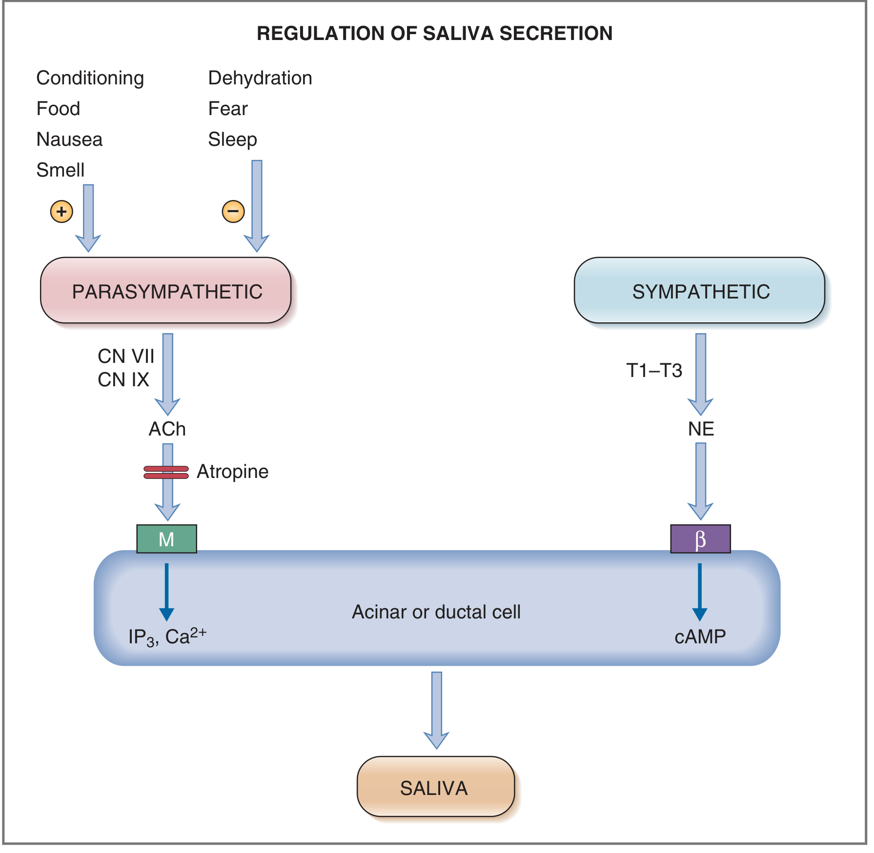
1. Parasympathetic Regulation (Dominant)
| Feature | Detail |
|---|---|
| Nerves | CN VII (facial) and CN IX (glossopharyngeal) |
| Nuclei | Superior and inferior salivatory nuclei (pons-medulla junction) |
| Postganglionic transmitter | Acetylcholine (ACh) |
| Receptor | Muscarinic (M) receptors on acinar and ductal cells |
| Intracellular messenger | IP3 → ↑ intracellular Ca2+ |
| Effect | Large volume of watery saliva; ↑ HCO3- and enzymatic content; myoepithelial cell contraction |
Stimuli that increase parasympathetic activity:
- Food in the mouth (taste and tactile stimuli - especially sour taste)
- Smell and sight of food
- Nausea
- Conditioned reflexes (Pavlov's dogs)
- Irritating food in stomach/upper GI
Stimuli that decrease parasympathetic activity:
- Fear
- Sleep
- Dehydration
2. Sympathetic Regulation
| Feature | Detail |
|---|---|
| Origin | Preganglionic: T1-T3 spinal segments |
| Ganglion | Superior cervical ganglion |
| Postganglionic transmitter | Norepinephrine (NE) |
| Receptor | Primarily β-adrenergic receptors on acinar/ductal cells |
| Intracellular messenger | ↑ cAMP (via adenylyl cyclase) |
| Effect | Smaller volume; thicker, more viscous saliva with more proteinaceous content |
Note: α-adrenergic receptors are also activated but β-adrenergic activation is considered dominant.
3. Blood Flow - Secondary Factor (0.5 mark)
Salivary glands have exceptionally high blood flow (>10x that of exercising skeletal muscle per gram of tissue). When parasympathetic nerves are activated:
- Blood vessels dilate, supporting secretion
- Activated salivary cells release kallikrein, which cleaves α2-globulin to form bradykinin - a potent vasodilator that further increases glandular blood flow
Diagram
Summary Box (Quick Recall)
| Parasympathetic | Sympathetic | |
|---|---|---|
| Nerve | CN VII, CN IX | T1-T3 via superior cervical ganglion |
| Neurotransmitter | ACh | Norepinephrine |
| Receptor | Muscarinic | β-adrenergic |
| Second messenger | IP3 / Ca2+ | cAMP |
| Volume | Large (watery) | Small (thick) |
| Dominant? | YES | No |
Sources: Costanzo Physiology 7th Ed., p. 361-364 | Guyton & Hall Textbook of Medical Physiology, p. 800-801 | Ganong's Review of Medical Physiology 26th Ed., p. 459
Can you include diagrams
Let me fetch all the relevant diagrams from the textbooks for you.
Loading Image
Loading Image
Loading Image
Loading Image
All four diagrams retrieved. Here is the complete SAQ with all textbook diagrams embedded:
SAQ: Salivary Secretion and Its Regulation (6 Marks)
Introduction (0.5 mark)
Saliva is produced by three pairs of salivary glands - parotid, submandibular, and sublingual - at a rate of 1,000-1,500 mL/day. It is hypotonic relative to plasma and serves digestion (amylase, lingual lipase), lubrication (mucins), buffering (HCO3-), and antibacterial protection (IgA, lysozyme).
Mechanism of Salivary Secretion - Two-Step Process (1.5 marks)
Step 1 - Acinar (Primary) Secretion
- Acinar cells produce an isotonic, plasma-like primary saliva containing water, electrolytes, enzymes, and mucus.
Step 2 - Ductal Modification
- As saliva flows through the striated ducts, ductal cells modify it:
- Na+ and Cl- are reabsorbed (active transport)
- K+ and HCO3- are secreted into the lumen
- Ducts are impermeable to water → loss of NaCl renders saliva hypotonic
- At low flow rates: more NaCl reabsorbed → very hypotonic saliva
- At high flow rates: less reabsorption time → approaches plasma tonicity (still slightly hypotonic)
Diagram 1 - Mechanism of Salivary Secretion (Costanzo Physiology)
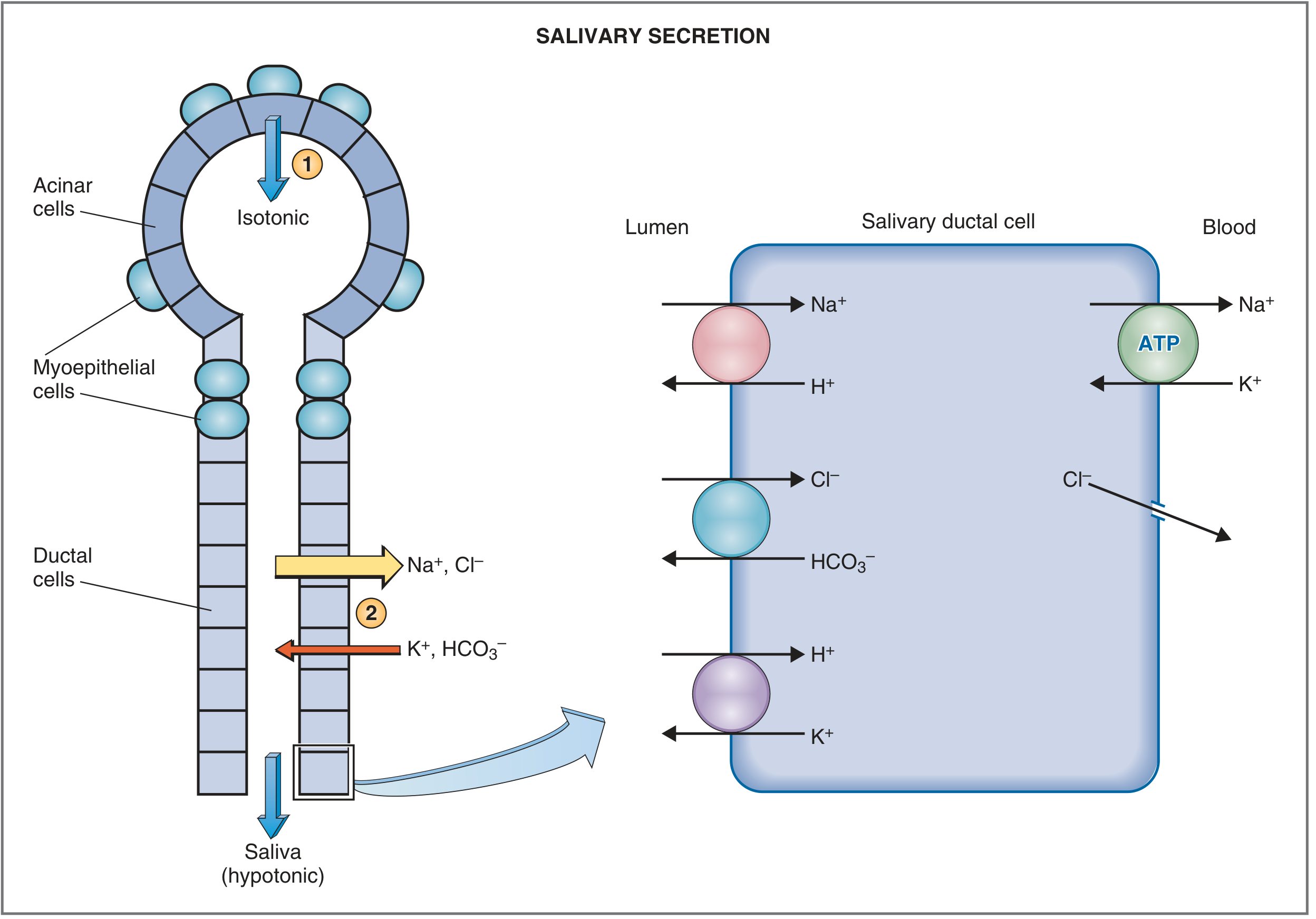
Fig. 8.12 - Costanzo Physiology 7th Ed. Acinar cells produce isotonic primary saliva (Step 1); ductal cells reabsorb Na+/Cl- and secrete K+/HCO3- to yield hypotonic final saliva (Step 2).
Regulation of Salivary Secretion (3 marks)
Two Unique Features
- Salivary secretion is exclusively under autonomic neural control - unlike other GI secretions which are also hormonally regulated.
- Both parasympathetic AND sympathetic stimulation increase secretion - this is unusual since the two divisions normally have opposing effects. Parasympathetic control is dominant.
1. Parasympathetic Regulation (Dominant)
| Feature | Detail |
|---|---|
| Cranial nerves | CN VII (facial - via chorda tympani) and CN IX (glossopharyngeal) |
| Salivatory nuclei | Superior and inferior salivatory nuclei at pons-medulla junction |
| Ganglia | Submandibular ganglion (for submandibular + sublingual); Otic ganglion (for parotid) |
| Neurotransmitter | Acetylcholine (ACh) |
| Receptor | Muscarinic (M) receptors |
| Second messenger | IP3 → ↑ intracellular Ca2+ |
| Effect | Large volume of watery saliva; ↑ HCO3- and enzymatic content; myoepithelial contraction |
Stimuli that INCREASE parasympathetic activity (+):
- Food (taste/tactile stimuli - especially sour taste: 8-20x basal rate)
- Smell and sight of food
- Nausea / GI irritation
- Conditioned reflexes (Pavlov's experiment)
Stimuli that DECREASE parasympathetic activity (-):
- Fear, sleep, dehydration, fatigue
Diagram 2 - Parasympathetic Neural Pathways (Guyton & Hall)
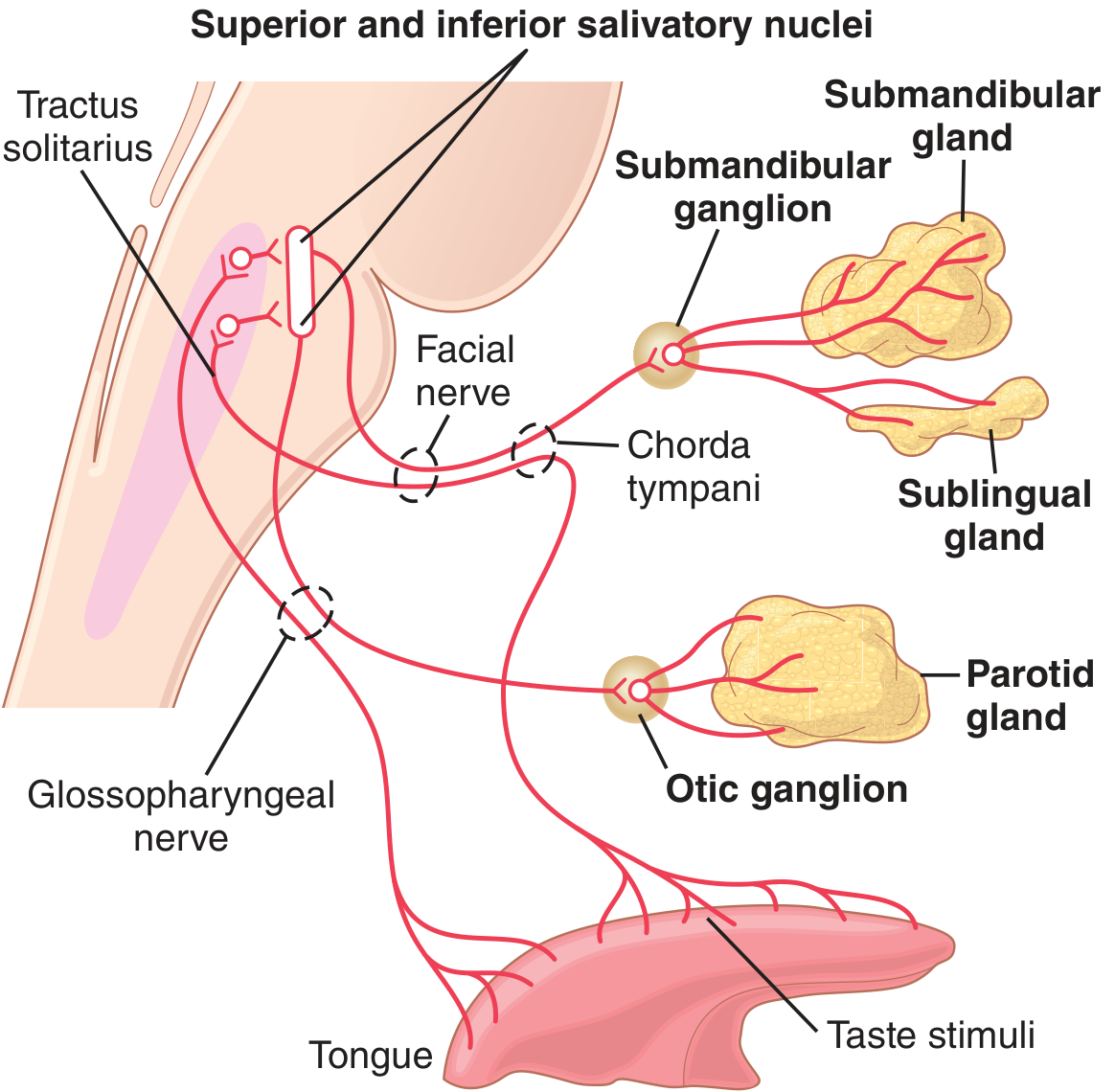
Fig. 65.3 - Guyton & Hall Textbook of Medical Physiology. Parasympathetic pathways from salivatory nuclei via CN VII and CN IX to submandibular, sublingual, and parotid glands.
Diagram 3 - Regulation by Higher Centers (Ganong's)
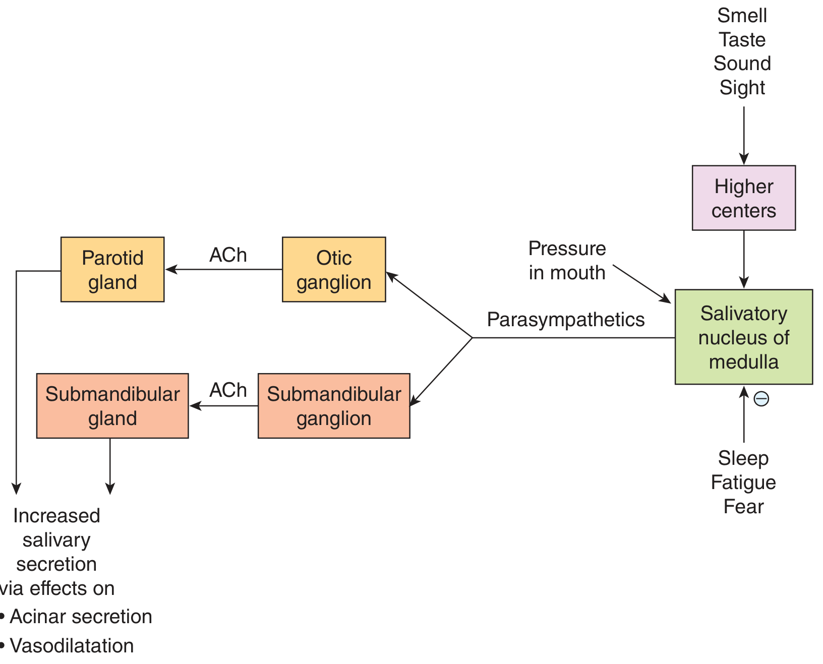
Fig. 25-4 - Ganong's Review of Medical Physiology 26th Ed. Shows higher centers (smell, taste, sight, sound) stimulating the salivatory nucleus, and how sleep/fear/fatigue inhibit it.
2. Sympathetic Regulation
| Feature | Detail |
|---|---|
| Origin | Preganglionic: T1-T3 spinal segments |
| Ganglion | Superior cervical ganglion |
| Postganglionic path | Travels along blood vessel walls to glands |
| Neurotransmitter | Norepinephrine (NE) |
| Receptor | Primarily β-adrenergic receptors (also α-adrenergic) |
| Second messenger | ↑ cAMP (via adenylyl cyclase) |
| Effect | Smaller volume; thick, viscous, proteinaceous saliva |
Diagram 4 - Autonomic Regulation Summary (Costanzo Physiology)
Fig. 8.14 - Costanzo Physiology 7th Ed. Complete summary of autonomic regulation. Note: Atropine (muscarinic blocker) inhibits the parasympathetic pathway.
3. Vascular (Secondary) Factor (0.5 mark)
- Salivary glands have the highest blood flow per gram of any organ (>10x that of exercising skeletal muscle)
- Parasympathetic stimulation triggers vasodilation
- Activated salivary cells secrete kallikrein → cleaves α2-globulin → forms bradykinin (potent vasodilator) → further increases blood flow to support secretion
Quick Comparison Table
| Parasympathetic | Sympathetic | |
|---|---|---|
| Nerve | CN VII, CN IX | T1-T3 → superior cervical ganglion |
| Neurotransmitter | ACh | Norepinephrine |
| Receptor | Muscarinic (M) | β-adrenergic |
| Second messenger | IP3 / Ca2+ | cAMP |
| Saliva volume | Large | Small |
| Saliva type | Watery | Thick / viscous |
| Dominant? | YES | No |
Sources: Costanzo Physiology 7th Ed. pp. 361-364 | Guyton & Hall Medical Physiology pp. 800-801 | Ganong's Review of Medical Physiology 26th Ed. p. 459
This is a shared conversation. Sign in to Orris to start your own chat.