

Oral herpes management
herpes labialis treatment acyclovir valacyclovir
herpes labialis cold sore lip vesicles HSV-1
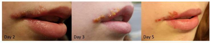
This clinical progression series consists of three photographs documenting the natural history of Herpes Labialis (cold sore) on the upper lip and oral commissure. The first panel (Day 2) illustrates the vesicular stage, characterized by clusters of small, fluid-filled, translucent blisters on an erythematous base along the vermillion border. The second panel (Day 3) depicts the transition to the weeping and crusting stage, where vesicles have ruptured and coalesced into a thicker, yellowish-brown serosanguinous crust. The third panel (Day 5) demonstrates the scab formation and early healing phase, showing a more defined, dry reddish-brown scab that has begun to contract at the edges, with some areas of re-epithelialization visible. These images serve as a primary educational resource for medical students and clinicians to recognize the dermatological morphology of labial herpes simplex virus (HSV-1) infection throughout its standard temporal evolution.
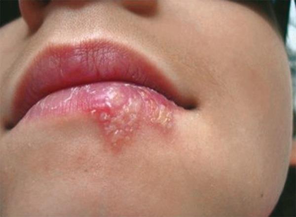
This clinical photograph shows a close-up of a patient's lower lip and perioral region, demonstrating a cluster of herpetic lesions consistent with Herpes Labialis (cold sores). The primary findings include a group of small, fluid-filled vesicles on an erythematous (reddened) base. These vesicles are concentrated on the vermilion border and extend onto the adjacent cutaneous surface of the lower lip. The vesicles exhibit varying degrees of opacity, with some appearing clear and others becoming cloudy as they progress. Mild perilesional inflammation (hyperemia) is visible. In contrast, the upper lip appears normal in color and texture with no active lesions. This image captures the vesicular stage of a recurrent herpes simplex virus (HSV-1) infection, illustrating typical morphology, including clustering, vesicle formation, and location at the mucocutaneous junction. It serves as an educational example for dermatological and oral pathology assessment, highlighting the physical signs used to diagnose and monitor the progression or healing of labial herpes.
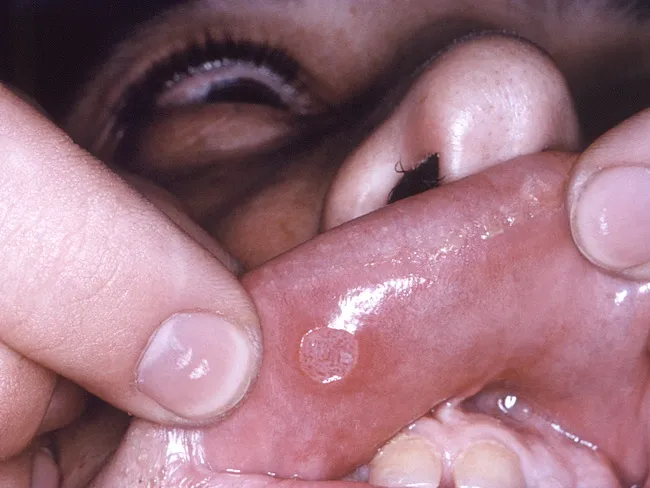
Clinical photograph of an active herpes simplex infection on the lip (herpes labialis). The image demonstrates a localized perioral mucocutaneous lesion. The primary lesion begins as erythematous papules that rapidly coalesce into grouped vesicles; vesicles are fragile, rupture easily, leaving shallow erosions on a reddened (erythematous) base. Some vesicles may appear pustular. The distribution is typically confined to the lips and perioral skin on the vermillion border, consistent with HSV-1 reactivation; seasonal or stress triggers; contagious; high risk of spread via kissing or shared objects. The lesion is often preceded by prodromal tingling or burning. On exam, erythema and swelling may be present; rupture leads to crusting in older lesions. The image is a close-up frontal view of the perioral region, captured with standard clinical photography settings; no contrast agents or staining required. Clinically, herpes labialis is diagnosed by appearance, but PCR or viral culture from vesicular fluid can confirm HSV-1 infection. Differential diagnoses include impetigo, aphthous ulcers, contact dermatitis, and herpes gladiatorum if extraneous sites; management includes antiviral therapy such as acyclovir or valacyclovir, topical analgesics, adequate hydration, and patient education about contagion. This image is useful for education, diagnosis practice, dermatology training, and clinical scenario databases.
Image Assessment
Background
- Causative agent: HSV-1 (occasionally HSV-2)
- Pathophysiology: Primary infection often occurs in childhood (gingivostomatitis); the virus establishes latency in the trigeminal ganglion. Reactivation is triggered by UV light, fever, stress, immunosuppression, or local trauma.
- Recurrences present as the classic prodrome of tingling/burning, followed by vesicle formation along the lip margin, which heals completely within ~10 days.
- The lesion is contagious - viral shedding occurs from active lesions through the crusting stage.
Treatment
1. Episodic (Outbreak) Treatment
| Drug | Dose | Duration | Notes |
|---|---|---|---|
| Valacyclovir | 2 g orally twice a day | 1 day only | FDA-approved; convenient single-day regimen |
| Famciclovir | 1500 mg single dose OR 750 mg twice daily | 1 day | Equally effective |
| Acyclovir | 400 mg orally 5 times/day | 4-5 days | Older regimen; still effective |
Oral antivirals are significantly more effective than topical agents. - Harrison's Principles of Internal Medicine 22E
- Penciclovir 1% cream - apply every 2 hours while awake for 4 days
- Docosanol 10% cream - apply 5 times/day; FDA-approved OTC option; reduces healing time by ~1 day
- Acyclovir 5% cream - apply 5 times/day
2. Suppressive Therapy (for frequent recurrences: ≥6/year)
| Drug | Dose |
|---|---|
| Acyclovir | 400 mg orally twice daily (long-term) |
| Valacyclovir | 500 mg once daily OR 1 g once daily |
- Also appropriate as prophylaxis just before known triggers (e.g., before intense UV exposure or a dental procedure).
3. Primary Infection (Gingivostomatitis)
| Drug | Dose | Duration |
|---|---|---|
| Acyclovir | 200 mg 5x/day OR 400 mg 3x/day | 7-10 days or until resolution |
| Valacyclovir | 1 g twice daily | 7-10 days |
| Famciclovir | 250 mg three times daily | 7-10 days |
| Acyclovir IV | For severely ill/immunocompromised | Hospital setting |
4. Immunocompromised Patients
- IV Acyclovir is required for severe or disseminated infection.
- Suppressive therapy is often continued long-term.
- Watch for acyclovir-resistant HSV (suspect if no response); treat with foscarnet or cidofovir.
5. Supportive Care
- Keep the area clean and dry
- Avoid kissing, sharing utensils, or touching the sore
- Apply sunscreen/lip balm with SPF to the area to reduce UV-triggered recurrences
- Analgesics (topical or oral) for pain relief
- Do NOT squeeze or rupture vesicles (increases spread risk)
Mechanism of Action (Antivirals)
- Selective phosphorylation by viral thymidine kinase (in infected cells only)
- Further phosphorylation by cellular kinases to the active triphosphate form
- Inhibition of viral DNA polymerase and chain termination
Differential Diagnoses to Exclude
| Condition | Distinguishing Feature |
|---|---|
| Angular cheilitis | Fissuring/scaling at commissure, no true vesicles; often fungal/bacterial |
| Impetigo | Rapidly ruptures to honey-colored crust; not grouped vesicles |
| Aphthous ulcers | Inside the mouth, on non-keratinized mucosa; not at vermilion border |
| Contact dermatitis | No vesicle clustering; history of allergen exposure |